Introduction
The presence of thyroid tissue in the tracheal lumen is normally only associated with locally advanced thyroid cancer, in which context it is a poor prognostic indicator.Reference McCarty, Kuhn, Williams, Ellenhorn, O'Brien and Preskitt1
However, there have been rare reports of implantation of thyroid tissue following blunt trauma,Reference Harach, Cabrera and Williams2 fine needle aspiration biopsyReference Ito, Tomoda, Uruno, Takamura, Kobayashi and Matsuzuka3, Reference Tamiolakis, Antoniou, Venizelos, Lambropoulou, Alexiadis and Ekonomou4 and endoscopic thyroid surgery.Reference Lee, Yun, Jeong, Nam, Chung and Park5
Ectopic thyroid tissue has also been reported at various sites between the foramen caecum and mediastinum,Reference De Felice and Di Lauro6 and even in the trachea.Reference Donegan and Wood7
We present a case of multinodular goitre arising in thyroid tissue within the trachea, in a patient who had undergone subtotal resection of the thyroid for follicular adenoma some 40 years earlier. The thyroid tissue appears to have been implanted at the time of this operation. To the best of our knowledge, intra-operative thyroid tissue implantation in the trachea and subsequent goitre development has not previously been described.
A literature search was conducted using the PubMed database, using key words ‘intratracheal’ and ‘ectopic thyroid’. The bibliographies of relevant articles were also searched. English language articles were selected.
Case report
A 79-year-old, non-smoking woman presented to our department with a 12-month history of dyspnoea and a six-month history of increasing hoarseness and stridor. She complained of occasional right-sided throat pain but denied odynophagia and dysphagia. She had been treated for asthma by her general practitioner.
Forty years earlier, the patient had been treated for goitre with a subtotal thyroid resection performed under local anaesthesia. Histology reports from that time described a well defined, encapsulated follicular adenoma in the upper half of the left lobe of the thyroid, together with a nodular colloid goitre. A number of aspirations of what may have been a seroma took place post-operatively, followed by re-exploration of the wound and removal of a suture.
During the current presentation, clinical examination showed a midline neck swelling immediately above the previous thyroidectomy scar, together with moderate, biphasic stridor.
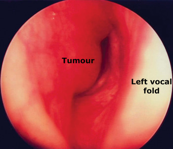
Flexible nasoendoscopy revealed normal vocal folds but 80–90 per cent narrowing of the upper tracheal airway due to intraluminal tissue on the right (Figure 1).
Endoscopic view of the right-sided intratracheal tumour.

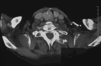
Spirometry, thyroid function tests and oesophagoscopy were all normal. Computed tomography scanning showed tumour in the region of the right lobe of the thyroid, which extended into and narrowed the trachea (see Figure 2).
Axial computed tomography scan demonstrating tumour in the region of the right lobe of the thyroid, which extends into and narrows the trachea. R = right

The patient underwent microlaryngoscopy, biopsy and CO2 laser debulking of the intraluminal tumour.
Histologically, the biopsy featured follicles of varying size lined by follicular epithelium and stromal fibrosis, consistent with a diagnosis of multinodular goitre. There was no evidence of malignancy.
Definitive excision of the right thyroid lobe tumour and cricotracheal resection with anastomosis were then performed. A modified Gluck Sorensen incision was utilised for surgical exposure. Thyroid tissue was progressively ‘shaved’ off the trachea, using a scalpel, until the trachea could be clearly identified. A clear plane between the thyroid and the oesophagus was noted. The trachea was divided inferiorly between the second and third rings, and superiorly to incorporate 3–4 mm of the lower border of the cricoid on the right, and through the cricotracheal membrane on the left. Both recurrent laryngeal nerves were preserved. Drill holes were placed in the cricoid cartilage, and the trachea was then anastomosed. Posteriorly interrupted 3.0 Novafil sutures were placed with the head in a flexed position, and anterior closure was completed with a continuous 3.0 Novafil suture. A Grillo suture was placed from the chin to the chest. The patient was extubated post-operatively and made an uneventful recovery.
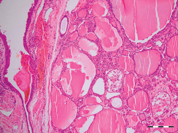
On macroscopic examination, the tumour was found to have arisen from the trachea. Histologically, the tumour was composed of nodular thyroid tissue, with Hurthle cell change and colloid-containing follicles varying in size and shape. There was no evidence of vascular invasion (see Figure 3). The findings were consistent with a diagnosis of multinodular goitre.
High power photomicrograph of benign thyroid tissue in the tracheal wall. (H&E; bar = 200 µm)

The patient made an unremarkable recovery. She was followed up for two years post-operatively and remained well, with no symptoms of airway obstruction. Vocal fold movements were symmetrical and her voice was normal. The upper trachea remained free of stenosis and recurrence.
Discussion
With the exception of malignancy, thyroid tissue is only rarely found in the trachea. The most commonly reported cause is an ectopic thyroid, when the thyroid fails to descend to its normal position during embryological development. Intratracheal ectopic thyroid was initially described by Ziemssen in 1875, and was first managed surgically by Heise in 1888, using tracheal fissure and curettage.
Ziemssen and von BrunsReference Beck8 proposed the malformation theory to explain the presence of ectopic thyroid: the developing thyroid is divided by the developing trachea and its cartilages, thereby creating nests of cells in the lumen. The rival ingrowth theory, proposed by Pultauf, suggests that the late fetal or postnatal thyroid tissue directly invades the laryngotracheal cartilage due to the developmental failure of mesenchymal tissue between the thyroid and trachea.
Ectopic thyroid tissue size is thought to be related to a number of factors, including diet, hormone levels and previous total thyroidectomy.Reference Byrd, Thompson and Wieneke9 There is a higher incidence among females.Reference Brandwein, Som and Urken10 Ectopic tissue can remain subclinical for years;Reference Dowling, Johnson, Collier and Dillarg11 patients normally present between the ages of 20 and 50 yearsReference Brandwein, Som and Urken10 with stridor, cough, dyspnoea or hoarseness.Reference Byrd, Thompson and Wieneke9
The majority of ectopic thyroid specimens are benign or display adenomatous change. However, malignant change can occur in up to 11 per cent of cases,Reference Dowling, Johnson, Collier and Dillarg11 most commonly papillary carcinoma.Reference Hari, Brown and Thompson12 Seventy-five per cent of cases are associated with an external goitre.Reference Dowling, Johnson, Collier and Dillarg11 Neoplastic change within endotracheal ectopic thyroid tissue remains rare, and represents less than 1 per cent of all primary endotracheal tumours.Reference Byrd, Thompson and Wieneke9
In this case report, the thyroid tissue was probably directly implanted into the tracheal lumen during the patient's initial treatment for follicular adenoma. While details of that surgical procedure were not available, implantation probably occurred either during surgery or during subsequent seroma aspirations. The resultant remnant remained quiescent for 40 years before enlarging and becoming symptomatic.
Cases of thyroid tissue implantation following blunt trauma,Reference Harach, Cabrera and Williams2 fine needle aspiration biopsyReference Ito, Tomoda, Uruno, Takamura, Kobayashi and Matsuzuka3, Reference Tamiolakis, Antoniou, Venizelos, Lambropoulou, Alexiadis and Ekonomou4 and endoscopic thyroid surgeryReference Lee, Yun, Jeong, Nam, Chung and Park5 have been reported, but are rare. It is postulated that rupture of the thyroid tissue leads to its implantation.Reference Harach, Cabrera and Williams2
• Intratracheal thyroid tissue is rare, and its pathogenesis is unexplained
• This case of intratracheal thyroid tissue presented as a multinodular goitre, 40 years after subtotal thyroidectomy for follicular adenoma
• The tissue was probably implanted during this operation or subsequent needle aspirations, remained quiescent for decades, then developed into a goitre
• Thyroid surgery must involve meticulous tumour control and careful maintenance of normal anatomy
There are only five documented cases of intratracheal thyroid tissue associated with previous thyroidectomy.Reference Byrd, Thompson and Wieneke9, Reference Dowling, Johnson, Collier and Dillarg11, Reference Osammor, Bulman and Blewitt13–Reference Abboud, Tabchy, Atallah, Aouad, Matar and Ghorra15 These patients presented four to 21 years after their original thyroid surgery, with dyspnoea or stridor. The tissue was resected or biopsied in all cases, and found to contain benign thyroid tissue. The authors attribute the presence of the intratracheal thyroid to ectopic tissue which may have undergone ‘compensatory hyperplasia’Reference Dowling, Johnson, Collier and Dillarg11 subsequent to thyroid surgery, perhaps in response to increased thyroid-stimulating hormone levels.Reference Karakullukcu, Guvenc, Cansiz, Oktem and Oz14
Intra-operative thyroid tissue implantation in the trachea with subsequent symptomatic goitre development has not previously been described. The current case illustrates the need for careful tissue handling during implantation surgery, and precise technique during needle aspiration biopsy. Maintaining the integrity of normal anatomical structures during these procedures will help prevent the rare complication of thyroid implantation.