Introduction
Ascites syndrome, a metabolic disorder, is becoming one of the most noticeable, non-infectious causes of losses in the broiler industry worldwide via either increased mortalities ranging from 5 to 7% or from large scale condemnation of carcasses at slaughter (De Smit et al., Reference DE SMIT, TONA, BRUGGEMAN, ONAGBESAN, HASSANZADEH, ARCKENS and DECUYPERE2005). In developing countries the demand for meat and eggs is increasing continuously to meet the consumer demands, and more birds are kept in confined areas leading to increased pressure on poultry health status.
Genetically, the modern broiler is very prone to develop this illness. This is probably due to the bird's exceedingly fast growth rate and high oxygen demand. Selection for rapid growth and efficient feed conversion has resulted in a broiler with such a high rate of metabolism that its heart and lungs are barely capable of producing enough oxygen to sustain the body. Inadequate oxygen levels trigger a series of events, including peripheral vasodilatation, increased cardiac output and pulmonary arterial pressure, and right ventricular hypertrophy (Wideman and Bottje, Reference WIDEMAN and BOTTJE1993; Bottje and Wideman, 1995). The right ventricular hypertrophy that occurs in response to the increased work-load by the right ventricle leads pulmonary hypertension (Julian, Reference JULIAN1987). Broilers susceptible to pulmonary hypertension undergo patho-physiological progression leading sequentially to right-side congestion, pressure-induced cirrhosis of the liver, and transudation of fluid into the abdominal cavity (Peacock et al., Reference PEACOCK, PICKETT, MORRIS and REEVES1989, Reference PEACOCK, PICKETT, MORRIS and REEVES1990; Julian, Reference JULIAN1993).
Etiology
There are many factors that cause ascites, they are listed as follows:
• High and low altitude: Increased incidence at high altitude is related to the elevated oxygen requirement to sustain rapid growth and the inability of heart and lungs to deliver adequate amount of oxygen to the tissues in order to maintain the nutritional and growth rate potential. Ascites is observed at low altitude where it is associated with poor ventilation and respiratory diseases (Teuscher et al., Reference TEUSCHER, LOPEZ and ALVAREZ1971; Huchzermeyer, Reference HUCHZERMEYER1986; Olkowski and Classen, Reference OLKOWSKI and CLASSEN1998).
• Cold: Cold is an important factor. Cold ambient temperatures tend to increase T3 concentration, which is required for the generation of additional metabolic heat to maintain body temperature in the cooler environment. The subsequent increase in metabolic rate results in an increase in blood pressure as the heart attempts to maintain oxygen supply to the organs and muscles, thus leading to pulmonary hypertension and right ventricular failure (Julian et al., Reference JULIAN, MC MILLAN and QUINTON1989; Acar et al., Reference ACAR, SIZEMORE, LEACH, WIDEMAN, OWEN and BARBOLO1995).
• Heat: Heat also causes an increased demand for oxygen, and thereby increases the incidence of ascites (Tattori et al., Reference TATTORI, YAMAGUCHI, TAKE, UCHIDA and TAETEYAMAS1995).
• Age: Broilers from three to seven weeks of age are more susceptible to ascites (Kamindjolo et al., Reference KAMINDJOLO, WAMUKOYA and NYAGA1978; Banday and Maqbool, Reference BANDAY and MAQBOOL1994).
• Sex: Males are more susceptible than females (Coello et al., Reference COELLO, ODOM and WIDEMAN1985; Wideman et al., 1997).
• Breed: Fast growing breeds require more oxygen to sustain the rapid growth rate as compared to slow growing breed and they have a small lung volume: body growth ratio, which causes hypoxemia so predisposing them for ascites syndrome (Julian, Reference JULIAN1989).
• House environment: Ammonia formation and dust increases the incidence of ascites. Diseases causing micro-organisms can be transferred through dust particles, causing irritation or infection to lungs and leads to reduce oxygen transfer between the birds and the environment (McGovern et al., Reference MCGOVERN, FEDDES, ROBINSON and HANSON1998).
• Light: Continuous lighting increases and intermittent lighting reduces the incidence of ascites in broilers (Gordon, Reference GORDON1997; Buys et al., Reference BUYS, BUYSE, LADMAKHI and DECUYPERE1998).
• Nutritional factors: High energy diets and pelleted feed increases the incidence of ascites syndrome. Diet containing T3, high sodium and low phosphorus, vitamin C, vitamin E and selenium induces ascites in broilers (Dale, Reference DALE1987; Silva et al., Reference SILVA, DALE and LUCHESI1988; Ekanayake et al., Reference EKANAYAKE, SILVA, PRIYANKARAGE, ASEKARA, HORADAGODA, ABEYNAYAKE and GUNARATNE2004).
• Sodium chloride: Acute salt poisoning is a cause of ascites. This is because it leads to the reduction of erythrocyte deforming ability and this may affect blood flow dynamics and cause pulmonary hypertension thus results in ascites (Julian, Reference JULIAN1987; Mirsalimi et al., Reference MIRSALIMI, JULIAN and O'BRIEN1992).
• Diseases: Aspergillus spp. (Zafra et al., Reference ZAFRA, PÉREZ, PÉREZ-ECIJA, BORGE, BUSTAMANTE, CARBONERO and TARRADAS2008), infectious bronchitis virus, E. coli and avian leukosis virus induce ascites (Huchzermeyer, Reference HUCHZERMEYER1986; Hihara et al., Reference HIHARA, MAEDA, NAKAMURA, ISHINO, TSUKAMOTO, YUSASA and SHIRAJ1998; Yamaguchi et al., Reference YAMAGUCHI, TOTTORI, UCHIDA, TATEYAMA and SUGANO2000; Stedman and Brown, Reference STEDMAN and BROWN2002).
• Toxins: Hepatotoxin, mycotoxins and high levels of furazolidone in feed causes the ascites in broilers (Bhagat et al., Reference BHAGAT, NAGI and MOUSTAFA1990).
Clinical signs
The peak incidence of ascites occurs in the 5th or 6th weeks of the growing period. Mortality is greatest after four weeks. Symptoms do not develop until right ventricular failure occurs. Affected birds are smaller than normal, and show signs of cyanosis (a blue discolouration of the skin) especially around the comb and wattles and depressed with ruffled feathers. Subcutaneous oedema is present and the veins are dilated and prominent resulting in unsteady gait and sudden death. The chicks affected for several days show dilated abdomens, rapid respiration, bradycardia, potbellied appearances and died due to hypoxia (Wang et al., Reference WANG, LUDENG, XIZHEN, WANG and WANG1995; Olkowski and Classen, Reference OLKOWSKI and CLASSEN1998; Shome et al., Reference SHOME, PADHI, SHOME, SENANI, SAHA and RAI2000).
Necropsy findings
gross lesions
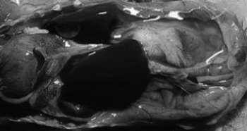
Opening the abdominal cavity of an ascitic bird reveals the presence of a large amount of clear yellow fluid, called ascitic fluid (Figure 1). More than 300 ml of fluid with or without fibrin clots may be present. This explains the abdominal distension (Swire, Reference SWIRE1980; Shome et al., Reference SHOME, PADHI, SHOME, SENANI, SAHA and RAI2000). The heart is markedly enlarged. The enlargement is due to dilatation of both right atrium and right ventricle and also the enlargement of the right ventricular wall (Kamindjolo et al., Reference KAMINDJOLO, WAMUKOYA and NYAGA1978; Olkowski and Classen, Reference OLKOWSKI and CLASSEN1998). Hydropericardium and pericardium with adhesions are noted (Kamindjolo et al., Reference KAMINDJOLO, WAMUKOYA and NYAGA1978). The lungs are extremely congested and oedematous. Ectopic cartilaginous and osseous nodules in lungs of young broilers are seen on post-mortem examination (Maxwell, Reference MAXWELL1988). The liver is swollen (Figure 2), congested and shrunken appearance with fibrin adherence apparent on its irregular surface. In some cases oedema under Glisson's capsule may be noted (Kamindjolo et al., Reference KAMINDJOLO, WAMUKOYA and NYAGA1978; Biswas et al., Reference BISWAS, BHOWMIK and DALAPATI1995). The kidney is swollen, congested and enlarged with adherence of fibrin clots (Kamindjolo et al., Reference KAMINDJOLO, WAMUKOYA and NYAGA1978; Shome et al., Reference SHOME, PADHI, SHOME, SENANI, SAHA and RAI2000).
Distended abdomen with ascetic fluid and marked passive congestion as evidenced by deep red colour of the musculature.

Opened abdominal cavity of an ascetic broiler with enlarged heart and the abundance of ascetic fluid.

Microscopic lesions: (Kamindjolo et al., Reference KAMINDJOLO, WAMUKOYA and NYAGA1978; Maxwell, Reference MAXWELL1988; Maxwell et al., 1989; Rath, Reference RATH2000).
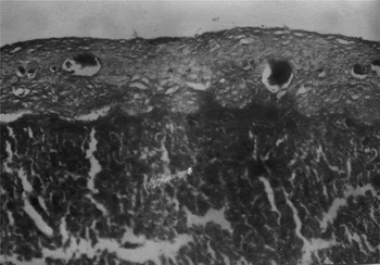
Liver lesions include blood stasis and dilatation of central veins and near the hepatic sinusoids. There is congestion and vacuolar degeneration of hepatic cells (Figures 3 and 4).
Section of liver showing thickened capsule with presence of collagen fibres.

Section of liver showing congestion and vacoular degeneration of hepatic cells.

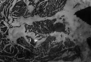
Microscopic examination of heart tissue from ascetic birds shows myofibril disorientation and degeneration with extensive mitochondrial hyperplasia (Figure 5).
Section of heart showing interstitial oedema and haemorrhage.

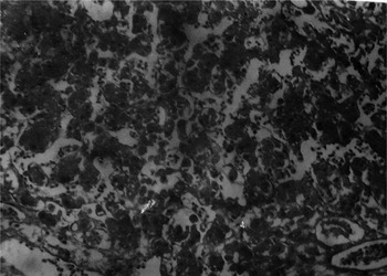
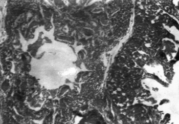
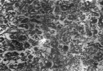
In the lungs, the parabronchi are greatly dilated with areas of sub-epithelial fibrosis and presence of more surfactant material than normal. There is marked congestion, oedema and thickening of septal wall (Figure 6 and Figure 7).
Section of lung showing oedema and congestion.

Section of lung showing marked congestion, oedema with thickened septal wall.

The kidney shows the extensive tubular necrosis with haemorrhages and protenaceous and haematogenous casts in the distal convoluted and collecting tubules of the kidney (Figure 8).
Section of kidney showing tubular epithelial cell degeneration and desquamation along with glomerular degeneration.

Treatment and control
There is no effective treatment for ascites, and once birds show symptoms, death occurs fairly quickly. Frusemide, a diuretic, can be used to reduce the mortality in ascites. Vitamin C, vitamin E and selenium can be used as feeding strategies to reduce mortality as they decrease the free radicals that are generated in ascites (Ladmakhi et al., Reference LADMAKHI, BUYS, DEWIL, RAHIMI and DECUYPERE1997; Vegad, Reference VEGAD2004). Several other measures can be taken to reduce the incidence of ascites syndrome in flocks. Broilers with faster growth rates are more likely to develop ascites due to the increased demand that this fast growth placed on the heart. However, with appropriate selection strategies, the predisposition of fast growing broilers to ascites can be reduced (Druyan et al., Reference DRUYAN, SHINDER, SHLOSBERG, CAHANER and YAHAV2009).
Ascites can be prevented through feed restriction, or through the use of very low energy feed throughout the entire cycle. Therefore, feed restriction slows body growth, allowing the cardiopulmonary organs (heart and lungs) to keep up with the oxygen demands of the birds (Tottori et al., Reference TOTTORI, YAMAGUCHI, MURAKAWA, SATO, UCHIDA and TATEYAMA1997). This can be done by following ways:
• Feed form (Mash vs. Pellets): Incidence of ascites can be reduced by reducing the intake of nutrients. Pelleted feed is more nutrient dense, and allows birds to increase nutrient intake. Mash feed can be fed during the starter period (from day old to 21 or 28 days of age) to reduce ascites (Shlosberg et al., Reference SHLOSBERG, BERMAN, BENDHEIM and PLAVNIK1991).
• Composition of the feed: Feed having less energy can be given to reduce the incidence of ascites as it reduces the oxygen requirement.
• Limited access to feed: Ascites can be reduced by reducing the time of feeding by using the intermittent lighting (reduce the level of activity) and removing the feed from the birds at night.
• Skip a day feeding: Skip-a-day programs used early in the starting period reduce negative effects on subsequent body weight gain (Arce et al., Reference ARCE, BERGER and COELLO1992). The most effective treatment reported for the control of ascites was a skip-a-day feed restriction program used in Kenya. The skip-a-day program was applied during the starter period (no feed on days 7, 9, 11, and 13) in association with a low protein starter capon mash. This program has dramatically reduced the annual incidence from >30% to 5% (Maxwell and Robertson, Reference MAXWELL and ROBERTSON1997).
• The most influential environmental factor affecting ascites in broilers is the oxygen content of air brought into the poultry house. Ascites symptoms are more acute at high altitudes as the air has a lower partial pressure of oxygen than that at sea level. Exposure to a lower partial pressure of oxygen will lead to an increased workload on the heart. In this situation it is critical to ventilate correctly and provide as much oxygen to the flock as possible.
• Maintaining adequate brooding temperatures are critical to the prevention of ascites. Exposure to cold periods forces the birds to use energy (which requires oxygen) in order to maintain body temperature. This predisposes a flock of broilers to ascites, although the symptoms show up later in production. Correct and monitored temperature during brooding, along with a good minimum ventilation program from placement, will help reduce and in some cases eliminate any ascites problems seen later in the grow-out period (Sato et al., Reference SATO, TEZUKA, SHIBUYA, WATANABE, KAMATA and SHIRAI2002).
• Avoid dust and ammonia using the poultry litter treatment (Terzich et al., Reference TERZICH, QUARLES, GOODWIN and BROWN1998). They are major lung irritant and damage the lungs, resulting in decreased oxygen transfer between the bird and the environment and thus contribute to ascites.
• Intermittent lighting, or using lighting programmes with increasing photoperiods, has proved successful in reducing the incidence of ascites without affecting broiler performance. The benefits of reducing day length are due to slower early growth rates (Buys et al., Reference BUYS, BUYSE, LADMAKHI and DECUYPERE1998; Hassanzadeh et al., Reference HASSANZADEH, FARD, BUYSE and DECUYPERE2003).
• Feed should not contain excess sodium as it increases blood volume, which leads to pulmonary hypertension and thus contributes to the development of ascites.
• Contamination of feed with toxins should be avoided.
Conclusions
Ascites is a complex problem caused by interactions between physiological, environmental and management factors. The incidence of ascites can be reduced by ensuring the nutritional, medicinal and good management strategies.