

Introduction
The main characteristics of embryonic stem (ES) cells are self-renewal ability and pluripotency (Brevini et al., Reference Brevini, Antonini, Cillo, Crestan and Gandolfi2007). Because of these characteristics, ES cells play an important role in cell therapy, regenerative medicine, tissue repair and use as a cell biology model (Rizzino, Reference Rizzino2009). The shortage of human organs that can be used for allotransplantation has motivated a search for alternative sources. The pig is a potentially useful model because of its similarities to humans in terms of organ size, immunology and whole-animal physiology (Li et al., Reference Li, Zhang, Hou, Jiao, Zheng and Wang2003). Xenotransplantation of pig organs is an attractive option because of the compatible size, physiology and potentially large supply of organs (Lai & Prather, Reference Lai and Prather2002). The establishment of a pig ES cell line has become very important. Previous reports have described the isolation of ES cells before implantation in mammals such as dogs (Hatoya et al., Reference Hatoya, Torii, Kondo, Okuno, Kobayashi, Wijewardana, Kawate, Tamada, Sawada, Kumagai, Sugiura and Inaba2006), monkeys (Mitalipov et al., Reference Mitalipov, Kuo, Byrne, Clepper, Meisner, Johnson, Zeier and Wolf2006), bovines (Verma et al., Reference Verma, Gautam, Singh, Manik, Palta, Singla, Goswami and Chauhan2007), cats (Yu et al., Reference Yu, Jin, Yin, Cho, Jeon, Lee and Kong2008) and swine (Chen et al., Reference Chen, Shiue, Bertoloni, Medrano, BonDurant and Anderson1999). ES cell lines have only been established from mice and humans. The mouse ES cell line is not a good candidate because of the large differences between mice and humans.
Reprogramming of somatic cell nuclei towards pluripotency has been achieved by nuclear transfer into enucleated oocytes and introduction of four defined factors to generate induced pluripotent stem (iPS) cells (Ezashi, Telugu et al., Reference Ezashi, Telugu, Alexenko, Sachdev, Sinha and Roberts2009). The iPS cells were derived from porcine fetal fibroblasts by lentivirus transduction of four human (h) genes: OCT4, SOX2, KLF4 and c-MYC. These cells derived from somatic cell nuclear transfer blastocysts and porcine fetal fibroblasts are called ES-like cells. These ES-like cells exhibited normal ES cell morphology, showed positive alkaline phosphatase activity, expressed key stem-cell markers and differentiated into multiple cell types in vitro and in vivo. Embryoid bodies were also formed when the cells were cultured in suspension (Pozzobon et al., Reference Pozzobon, Ghionzoli and De Coppi2010).
The mouse nuclear transfer embryonic stem cell (ntESc) line was established from different strains and tissues (Wakayama et al., Reference Wakayama, Ohta, Kishigami, Thuan, Hikichi, Mizutani, Miyake and Wakayama2005a) and most cells of the ntESc line (up to 80%) showed normal karyotypes and gene line transmission, which confirmed their pluripotency. Mice were then cloned from ntESc, but the production of cloned mice from ntESc nuclei did not improve from the cloning of somatic cells (Wakayama et al., Reference Wakayama, Mizutani, Kishigami, Thuan, Ohta, Hikichi, Bui, Miyake and Wakayama2005b). The establishment of the ntESc line provides a better technique for cloning mice from individual donors because of the multiplication ability of this cell line. In this study, we established pig ES-like cell lines from nuclear transfer blastocysts (pntESc) of different days to determine the best day to isolate cells.
Materials and methods
Nuclear donor cell preparation
A neomycin resistance gene containing a phosphoglycerate kinase promoter was inserted into an enhanced green fluorescent protein (EGFP)-containing vector, pCX-EGFP. The vector was then transfected into the fibroblast cells isolated from the pig fetuses of the 33rd day. After approximately 7 days of drug selection, surviving cells continued to be maintained in the cell culture solution and a GFP-expressing cell line was established. Aliquots of cells from each passage were frozen using frozen medium (90% fetal bovine serum (FBS) + 10% dimethyl sulphoxide (DMSO)).
In vivo oocyte production and nuclear transfer
The production of oocytes and nuclear transfer were performed as described by Lai & Prather (Reference Lai and Prather2003). After culture, the blastocysts were collected.
Differential staining
Blastocysts from different days (D5, D7, and D9) were stained using the method described by AA Fouladi-Nashta. First, embryos were incubated in freshly prepared 0.2% Triton X-100 in phosphate-buffered saline (PBS) containing 2 mg/ml bovine serum albumin (BSA) for 20 s and immediately washed twice in PBS–BSA medium. Then, the embryos were transferred into PBS–BSA medium containing 30 μg/ml propidium iodide (PI), incubated in the dark for 5 min in a 39°C warm chamber and washed twice in PBS–BSA medium. Lastly, embryos were incubated in 4% paraformaldehyde (PFA) containing 10 μg/ml bisbenzimide (Hoechst 33342; Hoechst, Sigma) for 30 min at RT, followed by two washes in PBS–BSA medium. The embryos were examined under UV light using a Nikon Diaphot inverted microscope (Nikon Corp.).
RNA isolation and qPCR
The total RNA from blastocysts from different days (D5, D7, D9) was isolated using the RNeasy Micro Kit (Qiagen) following the manufacturer's protocol. Contaminating genomic DNA was removed by a 15-min on-column digest with DNase I (RNeasy). The total RNA was eluted from the glass silica columns with 14 μl of water. The RNA was reversed transcribed using a BioRT First Strand Synthesis Kit (Bioer Technology). To evaluate pluripotency signalling in the porcine embryo, we performed comparative real-time RT-PCR on porcine embryos from D5, D7 and D9. We analysed the expression of OCT4, NANOG, SOX2 and CMYC in 20 embryos each from D5, D7 and D9.
ES-like cell isolation and cultivation
Blastocysts from different days (D5, D7, D9) were collected after 120 h, 168 h and 216 h culture and the zona pellucida (if present) of the blastocysts was removed with 0.2% pronase. Zona-free blastocysts were cultured in 200 μl of ES medium in a 4-well plate (NUNC) containing a feeder layer and examined every day. The ES medium was changed daily. The ES medium contained D/F medium supplemented with 0.1 mM β-mercaptoethanol (Amresco), 100 IU/ml penicillin, 0.05 mg/ml streptomycin, 0.1 mM MEM non-essential amino acids (Gibco), 2 mM l-glutamine, 10 ng/ml recombinant human-fibroblast growth factor-basic (rh-bFGF) (Sigma) and 20% KSR (Gibco). The D/F medium had the same volume of DMEM (Gibco) and Ham's F10 (Gibco). The culture medium was changed two days after the blastocysts were transferred to the ES medium and every day following the first 2 days. The growth and development of embryos and colonies were examined daily. Approximately 2–5 days after the blastocysts were seeded, ES-like clones formed. These colonies were partially disaggregated with the aid of two fine needles under a micropipette. The disaggregated colony cells were re-seeded onto new MEF feeder layers in a 96-well plate.
Alkaline phosphatase (AP) staining
Expression of AP activity in ES-like colonies was detected by AP staining using the Vector Red alkaline phosphatase Substrate Kit I (cat.No.SK-5100). The working solution was prepared before use by first adding two drops of Reagent 1 to 5 ml of 100 mM Tris–HCl, pH 8.2–8.5 buffer and mixing well, then adding two drops of Reagent 2 and mixing well and finally adding two drops of Reagent 3 and mixing well. It is important to make this working solution in 100–200 mM Tris–HCl, pH 8.2 – pH 8.5. After the ES medium was removed and washed twice using PBS (Gibco), the working solution was added. The development times should be determined by the investigator, but generally 20–30 min provides a good staining intensity. Improved staining may be obtained by developing the substrate in the dark.
Statistical analysis
Experiments were replicated three times. The data were analysed by the chi-squared test and considered statistically significant when the p-value was less than 0.05.
Results
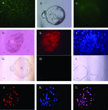
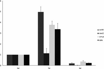
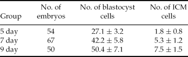
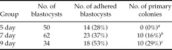
After GFP-positive cells (Figure 1) were injected into the enucleated oocytes, we cultured the embryos from each group (D5, D7 and D9). The blastocysts, which were GFP positive (Figure 2), were collected at each time point. A total of 171 embryos were used for differential staining (Figure 1). Blastocyst cells and inner cell mass (ICM) cells was counted (Table 1) and the total RNA from blastocysts from the different time points was isolated from 157 embryos. Comparative real-time PCR analysis of D5, D7 and D9 blastocysts was performed with three replicates. The expression of the four factors (OCT4, NANOG, SOX2 and CMYC) increased from D5 to D7, whereas the expression decreased from D7 to D9. We tried to isolate ES-like cells from these embryos. A total of 62 embryos (D7) and 34 embryos (D9) were used for ES-like cell isolation. We observed that 23 of the D7 embryos adhered to the feeder layer and small colonies formed from 10 of the 23 adhered blastocysts. The ratio of colony formation of D7 was higher (10/34) from the D9 embryos (Table 2). ES-like cells from D7 blastocysts grew slowly and expressed alkaline phosphatase, whereas the cells from D9 blastocysts grew rapidly but did not express alkaline phosphatase. ES-like cells were not isolated from the D5 blastocysts.
Morphology of embryonic stem (ES)-like cells from pig nuclear transfer blastocysts from different days and differential staining. (A) Green fluorescent protein (GFP)-positive donor cell line (×100). (B) Blastocyst formation after culture in the PZM-3 medium for 7 days (×600). (C) GFP-positive blastocyst (×600). (D) ES-like cells from D7 blastocyst (×600). (E) Positive AP staining of ES-like cells from D7 blastocyst. (F) Hoechst staining of the ES-like cells from D7 blastocyst. (G) ES-like cells from D9 blastocyst (×40); (H) Negative AP staining of ES-like cells from D9 blastocyst. (I) Cells from D9 blastocyst after 15 days of culture (×40). (J) Propidium iodide (PI) staining of the blastocyst (×600). (K) Hoechst staining of the blastocyst. (L) Overlay of (J) and (K). The blue cells are inner cell mass (ICM) cells.

Real-time reverse transcription (RT)-PCR analysis of relative transcript concentration of the four pluripotent factors (OCT4, SOX2, KLF4 and c-MYC).

Cell number of in vitro development of embryos
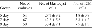
Experiments were replicated five times; p < 0.05.
ICM, inner cell mass.
Isolation and culture of ES-like cells from blastocysts of different days

a, b, cValues within the same column with different superscripts are significantly different (p < 0.01).
Discussion
Embryonic stem cells are a promising tool for cell therapy, regenerative medicine and tissue repair. ESCs constitute an invaluable model for basic research in developmental biology, nuclear reprogramming and the differentiation process. These cells are very unique because of their unlimited self-renewal ability and high plasticity, which allows them to differentiate into all embryonic tissues. The pig is similar to humans in organ size, immunology and whole-animal physiology. Thus, the isolation of pig embryonic stem cells (pESCs) is an important procedure, but there has not been a report about the successful isolation of a real pESCs line until recently.
In this study, we aimed to establish embryonic stem (ES)-like cell lines from nuclear transfer blastocysts. ES-like cell lines from certain animals already exist. H-S. Kim et al., produced 12 primary colonies from 65 nuclear transfer blastocysts (Kim, Son et al., Reference Kim, Son, Kim, Lee, Park, Kang, Lee, Hwang and Lee2007) and J. A. Byrne et al., successfully isolated two ES cell lines from the somatic cell nuclear transfer (SCNT) rhesus macaque blastocysts (Byrne, Pedersen et al., Reference Byrne, Pedersen, Clepper, Nelson, Sanger, Gokhale, Wolf and Mitalipov2007). We also aimed to find out the reason why it is so difficult to establish an ES cell line from pig nuclear transfer blastocysts. Certain conditions are necessary to maintain ES cell growth (Vackova, Ungrova et al., Reference Vackova, Ungrova and Lopes2007). The first condition is the right culture medium. In the culture medium for mouse ES cells (mesc), the cytokine leukemia inhibitory factor (LIF) integrates signals into mouse embryonic stem (ES) cells to maintain pluripotency (Niwa, Ogawa et al., Reference Niwa, Ogawa, Shimosato and Adachi2009), whereas FGF plays a role in the culture of human ES cells (hESc). As reported by Ramiro Alberio and colleagues, the addition of heterologous LIF does not prevent differentiation of pig embryonic stem cells; thus, LIF signalling is not critical for maintaining pESc (Alberio, Croxall et al., Reference Alberio, Croxall and Allegrucci2010). Comparing the expression profiles of ESC pluripotency-associated factors with the known expression profiles of mESC and hESC suggests that the undifferentiated porcine ICM and epiblast may be most similar to hESC (Blomberg, Schreier et al., Reference Blomberg, Schreier and Talbot2008). In our study, the ES medium was supplemented with hFGF and LIF was added in the control group. We found that the clone formation rate of the hFGF group was higher (data not shown). In addition, we cultured the blastocysts in DMEM + Ham's F10 (D/H medium), DMEM + F12 (D/F medium) and DMEM alone and with hFGF added into these three kinds of medium. We found that the clone formation rate was the highest in the D/H medium and was lower in the DMEM medium, whereas no primary colonies were formed in the D/F medium (data not shown). We concluded that the initial cell number was another necessary condition. Mechanical dissection (Chen & Melton, Reference Chen and Melton2007) is a method that can protect the ICM cells from differentiation, with the main reason being that the cell mass can be divided into pieces but not into single cells; this method avoids the actions of a digestive enzyme. In our study, ES-like cells from D7 blastocysts grew slowly and expressed alkaline phosphatase, whereas the cells from D9 blastocysts grew rapidly but did not express alkaline phosphatase. The expression of alkaline phosphatase was conformed to the expression of the four pluripotency factors. These factors demonstrated that the cells from the D7 blastocysts were pluripotent, whereas cells from D9 blastocysts started to lose pluripotency. ES-like cells were not isolated from the D5 blastocysts. We conclude that the initial number of ICM cells plays an important role in cell growth. We counted the ICM and whole blastocyst cell number of the three different days by the differential staining method. We found that the number of ICM cells increased from 1.83 ± 0.8 to 5.37 ± 1.2 to 7.56 ± 1.5. Thus, the possibility of establishing stable pluripotent cell lines using D7 blastocysts is much greater than with D5 and D9 blastocysts. These findings are similar to work by Brevini, T. A. and colleagues (Brevini, Pennarossa et al., Reference Brevini, Pennarossa and Gandolfi2010).
In summary, we determined the ICM and the whole blastocyst cell number of blastocysts from different days and the expression state of the four pluripotency factors, which increased from D5 to D7 and decreased from D7 to D9. We isolated ntES cell lines from D7 and D9 blastocysts. It is possible that the reprogram process was not complete or the initial cell number was too low. The cells from the D7 blastocysts grew slowly but expressed alkaline phosphatase and the cells from D9 blastocysts grew rapidly but did not express alkaline phosphatase. We did not isolate any ES-like cells from the D5 blastocysts. These results indicate that the possibility of establishing stable pluripotent cell lines using D7 blastocysts is much more plausible than it is using D5 and D9 blastocysts. There is still a lot of work to be done to improve the quality of blastocysts.
Acknowledgements
We thank Dr Na Song for her help with qPCR and we are grateful for Mr Wanhua Xie's work on nuclear transfer.