Noonan syndrome is a genetically heterogeneous disorder with an incidence ranging from 1 in 1000 to 1 in 2500 live births. Reference Mendez and Opitz1 Cardiovascular disorders are known to occur in as high as 80–90% of patients with Noonan syndrome. Reference Prendiville, Gauvreau and Tworog-Dube2 Pulmonary stenosis, hypertrophic cardiomyopathy, atrial septal defects, and patent ductus arteriosus are more common, whereas mitral valve disease, aortic root dilatation, aortic valve stenosis, coarctation of aorta, and tetralogy of Fallot are much rarer. Reference Pierpont and Digilio3 Due to the rarity of mitral valve disease in this cohort, its clinical presentation, management, and prognosis are not well known. Reference Marino, Gagliardi and Digilio4 In our case, rapid progression of mitral valve dysplasia led to left atrial dilatation and recurrent atrial tachyarrhythmia, emphasising the need for close follow-up to allow prompt identification and prevent complications.
Case
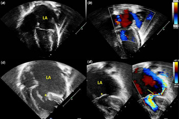
The presented patient was born at 32 weeks gestation with a perinatal history significant for polyhydramnios, bilateral hydro-nephrosis, and in-utero cocaine exposure. He was noted to have dysmorphic features which included low-set ears, frontal bossing, low nasal bridge with transverse crease, and a large anterior fontanelle in the neonatal period. Genetic analysis identified a pathogenic heterozygous mutation in RAF1 (c.770C > T, p. Ser257Leu) gene confirming the diagnosis of Noonan syndrome. An echocardiogram performed at 1 month of age was notable for a small patent ductus arteriosus, a patent foramen ovale, and a normal mitral valve (Fig. 1a and b). The size and function of the left ventricle was normal. A follow-up echocardiographic assessment at 3 months of age revealed mild dysplasia of the mitral valve leaflets with mild insufficiency. The patient did not return for a scheduled 3-month follow-up.
( a ) Apical four-chamber view at 1 month of age shows normal size left atrium. ( b ) Focused color Doppler imaging shows a competent mitral valve with no regurgitation across it. ( c ) Apical four chamber view at 18 months of age shows massively dilated left atrium with dysplastic posterior leaflet of mitral valve (plus sign) adherent to the inferolateral wall of the left ventricle. ( d ) Focused color Doppler imaging shows eccentric jet of mitral regurgitation (arrow) with prolapse of the anterior leaflet of mitral valve (asterisk).

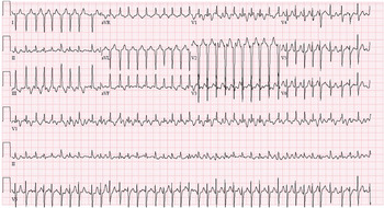
At 18 months of age, he presented to the emergency department secondary to increased work of breathing, increased precordial activity, fussiness, and decreased oral intake for 1 day. On presentation, his heart rate was 235 beats/minute, respiratory rate 52/minute, blood pressure 72/48 mmHg, and oxygen saturation 98% in room air. Chest radiogram showed cardiomegaly with upturned cardiac apex and pulmonary vascular congestion. Electrocardiogram was notable for a regular, narrow complex tachyarrhythmia (Fig. 2). Intravenous adenosine administration unmasked flutter waves, confirming the diagnosis of atrial flutter with 3:1 ventricular conduction. The child underwent synchronised electrical cardioversion, successfully converting him to sinus rhythm. An echocardiogram revealed a moderately dysplastic mitral valve with prolapse of the anterior leaflet causing severe mitral insufficiency (Fig. 1c and d). The posterior leaflet motion appeared to be limited and was adherent to the inferolateral wall of the left ventricle. The mitral valve leaflets were thickened with no apparent cleft present. The chordal attachments did not appear to obstruct the left ventricular outflow tract. The left atrium was massively dilated (Fig. 1c). He had mild left ventricular hypertrophy with posterior wall thickness measuring 8.5 mm (z score: +3.5) and interventricular septum measuring 6.7 mm (z score: +1.9) with mild left ventricular dilation visually (left ventricle internal diameter of 27 mm; z score: −0.28 in diastole and 13 mm; z score: −2.4 in systole). He was admitted to the paediatric intensive care unit for the management of heart failure symptoms. Diagnostic cardiac catheterisation was performed which showed small left-to-right shunts via a patent foramen ovale and tiny patent ductus arteriosus, yielding a net pulmonary-to-systemic blood flow ratio of 1.3. The left atrial and left ventricular end diastolic pressures were 12 and 15 mmHg, respectively. There was no significant gradient across the right or left ventricular outflow tracts. His symptom improved significantly and was discharged home on oral amiodarone, furosemide, and enalapril with close follow-up in the clinic. However, he had recurrent episodes of atrial flutter requiring hospital admission at 22 and 23 months of age. Presently, the cardiovascular surgical team is evaluating him for mitral valve replacement and Cox-Maze procedure.
Electrocardiogram shows regular narrow complex tachyarrhythmia with a ventricular rate of 230 beat per minute.

Discussion
The reported incidence of mitral valve disease in Noonan syndrome ranges from 2 to 6%. Reference Prendiville, Gauvreau and Tworog-Dube2,Reference Sarkozy, Dallapiccola and Conti5,Reference Colquitt and Noonan6 In the last 2 decades, a few studies have elucidated genotypic–phenotypic correlation with important prognostic implications. Reference Prendiville, Gauvreau and Tworog-Dube2 PTPN11 gene mutation is the most common gene affecting 40–50% of the cases whereas RAF1 gene mutations account for 3–17% of the cases with Noonan syndrome. In a case series, mitral valve anomaly was noted in about 13% of cases with a mutation in the RAF1 gene. Reference Pierpont and Digilio3 Genetic sequencing in our patient revealed a pathogenic mutation in the RAF1 gene.
Different mitral valve abnormalities have been reported in Noonan syndrome. In a post-mortem case series, grossly thickened and loose myxomatous mitral valve leaflets with shortened chordae and diminished interchordal space were noted. Reference Sreeram, Kitchiner and Smith7 Another series described partial atrioventricular canal defects and anomalous insertion of mitral valve over the interventricular septum. Reference Marino, Gagliardi and Digilio4 Annular dilation and prolapse of the mid portion of anterior mitral leaflet causing mitral regurgitation have also been reported. Reference Kang, Je and Lee11 Our patient had thickened, dysplastic mitral valve leaflets, with prolapse of the anterior mitral leaflet leading to severe regurgitation, as well as a posterior valve leaflet that was tethered to the lateral left ventricular wall.
Most cases of mitral valve disease in Noonan syndrome have been detected via routine echocardiographic surveillance or following presentation with symptoms of heart failure. Reference Ikeda, Onoue and Simizu8–Reference Kang, Je and Lee11 Some cases have also presented as sub-aortic obstruction due to anomalous insertion of the anterior mitral valve leaflet into the left ventricular outflow tract. Reference Marino, Gagliardi and Digilio4 Our case presented with atrial tachyarrhythmia due to an enlarged left atrium from severe mitral valve insufficiency. His left ventricle end-diastolic pressure was mildly elevated (15 mmHg). Therefore, his left ventricle hypertrophy was unlikely to cause severe left atrial dilatation. It was interesting to note that his mitral valve disease progressed rapidly as the echocardiogram performed at 1 month of age displayed no abnormalities of the mitral valve or left atrial dilation. Mild mitral valve dysplasia with mild accompanying insufficiency was noted 2 months following his initial echocardiogram. Unfortunately, the patient was lost to follow-up. A case of rapidly progressing enlargement of the left atrium and left ventricle due to worsening mitral regurgitation over 1 year was reported in an 18-year-old woman with Noonan syndrome. Reference Ikeda, Onoue and Simizu8 She was detected on routine follow-up and was asymptomatic at the time of diagnosis. However, that patient had mitral insufficiency characterised as “moderate” on her prior examination.
Our patient was discharged home on oral amiodarone after his initial hospital stay at 18 months of age. Despite adherence and dose escalation of amiodarone, he developed recurrent episodes of symptomatic atrial flutter at 22 and 23 months of age requiring cardioversion and hospitalisation. This was expected due to his massively enlarged left atrium providing a substrate for atrial flutter. He is presently under evaluation for mitral valve replacement and Cox-Maze procedure for recurrent atrial flutter. Due to the paucity of data with regards to the clinical presentation, progression, and management of mitral valve disease in Noonan syndrome, close monitoring is essential for early identification and timely intervention.
Acknowledgements
None.
Financial support
This research received no specific grant from any funding agency, commercial or not-for-profit sectors.
Conflict of interest
None.
Ethical standards
The authors assert that all procedures contributing to this work comply with the ethical standards. No human or animal experimentation was done.