INTRODUCTION
The Glyceridae Grube, Reference Grube1850 are burrower polychaetes, occurring from the intertidal down to the abyssal environment, and distributed worldwide (Rouse & Pleijel, Reference Rouse and Pleijel2001; Böggemann, Reference Böggemann2002). The family is subdivided in three genera: Glycera Savigny 1818, Hemipodia Kinberg 1865, and Glycerella Arwidsson Reference Arwidsson1899 (Böggemann, Reference Böggemann2002), which can be differentiated based on the characteristics commonly used in polychaete systematics, such as the shape of the parapodia, which are uniramous in Hemipodia and mostly biramous in Glycera and Glycerella; by the presence of falcigerous and spinigerous neurochaetae in Glycerella, while Glycera and Hemipodia only bear spinigerous neurochaetae, which vary morphologically throughout the body in these two genera (Parapar & Moreira, Reference Parapar and Moreira2010). Glycera and Glycerella also possess capillary notochaetae, which is absent in Hemipodia. They do not differ in the shape of their jaws, but the ailerons also show differences among genera: being rod-like shape in Glycerella and Hemipodia and sub-triangular or ‘Y’ shape in Glycera. Based on these and other characteristics, Böggemann (Reference Böggemann2002) in a phylogenetic study of the family, proposed Glycerella as a sister group of Glycera and Hemipodia.
In his review for the family Glyceridae, Böggeman (Reference Böggemann2002) reiterates Glycerella magellanica (McIntosh, Reference McIntosh1885) as the only valid species in the genus, considering both Glycerella atlantica Wesenberg-Lund, Reference Wesenberg-Lund1950 and Pseudolacydonia caeca Rullier, Reference Rullier1964 as junior synonyms. The species G. magellanica occurs in the North Atlantic, Caribbean Sea, Agulhas Basin (South Africa), south-west Indian Ocean, the Pacific north-west coast and Pacific–Antarctic Basin (near the west coast of South America), collected from shallow-water (45 m) down to deep-water (1,960 m) mainly in muddy sediments. Herein, the first record of G. magellanica is presented in the South Atlantic (off the Brazilian coast) filling a gap in the Atlantic distribution of this species. Also, this is the first record of this species associated with an anthozoan coral environment, in a dead fragment of the Caryophyllidae Solenosmilia variabilis (Duncan, 1873).
MATERIALS AND METHODS
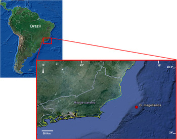
A single specimen was collected from the Campos Basin, on the south-east continental margin of Brazil, between 20.5° and 24°S (Figure 1). The material was collected under the scope of the research Deep-Sea Ecosystems Project (ECOPROF), conducted by the Research and Development Center of the Brazilian Energy Company, PETROBRAS, between 2008 and 2011. On each campaign, five samples containing five different species of anthozoan corals were collected: Lophelia pertusa (Linnaeus, 1758); Madrepora oculata Linnaeus, 1758; Enallopsammia rostrata (Pourtalès, 1878); Solenosmilia variabilis; and the hydrocoral Errina sp. The specimen was deposited in the Edmundo Ferraz Nonato collection located at the Biology Institute of the Federal University of Rio de Janeiro (IBUFRJ).
Sampling area (upper map) and occurrence area of Glycerella magellanica in ECOPROF project (lower map) (maps from Google Earth©).

Fragments of corals were collected using a remotely operated vehicle. The corals, including the associated fauna, were fixed in a combination of formaldehyde and borax, in a 10% concentration, without any anaesthetic process. In the laboratory, the polychaetes were sorted from the corals and studied under a stereomicroscope. Drawings were made with the aid of a camera lucida attached to the Zeiss V8 stereomicroscope and to a Wild M20 microscope. Three parapodia (anterior, median and posterior) were detached from the body and parts of the proboscis were mounted in permanent slides using the mounting medium Gray & Wess' (PVA) (Humason, Reference Humason1979) or Hoyer's medium.
RESULTS
SYSTEMATICS
Order PHYLLODOCIDA
Suborder GLYCERIFORMIA
Family GLYCERIDAE Grube, Reference Grube1850
Genus Glycerella Arwidsson, Reference Arwidsson1899
magellanica (McIntosh, Reference McIntosh1885) by monotypy
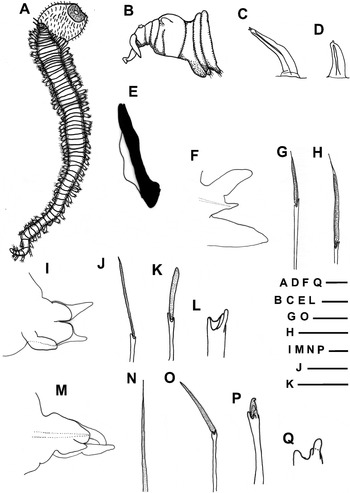
(Figures 2A–Q)
Hemipodus magellanicus McIntosh, Reference McIntosh1885, 349; pl. 42, figures11–15; pl. 22a, figures 12–15; pl. 35a, figures 5 & 7.
Glycerella atlantica Wesenberg-Lund, Reference Wesenberg-Lund1950, 24, pl. 5, figure 22; pl. 6, figure 28; O'Connor, Reference O'Connor1987, 21, figure 3a–c.
Pseudolacydonia caeca Rullier, Reference Rullier1964, 151, figure 8a–k.
Glycerella magellanica Böggemann, Reference Böggemann2002, 78, figures 124–126; Imajima Reference Imajima2005, 81, figure 35a–i; Parapar & Moreira, Reference Parapar and Moreira2009, 231, figures 1–5.
DIAGNOSIS (ACCORDING TO BÖGGEMANN, Reference Böggemann2002)
Body with numerous segments, elongated, tapering at both ends. Segments superficially triannulated. Prostomium consisting of four rings, appendages relatively long. Aileron rod-like. The first two parapodia uniramous consisting of neuropodium, ventral cirri and compound chaetae, dorsal cirrus from the second parapodium; the following parapodia biramous with dorsal and ventral cirri; notopodia and neuropodia indistinctly separated from each other; each parapodium with two prechaetal and two postchaetal lobes. Branchiae absent. Notopodia with simple capillary chaetae; neuropodia with compound chaetae of two types: dorsal spinigerous and ventral falcigerous.
MATERIAL EXAMINED
1 specimen (IBUFRJ 2125): located inside a dead base of the deep-sea coral Solenosmilia variabilis at 745 m depth; collected in the Campos Basin (Brazil) at 40°14′4578″W 22°31′197″S, on 17 March 2006.
DESCRIPTION
Body complete, broken into two parts near the most posterior region, measuring 16.77 mm long and with 77 chaetigers (Figure 2A). Brownish body. The anterior segments uniannulated, biannulated from the 9th chaetiger, triannulate mid-body from the 22nd chaetiger; anterior annulus the shortest and medium annulus the longest. Prostomium short and conical, consisting of 4 rings, posteriormost annulus with indication of a 5th incomplete ring finishing before the nuchal organ (Figure 2B). Four appendages in distal anterior part (a pair of dorsal antennae and a pair of ventral palpi; the two on the right lost) dorsoventrally flatted. The posteriormost annulus with one pair of half-moon shaped nuchal organs. Palps reaching the fifth annulus and antenna surpassing the fourth annulus. Proboscis with numerous digitiform papillae with a more or less distinctly straight, median, longitudinal ridge; some digitiform papillae slightly shorter, basally larger and distally more rounded than the majority (Figure 2C, D); all papillae presenting cilia at the top, the longer with two to three cilia and the shorter with one. Ailerons rod-like, slightly enlarged, seems rudimentary interramal plate (Figure 2E). The first two parapodia uniramous with one slender triangular prechaetal lobe and one more rounded and shorter postchaetal lobe (Figure 2F). The following parapodia biramous with two slender triangular to digitiform prechaetal lobes; notopodial lobe always slightly longer and wider than the neuropodial lobe (Figure 2I); both becoming slightly smaller in posterior parapodia (Figure 2M). The last three parapodia with their notopodial lobe broken. Two shorter postchaetal lobes; both lobes rounded anteriorly; notopodial lobe elongated and the following parapodia blunt triangular, slightly longer than rounded neuropodial lobe; in most posterior parapodia, notopodial lobe usually shorter and rounded than those from anterior and median parapodia. Dorsal cirri from the second parapodia, conical to oval; inserted on the body wall, above parapodial base. Ventral cirri in anterior parapodia relatively wide and conical (Figure 2I); in the posterior parapodia more slender triangular to digitiform, about as long as the neuropodial postchaetal lobe, and situated near the parapodial base. Branchiae absent. Parapodia with one noto- and one neuroacicula. Notochaetae simple, capillary, marginally slightly serrated at the border, surface rough on the whole extension on the dorsalmost (Figure 2N) and only on the distal end of the lowermost. Neurochaetae of two types: spinigerous on dorsal neuropodia (Figure 2G, H, J, O) and falcigerous on ventral neuropodia (Figure 2K, P); all chaetae with blade rough (Figure 2G, H, J, K, O, P). Spinigerous neurochaetae with blade very long and distally slender on the anterior parapodia (Figure 2 J), becoming slightly shorter, larger and distally curved on the posterior parapodia (Figure 2O). Falcigerous neurochaetae with blade long on the anterior parapodia (Figure 2K), becoming shorter and distally curved on the posterior parapodia (Figure 2P); shaft end of all neurochaetae with accessory lateral teeth on one side (Figure 2L, Q). Pygidial cirri lost.
Glycerella magellanica (McIntosh, Reference McIntosh1885): (A) complete specimen showing pharynx partially everted, dorsal view; (B) prostomium of the same; left lateral view; (C) anterior view of longer papillae; (D) anterior view of shorter papillae; (E) aileron; (F) first parapodium; (G) spinigerous neurochaeta of chaetiger 1; (H) detail of blade of the same neurochaeta; (I) posterior parapodium; (J) capillary notochaeta; (K) spinigerous neurochaeta; (L) falcigerous neurochaeta; (M) detail of the shaft end with secondary tooth; (N) middle parapodium; (O) spinigerous neurochaeta; (P) falcigerous neurochaeta; (Q) detail of the shaft end with secondary tooth. Scale bars: (A) 1.55 mm; (B) 350 µm; (C, E) 105 µm; (D, K, O, P) 40 µm; (F, N) 130 µm; (G, H) 74 µm; (I) 54 µm; (J, L) 15 µm; (M, Q) 4 µm.

DISCUSSION
This specimen seems morphologically similar to those examined and originally described by McIntosh (Reference McIntosh1885) and later by Böggemann (Reference Böggemann2002) during their re-description of the species. However the presence of a rudimentary interramal plate was observed on the side of the aileron, a feature that may have passed unnoticed by other authors, as observed in the drawings provided by Böggemann (Reference Böggemann2002) and by Parapar & Moreira (Reference Parapar and Moreira2009, Reference Parapar and Moreira2010). Böggemann (Reference Böggemann2002) also proposed a phylogenetic relationship for Glyceridae, in which the rod-like aileron is the basal characteristic within the family. Therefore this interramal plate, which is rudimentary in Glycerella, may disappear completely in Hemipodia and develop into a plate in Glycera.
Böggemann (Reference Böggemann2002) considers the chaetae as a weak characteristic to diagnose species of Glyceridae, because it shows little variation at an interspecific level. However, recent studies with Glycera and Hemipodia showed differences in the morphology of the chaetae along the body in the same specimen (Rizzo et al., Reference Rizzo, Steiner and Amaral2007; Parapar & Moreira, Reference Parapar and Moreira2010). A morphological variation among the neurochaetae was also observed throughout the body; they become smaller to the end of the body and present a clear indentation at the end of the falcigerous neurochaetae. In addition, Parapar & Moreira (Reference Parapar and Moreira2009) noted the the border of the blade on some specimens of G. magellanica can be serrated or smooth; the border of the blade of all chaetae examined in the studied specimen are smooth. However to explore these differences, which are only properly seen with the scanning electron microscope, may reveal interspecific differences which can be used in future phylogenetic studies, even though they would not be applicable in routine identification work (Parapar & Moreira, Reference Parapar and Moreira2010), among other things due to the small number of specimens.
The presence of a secondary tooth at the shaft end of the neurochaeta was observed as well. No reference was found for this characteristic, neither in the descriptions of McIntosh (Reference McIntosh1885), nor Böggemann (Reference Böggemann2002) nor Parapar & Moreira (Reference Parapar and Moreira2009, Reference Parapar and Moreira2010), and since there was only one specimen it was considered as a mere variation present in this specimen, or rather, that this may have been overlooked in previous work. Therefore, such a feature does not justify by itself creating a new species for the Brazilian specimen.
For the Brazilian coast, among the 16 species recorded belonging to the Glyceridae, 14 correspond to the genus Glycera and the other two to Hemipodia (Amaral et al., Reference Amaral, Nallin, Steiner, Forroni and Gomes Filho2012; Rizzo, unpublished data); therefore, this represents the first record of the genus Glycerella for the South Atlantic. According to Böggemann (Reference Böggemann2002) and Parapar & Moreira (Reference Parapar and Moreira2009), G. magellanica had been previously recorded, in all other global regions, to the Caribbean Sea (Barbados's coast) and to the Strait of Magellan (Chilean coast), with a gap occurring along the entire east coast of South America, which is partially filled with this new record.
This species was mostly collected below 100 m (Böggemann, Reference Böggemann2002); this may suggest that G. magellanica is more common in deeper waters. Also the species mainly occurs in muddy environments, with or without shell or coral debris (Böggemann, Reference Böggemann2002; Imajima, Reference Imajima2005; Parapar & Moreira, Reference Parapar and Moreira2009), and was never found in an anthozoan coral environment, as reported here, since this specimen was inside a dead portion of the Caryophyllidae S. variabilis. Parapar & Moreira (Reference Parapar and Moreira2009) previously reported this specimen in association with coral debris, however due to the sampling methodology used in their work (dredge), it was not possible to affirm if the specimens were among the coral debris or if they were carried to the debris during the dredging. Probably the paucity of studies in deeper water habitats, along the east coast of South America, might explain the absence of records of this species there. By increasing the numbers of studies in the Brazilian deep-sea it is expected the number of glycerid taxa occurring there may also increase and therefore the actual diversity of this family might be truly assessed.
ACKNOWLEDGEMENTS
We thank CENPES/PETROBRAS S.A. for providing the studied material and Dr. Helio R. Silva for reviewing this manuscript. CAPES (Coordenadoria de Aperfeiçoamento de Pessoal de Ensino Superior) funded the first author with a scholarship that allowed him to accomplish this study. The comments and suggestions provided by the referees are greatly appreciated.