Background
Fetal echocardiography plays an important role in the prenatal assessment of Tetralogy of Fallot, providing valuable information to predict postnatal critical obstruction to pulmonary blood flow necessitating early intervention. Reference Vetten, Auld and Han1 Among established predictors, retrograde (left to right, or aorta to pulmonary artery) flow across the ductus arteriosus and pulmonary valve size on fetal echocardiography are associated with an increased likelihood of needing early postnatal intervention to augment pulmonary blood flow. Reference Vetten, Auld and Han1–Reference Friedman, Balasubramanian and Tworetzky4 In a subset of Tetralogy of Fallot cases, the ductus arteriosus is absent, eliminating options such as prostaglandin therapy or ductal stenting to support pulmonary blood flow. This raises concerns for haemodynamic compromise at birth. Reference Maskatia, Glatz and Goldstein5,Reference Pahl, Muster, Ilbawi and DeLeon6 Existing literature on Tetralogy of Fallot-absent ductus arteriosus remains limited, with small cohorts and variation in inclusion criteria, making it challenging to ascertain its clinical significance. In this study, we compared early postnatal outcomes between Tetralogy of Fallot-absent ductus arteriosus and matched Tetralogy of Fallot-ductus arteriosus cases, and evaluated whether fetal and neonatal echocardiographic parameters can reliably predict the need for early intervention in Tetralogy of Fallot-absent ductus arteriosus and in Tetralogy of Fallot-ductus arteriosus.
Methods
We conducted a retrospective matched cohort study of fetuses diagnosed with Tetralogy of Fallot between January 2000 and July 2024 using the Texas Children’s Hospital fetal echocardiography database. Only fetuses with absent ductus arteriosus, confirmed by neonatal echocardiography in the first 24–48 hours of life, were included in the Tetralogy of Fallot-absent ductus arteriosus group. To assess whether absent ductus arteriosus influenced early postnatal intervention rates, we selected a matched control group of fetuses with Tetralogy of Fallot and a patent ductus arteriosus. Controls were matched in a 1:1 ratio to Tetralogy of Fallot-absent ductus arteriosus cases based on pulmonary valve Z-score within ± 10% and gestational age within ± 3 weeks at the time of fetal echocardiography. This matching aimed to control for the degree of pulmonary outflow tract obstruction, a known predictor of early intervention. Reference Rodenbarger, Thorsson and Stiver2,Reference Quartermain, Glatz and Goldberg7–Reference Park, Won and Kim9 We excluded Tetralogy of Fallot cases with pulmonary atresia, absent pulmonary valve, or major aortopulmonary collateral arteries to ensure a uniform cohort of Tetralogy of Fallot with antegrade pulmonary outflow. This study was approved by the Institutional Review Board at Baylor College of Medicine (Protocol H-50486, last approved on 8/23/2021), with a waiver of informed consent due to its retrospective nature.
Data collected included fetal and neonatal demographics (gestational age, sex). We obtained fetal and neonatal echocardiographic measurements of peak velocities across the pulmonary valve and aortic valve, and Z-scores Reference Sluysmans, Colan, Lai, Cohen, Geva and Mertens10,Reference Colan, Lai, Cohen, Geva and Mertens11 of pulmonary valve, aortic valve, ascending aorta, main pulmonary artery, right pulmonary artery, and left pulmonary artery from the echocardiographic reports. Pulmonary valve/aortic valve and main pulmonary artery/ascending aorta ratios were calculated as percentages to assess for right ventricular outflow tract size within each subject. We recorded aortic arch sidedness and the origin of the ductus arteriosus (from either the aorta or subclavian artery) in the group with Tetralogy of Fallot-ductus arteriosus. Additionally, we recorded the higher value of peak velocity in the right ventricular outflow tract or pulmonary valve at the time of early postnatal intervention or the next available follow-up echocardiogram.
Early postnatal intervention was defined as any surgical or transcatheter procedure to augment pulmonary blood flow, including shunt placement, ductal stenting, pulmonary valvuloplasty, right ventricular outflow tract stenting, or a complete surgical repair performed within the first six weeks of life. This cutoff was decided based on our institutional approach that complete repair is typically performed in patients outside of the neonatal period and weighing >4 kg. Reference Morales, Zafar and Heinle12
Genetic testing results were abstracted from the medical record when available, including 22q11.2 microdeletion analysis and chromosomal microarray findings.
Standard descriptive statistics were used to summarise participant characteristics. For comparison of continuous variables between the two matched cohorts, we used Wilcoxon matched-pairs signed-rank test, whereas Wilcoxon signed-rank test was used for comparison between groups with and without early intervention in each cohort. Categorical variables were assessed using Chi-squared or Fisher’s exact test, where appropriate.
Logistic regression analysis was used to evaluate the association between fetal or neonatal echocardiographic parameters significantly associated with the need for early intervention in each cohort, Tetralogy of Fallot-absent ductus arteriosus, and Tetralogy of Fallot-ductus arteriosus. Odds ratios with 95% confidence intervals were estimated for each model. Model performance was assessed using the likelihood ratio chi-square test and Pseudo R 2. Discriminatory ability was evaluated through receiver operating characteristic curve analysis, with the area under the curve calculated for each model. Sensitivity and specificity were determined across a range of predicted probability thresholds. The Youden Index (sensitivity + specificity − 1) was used to identify the optimal cutpoint that maximised combined sensitivity and specificity.
Significance was defined as two-tailed p value < 0.05. All statistical analyses were conducted using Stata (version 14.2, StataCorp, College Station, TX).
Results
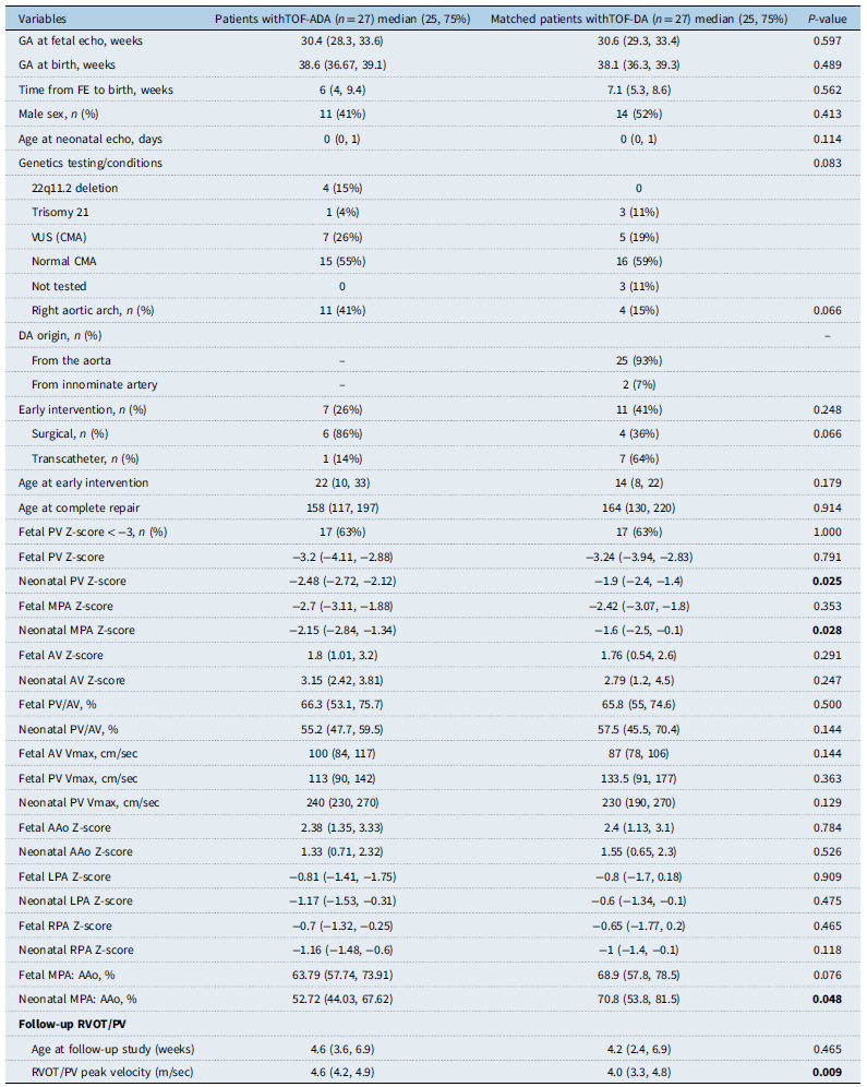
From the fetal echocardiography database at Texas Children’s Hospital (2000–2024), an initial cohort consisting of 409 fetuses diagnosed with Tetralogy of Fallot was queried and reviewed by N.B. and A.P. We excluded 156 cases that had absent pulmonary valve syndrome, pulmonary atresia, and major aortopulmonary collateral arteries, resulting in 253 cases. Among these, 207 fetuses had a documented ductus arteriosus and 46 had an absent or undefined ductus arteriosus. Postnatal data were unavailable in eight cases. Ductus arteriosus was present in 11 patients, including three with right aortic arch and an anterior ductus arteriosus from the left subclavian artery and seven patients with left aortic arch and a very small ductus arteriosus. Absent ductus arteriosus was confirmed postnatally in 27/245 (11%) patients (Figure 1), who were included in the Tetralogy of Fallot-absent ductus arteriosus cohort. A matched group of 27 fetuses with Tetralogy of Fallot-ductus arteriosus was selected based on fetal pulmonary valve Z-scores and gestational age (Figure 2). We successfully matched the two cohorts with no statistical difference in pulmonary valve or aortic valve on fetal echocardiography (FE); the rates of fetal pulmonary valve Z-scores < −3 were similar between the two cohorts (Table 1). It is interesting to note that the neonatal measurements of pulmonary valve and main pulmonary artery, as well as main pulmonary artery/ascending aorta ratio, were significantly lower in Tetralogy of Fallot-absent ductus arteriosus compared to Tetralogy of Fallot-ductus arteriosus. A right aortic arch was more frequent among patients with Tetralogy of Fallot-absent ductus arteriosus, though the difference was not statistically significant. In terms of genetic findings, 22q11.2 deletion was identified in four patients with Tetralogy of Fallot-absent ductus arteriosus (vs none in Tetralogy of Fallot-ductus arteriosus), while trisomy 21 was present in one patient with Tetralogy of Fallot-absent ductus arteriosus and three patients with Tetralogy of Fallot-ductus arteriosus.
Flowchart identifying fetuses with TOF-ADA, confirmed on postnatal echocardiography, from all fetuses with TOF at Texas Children’s Hospital (2000–2024). ADA = absent ductus arteriosus; DA = ductus arteriosus; LAA = left-sided aortic arch; LSCA = left subclavian artery; RAA = right-sided aortic arch; TOF = Tetralogy of Fallot.

Early postnatal intervention in a matched cohort of fetuses with confirmed TOF-ADA and TOF-DA. ADA = absent ductus arteriosus; DA = ductus arteriosus; mBTT = modified Blalock-Thomas-Taussig shunt; PV = pulmonary valve; RVOT = right ventricular outflow tract; TOF = Tetralogy of Fallot.

Characteristics of patients with TOF-ADA and TOF-DA

Abbreviations: AAo = ascending aorta; AV = aortic valve; CMA = chromosomal microarray; GA = gestational age; LPA = left pulmonary artery; MPA = main pulmonary artery; PV = pulmonary valve; RPA = right pulmonary artery; RVOT = right ventricular outflow tract; TOF-ADA = Tetralogy of Fallot with absent ductus arteriosus; TOF-DA = Tetralogy of Fallot with ductus arteriosus; VUS = variant of unknown significance.
Of 27 patients in each group, early intervention occurred in seven Tetralogy of Fallot-absent ductus arteriosus (26%) and 11 Tetralogy of Fallot-ductus arteriosus (41%) cases (p = 0.25). Surgical intervention was more frequent in Tetralogy of Fallot-absent ductus arteriosus (86 vs 36%, p = 0.066), while transcatheter procedures predominated in Tetralogy of Fallot-ductus arteriosus. The two groups had similar median ages at early intervention and at complete Tetralogy of Fallot repair.
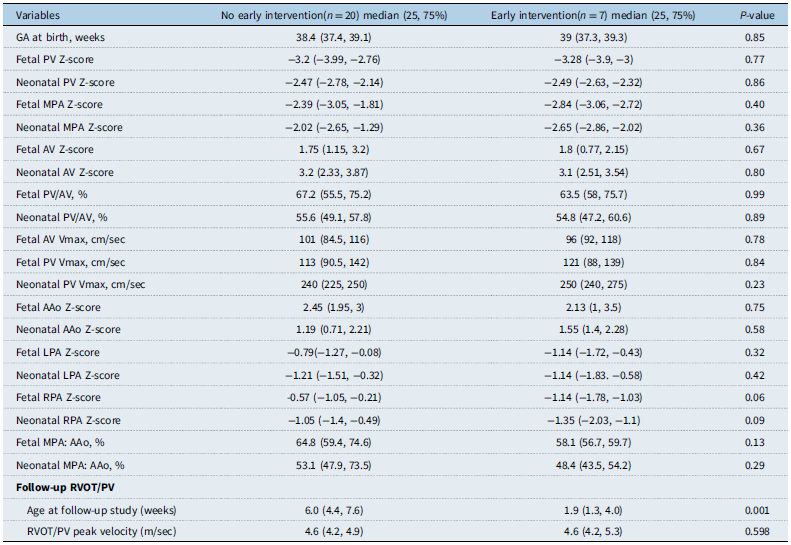
In Tetralogy of Fallot-absent ductus arteriosus group, none of the fetal or neonatal echocardiographic parameters was associated with the need for early intervention (Table 2). These findings suggest that conventional echocardiographic predictors of early intervention applied in Tetralogy of Fallot-ductus arteriosus may not hold predictive value in fetuses with absent ductus arteriosus, highlighting the challenge of risk stratification in this subset. In addition to analysing the first neonatal echocardiogram, we have now included right ventricular outflow tract peak velocity data at the time of intervention for the early intervention group and at first follow-up echocardiogram for the non-intervention group. While peak velocity was statistically lower in those who did not require early intervention, there was substantial overlap between groups, limiting its clinical utility.
Fetal and neonatal echocardiographic variables related to early intervention in 27 fetuses with TOF-ADA

Abbreviations: AAo = ascending aorta; AV = aortic valve; GA = gestational age; LPA = left pulmonary artery; MPA = main pulmonary artery; PV = pulmonary valve; RPA = right pulmonary artery; RVOT = right ventricular outflow tract; TOF-ADA = Tetralogy of Fallot with absent ductus arteriosus; TOF-DA = Tetralogy of Fallot with ductus arteriosus.
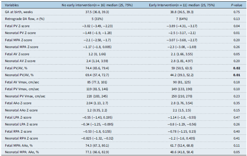
In the Tetralogy of Fallot-ductus arteriosus group, several echocardiographic parameters were associated with the need for early intervention. Lower fetal pulmonary valve Z-scores, lower pulmonary valve/aortic valve ratios, and lower neonatal pulmonary valve Z-scores were significantly associated with early intervention. Retrograde ductal flow was also more frequently observed in those requiring early intervention, although this did not reach statistical significance (Table 3). Logistic regression analysis identified fetal pulmonary valve/aortic valve ratio as a significant predictor of early intervention in Tetralogy of Fallot-ductus arteriosus group, with an odds ratio of 1.15 (95% CI: 1.03–1.28; p = 0.013). The model demonstrated moderate explanatory power (Pseudo R 2 = 0.30), and receiver operating characteristic curve analysis showed good discrimination with an area under the curve of 0.858 (95% CI: 0.713–1.000). The optimal threshold based on the Youden Index was 44.66%, achieving a sensitivity of 81.82% and specificity of 80% (Figure 3A). Additionally, neonatal pulmonary valve/aortic valve ratio was also significantly associated with early intervention in this Tetralogy of Fallot-ductus arteriosus group. In this model, the odds ratio was 1.22 (95% CI: 1.05–1.41; p = 0.009), with stronger explanatory power (Pseudo R 2 = 0.50) compared to the fetal model. Receiver operating characteristic analysis demonstrated excellent discrimination (area under the curve = 0.932; 95% CI: 0.842–1.000). The optimal cutpoint was identified at 58.04%, corresponding to a sensitivity of 81.82% and specificity of 93.75%, and yielding the highest Youden index observed in the analysis (Figure 3B).
Fetal and neonatal echocardiographic variables related to early intervention in 27 matched fetuses with TOF-DA

AAo = ascending aorta; AV = aortic valve; DA = ductus arteriosus; GA = gestational age; LPA = left pulmonary artery; MPA = main pulmonary artery; PV = pulmonary valve; RPA = right pulmonary artery; TOF-ADA = Tetralogy of Fallot with absent ductus arteriosus; TOF-DA = Tetralogy of Fallot with ductus arteriosus.
Discussion
This study adds to the limited data on the clinical relevance of absent ductus arteriosus in fetuses with Tetralogy of Fallot. Prior reports were limited to series of 6 to 11 confirmed fetal cases from single centers Reference Vetten, Auld and Han1,Reference Stern, Wadekar and Mertens13 or 17 cases in the multicenter series by Maskatia et al. Reference Maskatia, Glatz and Goldstein5 The present study comprised 27 postnatally confirmed cases of fetal Tetralogy of Fallot-absent ductus arteriosus and represents one of the largest series to date. Data regarding risk factors and the need for early postnatal intervention remain sparse, leaving the condition relatively unexplored. These considerations highlight both the novelty and the clinical relevance of the current findings.
In this study, absent ductus arteriosus was present in 11% of Tetralogy of Fallot cases with antegrade pulmonary flow. Reported prevalence of Tetralogy of Fallot-absent ductus arteriosus varies widely in the literature, ranging from 7 to 40%. Reference Grzyb, Koleśnik, Bokiniec and Szymkiewicz-Dangel14,Reference Issapour, Dasgupta and Zhang15 This discrepancy likely reflects differences in study design, patient selection, imaging criteria, and particularly the timing of confirmatory echocardiograms, as absent ductus arteriosus may be more apparent on follow-up imaging. Our findings fall within this spectrum and underscore the need for standardised approaches to diagnosis. Among these 27 patients, 26% of patients with Tetralogy of Fallot-absent ductus arteriosus required early intervention, a rate higher than the 17% previously reported in unselected cohort of Tetralogy of Fallot with pulmonary stenosis. Reference Morales, Zafar and Heinle12 This difference may reflect more severe obstruction in Tetralogy of Fallot-absent ductus arteriosus or differences in study design.
An important key strength of this study lies in its use of matched-pair design controlling for fetal pulmonary valve Z-score, a known predictor of early intervention. Reference Vetten, Auld and Han1,Reference Rodenbarger, Thorsson and Stiver2,Reference Issapour, Dasgupta and Zhang15 Unlike prior studies that did not account for the heterogeneity in Tetralogy of Fallot severity, Reference Stern, Wadekar and Mertens13,Reference Issapour, Dasgupta and Zhang15 we attempted to isolate the impact of ductal status. We found that absent ductus arteriosus did not independently predict the need for early intervention compared to a matched cohort of Tetralogy of Fallot-ductus arteriosus with similar pulmonary valve size on FE. While it is known that fetal main pulmonary artery size and growth pattern may be predictive of postnatal pulmonary outflow obstruction, Reference Hornberger, Sanders and Sahn8,Reference Park, Won and Kim9,Reference Kaguelidou, Fermont, Boudjemline, Le Bidois, Batisse and Bonnet16 the growth of the pulmonary valve and main pulmonary artery in patients with versus without ductus arteriosus was not previously known. Issapour et al. noted main pulmonary artery and main pulmonary artery/ascending aorta ratio were smaller in absent ductus arteriosus cases, compared to non-matched Tetralogy of Fallot-ductus arteriosus with no significant difference in postnatal outcomes (n = 35 Tetralogy of Fallot-ductus arteriosus versus 23 Tetralogy of Fallot-absent ductus arteriosus). Reference Issapour, Dasgupta and Zhang15 Our study findings showed that Tetralogy of Fallot-absent ductus arteriosus patients had significantly smaller pulmonary valve and main pulmonary artery dimensions at birth, despite comparable fetal Z-scores, suggesting possibly impaired growth of the pulmonary outflow tract in absent ductus arteriosus, although variations in measurements may also contribute.
Unlike Maskatia et al., Reference Maskatia, Glatz and Goldstein5 who studied symptomatic neonates requiring emergent palliation and found absent ductus arteriosus predictive of worse mortality, our cohort included all Tetralogy of Fallot-absent ductus arteriosus cases confirmed postnatally, regardless of clinical severity. Importantly, none of the patients in our cohort presented with haemodynamic instability, and both deaths in the absent ductus arteriosus group were unrelated to cardiac causes. These findings support that absent ductus arteriosus does not uniformly confer high risk, although most Tetralogy of Fallot-absent ductus arteriosus patients had smaller pulmonary outflow dimensions compared to historical Tetralogy of Fallot with pulmonary stenosis (TOF-PS) data. Reference Vetten, Auld and Han1,Reference Issapour, Dasgupta and Zhang15
While fetal pulmonary valve and main pulmonary artery Z-scores were similar between matched Tetralogy of Fallot-absent ductus arteriosus and Tetralogy of Fallot-ductus arteriosus groups, neonatal measurements showed significantly smaller pulmonary valve and main pulmonary artery diameters in Tetralogy of Fallot-absent ductus arteriosus, highlighting a potential difference in prenatal growth patterns. Previous studies have shown that fetal main pulmonary artery size and growth trajectory are associated with postnatal pulmonary outflow obstruction, but the impact of ductal status on growth had not been well established. Issapour et al. similarly observed smaller main pulmonary artery and main pulmonary artery/ascending aorta ratios in Tetralogy of Fallot-absent ductus arteriosus compared to unmatched Tetralogy of Fallot-ductus arteriosus, with no significant differences in early outcomes. Reference Issapour, Dasgupta and Zhang15
At our institution, TOF-PS management favours staged palliation over neonatal complete repair to preserve right ventricular function. Reference Morales, Zafar and Heinle12,Reference Fraser17 Historically, 17% of TOF-PS patients at our centre required shunt placement before complete repair. Reference Morales, Zafar and Heinle12 In our matched cohorts, early intervention occurred in 26% of Tetralogy of Fallot-absent ductus arteriosus and 41% of Tetralogy of Fallot-ductus arteriosus patients. This higher-than-expected rate in both groups likely reflects the inclusion of select fetuses with Tetralogy of Fallot-absent ductus arteriosus and matched Tetralogy of Fallot-ductus arteriosus who had more severe pulmonary outflow obstruction. Our findings suggest absent ductus arteriosus alone does not confer additional risk for early intervention when matched for anatomic severity. However, our study findings suggested less potential growth of the pulmonary outflow in fetuses with Tetralogy of Fallot-absent ductus arteriosus and the procedural approach differed: Tetralogy of Fallot-absent ductus arteriosus patients more often required surgical palliation (86%) compared to transcatheter approaches in Tetralogy of Fallot-ductus arteriosus (64%). These results underscore that pulmonary outflow obstruction remains the primary determinant of early intervention, with ductal status serving as a secondary anatomical variant that does not independently predict postnatal outcomes in this setting.
Notably, conventional echocardiographic markers predictive of early intervention in Tetralogy of Fallot-ductus arteriosus – such as low fetal pulmonary valve Z-score and pulmonary valve/aortic valve ratio – did not predict intervention in Tetralogy of Fallot-absent ductus arteriosus. This lack of association suggests that established fetal risk markers may not be applicable in this subset, complicating prenatal counselling. Additionally, in Tetralogy of Fallot-ductus arteriosus, fetal ductal flow direction was not significantly associated with early intervention, likely reflecting the narrow range of right ventricular outflow tract obstruction in the matched cohort. These findings further emphasise that pulmonary outflow calibre, rather than ductal status alone, remains the primary determinant of early postnatal management.
This retrospective, single-centre study is limited by its small Tetralogy of Fallot-absent ductus arteriosus sample size, which reduces power to detect differences in uncommon outcomes. Although matched-pair analysis controlled for fetal pulmonary valve Z-score and gestational age, residual confounding is possible. Echocardiographic measurements over a 24-year period may introduce interobserver variability, and timing differences in postnatal imaging could affect pulmonary outflow measurements. Systematic assessment and documentation for extracardiac anomalies were not uniformly performed across the study period, and thus these data were not included in the present analysis.
Conclusion
In this matched cohort study, absent ductus arteriosus in fetuses with Tetralogy of Fallot did not independently predict early postnatal intervention when matched for anatomic severity by fetal pulmonary valve Z-scores. However, Tetralogy of Fallot-absent ductus arteriosus was more often associated with significant pulmonary outflow obstruction, smaller neonatal pulmonary valve and main pulmonary artery dimensions, and potentially less growth of these structures compared to Tetralogy of Fallot-ductus arteriosus. The incidence of critically reduced pulmonary blood flow was comparable between groups. At our centre, when early intervention was required, Tetralogy of Fallot-absent ductus arteriosus needed surgical rather than transcatheter palliation due to absent ductus arteriosus, although institutional practices and regional differences in management strategies may influence this outcome and limit generalizability. Conventional fetal and neonatal echocardiographic predictors of early intervention in Tetralogy of Fallot-ductus arteriosus did not reliably apply in Tetralogy of Fallot-absent ductus arteriosus, complicating prenatal counselling. Larger, multicenter studies are needed to confirm these findings and identify reliable predictors of early intervention in this unique patient population.
Receiver operating characteristic (ROC) curves for fetal (left panel) and postnatal (right panel) pulmonary-valve-to-aortic-valve (PV/AV) ratios in predicting early intervention in TOF-DA. AUC = area under the curve; OR = odds ratio; TOF-DA = Tetralogy of Fallot with ductus arteriosus.

Acknowledgements
The authors are grateful to the patients and families who entrusted their care to our team. We thank the fetal cardiology and paediatric echocardiography teams at Texas Children’s Hospital for their dedication to high-quality imaging and data collection over the past two decades, which made this study possible.
Financial support
None.
Competing interests
None.
Ethical standard
This study was reviewed and approved by the Baylor College of Medicine Institutional Review Board (H-50497). Patient informed consent for participation was waived, given this was a retrospective chart review outcome analysis with anonymized publication data.