INTRODUCTION
Microsporidiosis is characterized as an evolutionarily conserved opportunistic infection in hosts ranging from nearly all vertebrates to invertebrates during immunocompromise (Canning and Lom, Reference Canning and Lom1986; Wittner and Weiss, Reference Wittner and Weiss1999). As fungus-like obligate intracellular parasites, microsporidia cause severe threats to human health, worldwide sericulture, beekeeping and aquaculture (Wittner and Weiss, Reference Wittner and Weiss1999; Bhat et al. Reference Bhat, Bashir and Kamili2009). In particular, Nosema bombycis, a microsporidian pathogen affecting the silkworm (Bombyx mori), inflicts severe worldwide economic losses in regions where sericulture is practiced. Unfortunately, no prospective vaccine or drug can efficiently prevent or control N. bombycis infection.
Cell surface proteins generally play major roles in signal transduction, cell adhesion and ion transport. Microsporidian spore walls generally consist of an electron-dense outer layer and an electron-lucent inner endospore layer (Xu et al. Reference Xu, Takvorian, Cali, Wang, Zhang, Orr and Weiss2006). Microsporidian surface proteins are exposed on the spore's electron-dense outer layer and may play a crucial role in spore adhesion to host cells (Southern et al. Reference Southern, Jolly, Lester and Hayman2007) by regulating water channels and increasing intrasporal pressure, leading to polar tube extrusion and penetration into the host cell (Ghosh et al. Reference Ghosh, Cappiello, McBride, Occi, Cali, Takvorian, McDonald and Weiss2006). Therefore, the systematic identification of the microsporidian surface proteome is important for understanding microsporidian invasion mechanisms and also for the discovery of prospective diagnostic markers and drug targets for the control of microsporidian diseases. For this purpose, a number of different approaches have been proposed to address the protein component of spore walls. Even so, the number of identified spore wall proteins (SWPs) is limited, and the number of identified surface proteins is even less. To date, only 4 microsporidian surface proteins have been discovered − one in Encephalitozoon cuniculi, one in Encephalitozoon intestinalis and two in N. bombycis − and these are involved in host–microsporidian interactions (Southern et al. Reference Southern, Jolly, Lester and Hayman2007; Li et al. Reference Li, Wu, Pan, He, Zhang, Hu and Zhou2009; Yang et al. Reference Yang, Dang, Peng, Ma, Jia, Qin, Long, Wu, Liu, Zhou, Pan and Zhou2014). Thus, many surface proteins remain to be identified. The reason for this low number is mainly attributable to existing technical barriers, including cytoplasmic protein contamination and the difficulty of protein extraction. The most important barriers are that the level of expression of a surface protein might be too low for it to be extracted or that its expression may be masked by other much more abundant proteins. Therefore, a valid strategy that specifically addresses the cell surface proteome is desirable for the separation and enrichment of microsporidian surface proteins.
Recently, cell surface biotinylation and affinity enrichment using immobilized beads have been widely applied in the identification of the surface proteome in many organisms. Biotin is a water-soluble, amine-reactive biotinylation reagent that is plasma membrane impermeable and that only reacts with protein molecules on the outer surface of the plasma membrane. The strong non-covalent biotin-avidin/streptavidin interaction is useful for purifying and detecting biotin-labelled proteins. Taking advantage of this approach, researchers have identified certain potential new surface adhesion factors that may participate in establishing and maintaining infection by pathogens, and they have discovered several suitable protein targets for the development of therapeutic strategies (de Miguel et al. Reference de Miguel, Lustig, Twu, Chattopadhyay, Wohlschlegel and Johnson2010; Che et al. Reference Che, Madrid-Aliste, Burd, Zhang, Nieves, Kim, Fiser, Angeletti and Weiss2011). In the present study, an effective biotinylation and affinity enrichment strategy was developed to isolate and identify the surface proteome of the microsporidian N. bombycis. Subsequently, a series of surface proteins were identified, many of which may be potentially suitable targets for the development of diagnostic and therapeutic strategies.
MATERIALS AND METHODS
Microsporidian spore culture and purification
The culturing and purification of mature spores were performed as previously described (Li et al. Reference Li, Pan, Li, Huang, Chen, Geng, Yang, Wang and Zhou2012). Briefly, the N. bombycis isolate CQ1 (Isolate 102059) was obtained from the China Veterinary Culture Collection Centre, Chongqing. Third-instar silkworm larvae were then infected to produce N. bombycis spores. These spores were purified by discontinuous Percoll gradient centrifugation, as previously described (Wu et al. Reference Wu, Li, Pan, Tan, Hu, Zhou and Xiang2008).
Spore surface protein biotinylation
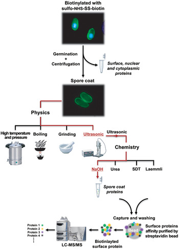
To identify surface proteins of N. bombycis, we developed a strategy that mainly includes spore surface protein biotinylation, protein extraction, streptavidin affinity purification, and LC-MS/MS analysis (Fig. 1). The spore surface protein biotinylation procedure was as follows. First, spores (109 spores mL−1) were washed three times with ice-cold phosphate-buffered saline (PBS), followed by incubation in 2 mg mL−1 EZ-Link Sulfo-NHS-SS-Biotin (Pierce, USA) solution on ice with gentle agitation for 4 h. The spores were then centrifuged (12 000 g , 5 min) and washed three times with ice-cold PBS. Excess biotin was blocked with 100 mm glycine for 30 min to terminate the reaction, and the spores were again washed three times with PBS.
Schematic description of the strategies used in this study for the identification of surface proteins in N. bombycis. Biotinylation was performed by incubating spores in a sulfo-NHS-SS-biotin solution that links to the free primary amines of surface proteins. The N. bombycis spores were then induced to germinate with K2CO3, and the biotinylated proteins were extracted by ultrasonication in NaOH and captured on streptavidin resin. After washing, the purified biotinylation proteins were eluted with 100 mm DTT, 70% formic acid and 30% acetonitrile. Biotinylated proteins were then identified by shotgun mass spectrometry using LC-MS/MS.

Protein extraction
Spores (109 spores mL−1) were germinated in 0·1 m K2CO3 (pH 8·0) at 28 °C for 40 min and then centrifuged (4 °C, 12 000 g , 10 min), after which the supernatant and pellet were each collected. Subsequently, 4 physical methods and 4 chemical lysis buffers were used to identify the optimal method for total protein extraction from N. bombycis (Fig. 1). Briefly, pellets were resuspended in PBS and then treated using one of the following methods: placement in an autoclave for 30 min (high temperature- and pressure-treated extract), incubation in boiling water for 5 min (boiled extract), grinding with quartz sand in liquid nitrogen (ground extract), or sonication on ice using three 1-s bursts (sonicated extract). After identifying the optimal physical method (sonicated extract), 4 chemical lysis buffers were used to extract total proteins. Briefly, following germination and centrifugation, pellets were resuspended in SDT lysis buffer (4% w/v SDS and 0·1 m dithiothreitol [DTT] in 0·1 m Tris-HCl pH 7·6) with the protease inhibitor phenylmethylsulphonyl fluoride (PMSF), urea extraction buffer (8 m urea, 300 mm NaCl, 0·5% NP40, 50 mm Na2HPO4, and 50 mm Tris pH 8·0) with PMSF, Laemmli lysis buffer (60 mm Tris-Cl pH 6·8, 2% SDS, 10% glycerol, and 5% β-mercaptoethanol) with PMSF, or 0·01 m NaOH extraction buffer with PMSF. Each sample was then sonicated on ice using three 1-s bursts. Finally, after centrifugation (4 °C, 12 000 g , 10 min), the supernatant was collected and stored at −80 °C before SDS-PAGE detection.
Streptavidin affinity purification
Biotinylated proteins were purified using a Cell Surface Protein Isolation Kit (Pierce, USA) with certain modifications. Briefly, 300 μL of streptavidin-agarose gel was washed three times with ice-cold PBS, mixed with the protein samples containing the EZ-Link Sulfo-NHS-SS-Biotin-labelled spore lysate, and incubated overnight on ice with end-over-end mixing using a rotator. The mixture was then centrifuged (1000 g , 1 min), and the gel slurry was washed five times with ice-cold PBS with protease inhibitors. Next, the biotinylated proteins were eluted from the streptavidin agarose with 200 μL of elution buffer (100 mm DTT, 70% formic acid and 30% acetonitrile) for 15 min at 95 °C. These biotinylated proteins were collected by centrifugation at 12 000 g for 10 min and separated by SDS-PAGE on 12% polyacrylamide gels.
LC-MS/MS and bioinformatics analysis
Protein identification was performed using the LC-MS/MS method (BIO-TECH Company, China) against our local N. bombycis genome database (http://microbe.swu.edu.cn/silkpathdb/). The dataset represents proteins that were identified based on at least three sequenced peptides that were detected with high mass accuracy. Further identification was conducted through bioinformatics analysis. Conserved domains were predicted by Pfam (Sonnhammer et al. Reference Sonnhammer, Eddy, Birney, Bateman and Durbin1998) and SMART (Schultz et al. Reference Schultz, Copley, Doerks, Ponting and Bork2000), and protein subcellular localization was predicted by WoLF PSORT (http://wolfpsort.seq.cbrc.jp/). Gene ontology (GO) analysis was also performed using an online server (http://geneontology.org/) and ExPASy (http://www.expasy.org/proteomics). Additionally, signal peptides (SignalP) and transmembrane helical regions (TMHMM) were predicted using online tools (http://www.cbs.dtu.dk/services), and glycosylphosphatidylinositol (GPI) anchor prediction was performed using the program big-PI Predictor (http://mendel.imp.univie.ac.at/sat/gpi/gpi_server.html). Sequence similarity searching was conducted using BLAST against our local database. Finally, amino acid sequence alignments were generated using the MUSCLE program (Edgar, Reference Edgar2004).
Gene cloning, recombinant protein expression and antibody production
Based on the mass spectrometry data, we chose SWP8 as a candidate surface protein. Gene cloning, recombinant protein expression and purification, and polyclonal antiserum production were performed as previously described (Li et al. Reference Li, Pan, Li, Huang, Chen, Geng, Yang, Wang and Zhou2012). Briefly, the SWP8 gene (GenBank accession number EF683108) was cloned using the forward primer 5′- CGCGGATCCATGTCTGATAATACAAACAATG-3′, containing a BamH I restriction site, and the reverse primer 5′-TTGCGGCCGCTTAAAGATCTCCGAACTCACCAT-3′, containing a Not I restriction site. Expression of the recombinant SWP8 protein was then induced with 1 mm isopropyl-β-D-thiogalactopyranoside (IPTG; BIO-TECH Company, China) at 16 °C for 48 h. A Ni-nitrilotriacetic acid (Ni-NTA) Superflow cartridge was used to purify the recombinant protein according to the manufacturer's instructions (Qiagen, Germany). Finally, antiserum was generated by immunising mice with the recombinant SWP8, as previously described (Li et al. Reference Li, Wu, Pan, He, Zhang, Hu and Zhou2009, Reference Li, Pan, Li, Huang, Chen, Geng, Yang, Wang and Zhou2012). The other SWP antisera used in this study were gifts from the State Key Laboratory of Silkworm Genome Biology, Southwest University, China.
Immunoblotting
Total proteins were extracted from N. bombycis as described in the Protein Extraction section. Briefly, spores (109 spores mL−1) were germinated in 0·1 m K2CO3 (pH 8·0) at 28 °C for 40 min and then centrifuged (4 °C, 12 000 g , 10 min). The resultant pellets were sonicated and combined via agitation in 0·1 m NaOH, followed by centrifugation (4 °C, 12 000 g , 10 min) and supernatant collection. The biotinylated proteins were then purified using a Cell Surface Protein Isolation Kit (Pierce, USA) and separated by SDS-PAGE on a 12% gel, after which they were transferred onto polyvinylidene difluoride (PVDF) membranes (Millipore, USA). These membranes were blocked with 5% PBS-bovine serum albumin (BSA) in TBST (25 mm Tris pH 7·5, 150 mm NaCl, and 0·1% Tween-20) for 2 h at room temperature. For surface protein analysis, the corresponding protein antisera as well as negative-control serum were used as primary antibodies, and peroxidase-labelled goat anti-mouse IgG (Roche, Switzerland) was used as the secondary antibody (1:5000 dilution). The membranes were incubated with these primary and secondary antibodies for 2 h each at room temperature. For biotinylated protein detection, the membranes were also incubated with HRP-labelled streptavidin (1:5000 dilution in TBST/5% BSA, Pierce). Finally, the blots were developed using the substrate diaminobenzidine tetrahydrochloride (DAB; BIO-TECH Company, China).
Indirect immunofluorescence assay
The detection of spores was performed by indirect immunofluorescence assay (IFA), as previously described (Li et al. Reference Li, Pan, Li, Huang, Chen, Geng, Yang, Wang and Zhou2012). Briefly, spores were fixed in 80% acetone, permeabilized with 70% ethanol containing 0·5% Triton X-100 for 10 min at room temperature, and incubated with polyclonal antisera or negative-serum dilutions in PBS-BSA at 37 °C for 60 min. Secondary fluorescein isothiocyanate (FITC)-conjugated goat anti-mouse IgG antibody (1:100 dilution, Sigma) or FITC-conjugated streptavidin (1:5000 dilution, Pierce) was then used to detect the bound primary antibodies. DNA was also stained with 4′,6-diamidino-2-phenylindole (DAPI) for 20 min, and the spores were examined with an Olympus BX50 fluorescence microscope.
RESULTS
Biotinylation of the spore surface
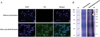
To ensure complete biotinylation of the surface proteins, an IFA and western blotting were performed to detect the efficiency of the EZ-Link Sulfo-NHS-SS-Biotin labelling. As shown in Fig. 2A, after incubation with EZ-Link Sulfo-NHS-SS-Biotin, a strong fluorescence signal was detected on the spore surface. In contrast, no significant intracellular labelling was detected in other fractions such as the polar tube and other organelles, and no fluorescence signal was detected in untreated spores, which served as a negative control. Consistent with these findings, in the labelled group, but not in the control group, a significant immunoblotting signal was present in the total protein lysates of the spores (Fig. 2B). Taken together, these results suggest that the spore surface proteins were successfully biotinylated, without unexpected contamination due to endogenous association with organelles.
Biotinylation of the spore surface using EZ-Link Sulfo-NHS-SS-Biotin. (A) Nosema bombycis spores were labelled with EZ-Link Sulfo-NHS-SS-Biotin and then incubated with FITC-conjugated streptavidin (Pierce, USA). Bright green fluorescence signals were observed in the spores incubated with EZ-Link Sulfo-NHS-SS-Biotin, whereas no green fluorescence signals were detected in the spores treated with PBS without EZ-Link Sulfo-NHS-SS-Biotin. Nuclei were stained with DAPI fluorescent dye. Magnification for all image areas, × 1000. Bar, 3 μm. (B) Spores’ biotinylated surface proteins were separated by SDS-PAGE, and analysed by western blotting. The obviously positive signal was observed in the total protein profiles hybridized with the HRP-labelled streptavidin against the biotinylated surface proteins.

Optimization of protein extraction
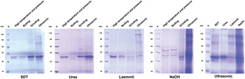
Considering the low abundance of surface proteins in whole-cell lysates, we tested different cell lysis strategies (Fig. 1). First, electrophoresis results showed that ultrasonication could significantly increase the protein yield (Fig. 3). Furthermore, new strategies using different lysis buffers combined with ultrasonic treatment were evaluated. The results indicated that more bands were detected for the samples treated with 0·1 m NaOH than for those treated with Laemmli, urea or SDT. Taken together, our results show that the optimal method for protein extraction from N. bombycis spores is ultrasonication combined with agitation in 0·1 m NaOH.
Total protein extraction from N. bombycis. Four physical conditions (high temperature and pressure, boiling, grinding and sonication) and 4 chemical lysis buffers (SDT lysis buffer, urea extraction buffer, Laemmli lysis buffer and NaOH extraction buffer) were used to explore the optimal method for total protein extraction from N. bombycis.

Identification of biotinylated proteins
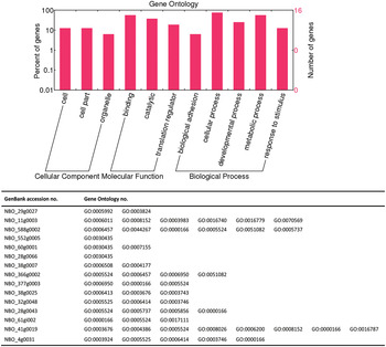
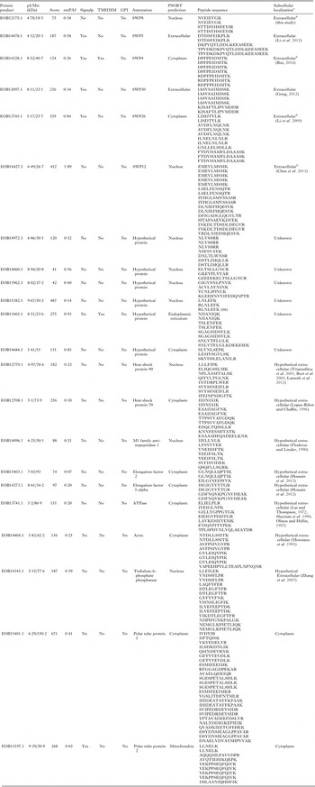
Following the determination of an optimal strategy, spores were labelled with EZ-Link Sulfo-NHS-SS-Biotin. The biotinylated proteins were then extracted and enriched by streptavidin pull-down. These enriched protein samples were subject to LC-MS/MS analysis. As the results show, 22 proteins were identified. The detailed information is summarized in Table 1 and Fig. 4.
GO of the N. bombycis surface proteins. The association of the GO categories ‘Cell component,’ ‘Biological process’ and ‘Molecular function’ with the surface proteins of N. bombycis.

LC-MS/MS and bioinformatics analysis of biotinylation protein from N. bombycis

SignalP, signal peptide; TMHMM, transmembrane helices in proteins; GPI, glycosylphosphatidylinositol anchor modified.
a A few literatures evidence supported for those proteins localized on the cell wall or outer membranes in some organisms, even though PSORT prediction and some published work mentioned as cytoplasm or nucleus proteins (reference in parentheses).
b Although PSORT predicted as cytoplasm or nucleus proteins, here revised as extracellular based on experimental evidence (reference in parentheses).
Five (SWP4, SWP5, SWP12, SWP26 and SWP30) of 22 biotinylated proteins were annotated as known SWPs (Wu et al. Reference Wu, Li, Pan, Tan, Hu, Zhou and Xiang2008; Li et al. Reference Li, Wu, Pan, He, Zhang, Hu and Zhou2009, Reference Li, Pan, Li, Huang, Chen, Geng, Yang, Wang and Zhou2012; Chen et al. Reference Chen, Geng, Long, Li, Li, Yang, Ma, Wu, Ma and Li2013; Rui, Reference Rui2014). Among them, only SWP4 possesses transmembrane helices, whereas the others contain a signal peptide. In addition, 8 putative surface proteins were identified based on the biotin labelling strategy. Two polar tube proteins, PTP2 and PTP3, were specifically identified, which have been reported to be binding partners of the spore wall protein SWP5 (Li et al. 2012). Moreover, 7 proteins were identified as hypothetical proteins without any known functional domain or motif, signal peptide or GPI anchor sites. Thus, these proteins could not be classified into any protein family.
GO analysis showed that 11 of 22 proteins could be categorized as associated with a biological process (Fig. 4). Briefly, 9 of the 11 proteins are putatively involved in biological adhesion, responses to stimuli, development or metabolic processes. In addition, 8 of the 11 proteins possess ATP-, GTP- and nucleotide-binding capacity and catalytic or translational regulation functions.
Validation of identified surface proteins
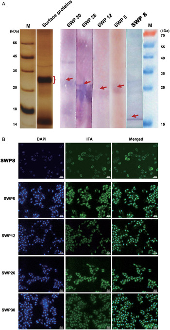
To validate the strategy used for surface protein identification, the hypothetical surface protein SWP8 was experimentally localized. This protein had been previously obtained from the N. bombycis spore coat, but no experimental evidence of subcellular localization had been obtained. In the present study, SWP8 was found to be localized on the spore surface (Fig. 5). In addition to SWP8, the localization of four other proteins (SWP5, SWP12, SWP26 and SWP30) was also analysed by immunoblotting. The results showed that they are spore surface proteins (Fig. 5A). Assessment by IFA also confirmed the previous reports on their localization (Li et al. Reference Li, Wu, Pan, He, Zhang, Hu and Zhou2009) (Fig. 5B). Collectively, our results suggest that SWP5, SWP8, SWP12, SWP26 and SWP30 are N. bombycis surface proteins.
Immunoblotting and IFA verification of surface proteins. Surface proteins were verified by western blotting and IFA. (A) Spores’ biotinylated surface proteins were recovered by affinity purification, separated by SDS–PAGE, and analysed by western blotting. The brackets mark where a band was excised for analysis by LC-MS/MS. An obviously positive signal (arrowhead mark) was observed in the surface protein profiles hybridized with antisera against the reported spore wall proteins SWP5, SWP12, SWP26 and SWP30. (B) IFA with mouse antisera against the spore wall proteins SWP5, SWP12, SWP26 and SWP30. Bright green fluorescence signals were observed in the spores incubated with these four antisera compared with the negative control, verifying that these four proteins were localized on the surface of the spore wall. Nuclei were stained with DAPI fluorescent dye. Magnification for all image areas, × 1000. Bar, 3 μm.

DISCUSSION
Microsporidian SWPs may be classified into endosporal proteins, cross wall proteins, exosporal proteins, and surface proteins that have their extracellular domain exposed on the spore surface. These surface proteins are considered to be crucial factors participating in initial adhesion to host cells (Hayman et al. Reference Hayman, Hayes, Amon and Nash2001). However, due to technical barriers, previous studies have mainly focused on identifying the components of the entire spore wall, rather than the surface proteins in particular. One reason for this gap is that the common approaches used cannot effectively extract proteins because of the electron-dense and chitinous structural barrier of the spore wall. Another reason is that the low abundance of surface proteins makes them difficult to separate from the total protein complex. Recently, a strategy using biotinylation of surface proteins and subsequent affinity purification was developed (Kischel et al. Reference Kischel, Guillonneau, Dumont, Bellahcene, Stresing, Clezardin, De Pauw and Castronovo2008; de Miguel et al. Reference de Miguel, Lustig, Twu, Chattopadhyay, Wohlschlegel and Johnson2010; Karhemo et al. Reference Karhemo, Ravela, Laakso, Ritamo, Tatti, Makinen, Goodison, Stenman, Holtta, Hautaniemi, Valmu, Lehti and Laakkonen2012). In the present study, based on this approach, an effective strategy for identifying the surface proteins of the microsporidian N. bombycis was established by combining cell surface biotinylation, protein extraction optimization, affinity purification and LC-MS/MS. Subsequently, 22 proteins were identified based on LC-MS/MS analysis.
Recently, the N. bombycis SWP26 protein was observed to be sparsely distributed on the exospore wall and involved in mediating spore-host adherence (Li et al. Reference Li, Wu, Pan, He, Zhang, Hu and Zhou2009). SWP30 has also been localized on the spore wall and associated with spore adherence to host cells (Lina, Reference Lina2014). In contrast, SWP4 (Rui, Reference Rui2014) and SWP12 (Chen et al. Reference Chen, Geng, Long, Li, Li, Yang, Ma, Wu, Ma and Li2013) are localized on the N. bombycis exospore. Interestingly, SWP5 has been observed not only on exospores but also in the region of the polar tube (Li et al. Reference Li, Pan, Li, Huang, Chen, Geng, Yang, Wang and Zhou2012). In the present study, these 5 proteins were also identified in our surface protein profile (Fig. 5). Moreover, we noted that SWP4, SWP8, SWP12 and SWP26 were predicted as nuclear or cytoplasmic proteins by bioinformatics due to the absence of a signal peptide amino acid sequence (Table 1). In addition to these proteins, another 7 hypothetical proteins were annotated as nuclear, endoplasmic reticulum or cytoplasmic proteins (Table 1). However, further analysis showed that NbHSWP8 (Wu et al. Reference Wu, Li, Pan, Tan, Hu, Zhou and Xiang2008), a hypothetical protein, is a surface protein located on the spore surface (Fig. 5), in contrast to the bioinformatics prediction that NbHSWP8 is a non-surface protein. This finding implies that bioinformatics analysis may not be sufficient to subcellularly localize all SWPs in N. bombycis. Taken together, our results suggest that the strategy developed in our study is effective and feasible.
Distinct from previous research, only 5 of 14 hypothetical SWPs were discovered based on our current surface protein LC-MS/MS data and experimentally identified as surface proteins. Recently, NbHSWP11, 1 of the 9 remaining hypothetical SWPs, was identified as a membrane component of the sporoblast and mature spore, but not as a component of the spore wall surface (Yang et al. Reference Yang, Dang, Peng, Ma, Jia, Qin, Long, Wu, Liu, Zhou, Pan and Zhou2014). Considering this finding, perhaps we could speculate that the other 8 of 14 hypothetical proteins are endosporal or intracellular proteins. However, we cannot ignore that the different protein extraction methods used in the current and previous research might account for the distinct protein libraries obtained, resulting in 8 other hypothetical proteins not being present in our LC-MS/MS data. Additionally, the level of expression of the remaining proteins on the spore surface might be too low to be detected, being masked by other much more abundant surface proteins (Arjunan et al. Reference Arjunan, Reinartz, Emde, Zanger and Schrader2009; de la Torre-Escudero et al. Reference de la Torre-Escudero, Valero, Perez-Sanchez, Manzano-Roman and Oleaga2012). Finally, the extracellular domains of the 8 putative proteins exposed on the spore surface likely cannot be biotinylated because they lack the ε-amine of lysine residues (de la Torre-Escudero et al. Reference de la Torre-Escudero, Valero, Perez-Sanchez, Manzano-Roman and Oleaga2012). Therefore, further protein subcellular localization investigations will be needed.
Additionally, a few intracellular proteins were identified (Table 1). In fact, this is a common phenomenon occurring in nearly all studies on surface protein identification. Although these proteins are putatively involved in intracellular functions, certain cell surface localization clues were also found based on their homologues in other organisms. For example, Hsp70 is localized on the Saccharomyces cerevisiae cell wall and plays roles in translocation across the membrane and as a molecular chaperone, and it varies in the profile of cell wall proteins (Lopez-Ribot and Chaffin, Reference Lopez-Ribot and Chaffin1996). Nevertheless, we cannot ignore the following phenomena. In the microsporidian invasion process, upon appropriate environmental stimulation, the polar tube suddenly extrudes and penetrates the plasma membrane of the host cell, after which the sporoplasm is transferred into the cytoplasm of the host cell, where the spores develop and complete the life cycle (Xu and Weiss, Reference Xu and Weiss2005). Nosema bombycis spore biotinylation and sporoplasm transfer into the external environment likely occur at the same time, resulting in intracellular proteins also being labelled by EZ-Link Sulfo-NHS-SS-Biotin during investigation. Furthermore, the strong interaction between cytoplasmic proteins and cross-membrane surface proteins should be considered. Thus, non-biotinylated cytoplasmic proteins likely cannot be entirely separated by NaOH lysis buffer and must be identified by mass spectrometry. In short, further investigations will be needed to confirm whether these general intracellular proteins are true cell wall components.
In conclusion, this study reports the development of a strategy for specifically identifying the surface proteome of the microsporidian N. bombycis. Our results provide certain clues about new targets for diagnostic research and drug design to control this microsporidian disease. Future research will focus on validating the surface localization of the remaining surface proteins and on identifying the key surface molecules responsible for N. bombycis–host interactions.
ACKNOWLEDGEMENTS
The authors are grateful to all who provided the means for us to acquire software free of charge, which we have used and cited in this article.
FINANCIAL SUPPORT
This work was supported by the National Science and Technology Plan Subject of China (863 Program, grant number 2012AA101301), the Natural Science Foundation of China (grant number 31101770 and 31402142), the Natural Science Foundation Project of Chongqing CSTC (grant number cstc2011jjA80019 and cstc2014jcyjA80039), the Science and Technology Project of the Chongqing Municipal Education Commission of China (grant number KJ100616) and the Natural Science Foundation Project of Chongqing Normal University of China.