Case report
A 14 -year-old boy presented with a 2-week history of abdominal pain which was treated with antacids. After experiencing acute symptoms of chest pain, increased work of breathing, and shortness of breath on walking and lying down he visited the emergency department. Chest X-ray demonstrated cardiomegaly and echocardiography showed a large pericardial effusion with suspicion of an intrapericardial mass. The patient was subsequently transferred to our intensive care unit with signs of cardiac tamponade and underwent pericardiocentesis, draining 2 L of haemorrhagic effusion (haemoglobin of the pericardial fluid was 7 g/L). Microscopic examination of the fluid showed inflammatory cells with a few reactive mesothelial cells, but no obvious malignant cells were found. No bacterial organisms were detected and the viral polymerase chain reaction of deoxyribonucleic acid screening was also negative.
Cardiac computed tomography (CT) confirmed a large intrapericardial mass compressing the right atrium and right ventricle. Intrapericardial teratoma was considered at that time.
Cardiovascular magnetic resonance (CMR) imaging was performed for tissue characterisation and to identify the relationship of the tumour to surrounding tissues.
Cardiovascular resonance imaging
Findings
CMR demonstrated a large, well-defined intrapericardial mass measuring ~8 × 13 × 13 cm. The mass was located behind the sternum and closely followed the course of the right coronary artery (RCA) with anterior compression of the right atrium, right ventricle, and vena cava superior. It was extended up to the level of the upper ascending aorta. The RCA was patent and passed through the mass with a larger branch entering the mass. The mass appeared tethered to the right atrium, right ventricle, and aortic root but the walls of these structures appeared intact and there was no intracardiac extension of the mass.
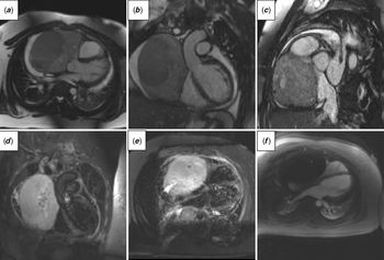
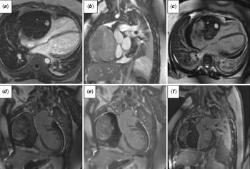
The mass had heterogeneous signal intensity on the steady-state free precession images (Fig 1a–c) and hyperintensity on T2-weighted turbo spin echo (TSE) images (Fig 1d and e). On T1-weighted TSE images the mass was isointense compared to the myocardium. T1 mapping revealed increased values ranging between 1530 and 1830 ms (normal values for myocardium at our 1.5 T scanner are less than <1050 ms). T2 mapping demonstrated long T2 values between 100 and 140 ms (normal values of myocardium at our 1.5 T are less than 56 ms). Rest perfusion showed hypoperfusion of the mass compared to the normal perfused myocardium, indicating low vascularity, except for few linear enhancements traversing the mass and originating close to the RCA (Fig 1f). On the early gadolinium enhancement images (Fig 2a–c), the mass was very hypointense compared to the surrounding tissue apart from a small hyperintense area which measured ~13 × 18 mm. This small area was also hyperintense on the late gadolinium enhancement images (Fig 2d–f). Apart from that, the mass appeared heterogeneous on the late gadolinium enhancement images with both areas of enhancement and hypointense areas. There was mild-to-moderate pericardial effusion (max. 2–2.5 cm), left-sided pleural effusion, and minimal right-sided pleural effusion.
(a–c) Balanced steady-state free precession sequence shows large heterogeneous mass compressing right atrium and right ventricle (a–c) and extending to the aortic root (b). (d and e) T2-weighted TSE images show hyperintense mass. (f) Rest perfusion revealed hypoperfusion of the mass compared to the normal perfused myocardium indicating low vascularity.

(a–c) On the early gadolinium enhancement images, the mass was very hypointense compared to the surrounding tissue apart from a small hyperintense area which measured ~13 × 18 mm. (d–f) The mass appeared heterogeneous on the late gadolinium enhancement images with both areas of enhancement and hypointense areas.

The volume of the right ventricle was normal with mildly reduced ejection fraction. Left ventricle had normal indexed volumes and ejection fraction. All valves had normal appearance.
Although the CMR features were not classic, atypical RCA coronary fistula with large haematoma and slow partial drainage into pericardium was suggested although a primary cardiac tumour such as angiosarcoma could not be excluded and therefore fluorodeoxyglucose positron emission tomography (FDG PET) was recommended.
Further imaging modalities
Whole-body FDG PET coupled with low-dose CT was subsequently performed and it showed no evidence of significant FDG uptake in the mass and there was no lymphadenopathy or lung abnormality noted. The coronary angiogram showed a dominant widely patent RCA with some tiny collaterals supplying the mass. Ultrasound-guided tissue biopsy and histology confirmed a mixed-type vascular malformation (lympho–venous), as suggested by CMR.
Subsequently, the patient was started on sirolimus therapy to decrease the size of the tumour.
Discussion
Primary pericardial masses are rare diseases with prevalence ranging between 0.001 and 0.007%.Reference Restrepo, Vargas, Ocazionez, Martinez-Jimenez, Betancourt Cuellar and Gutierrez1 On the other hand, the prevalence of direct invasion of a malignant process or metastatic disease into pericardium is 100–1000 times more frequent.Reference Restrepo, Vargas, Ocazionez, Martinez-Jimenez, Betancourt Cuellar and Gutierrez1 There are three main types of pericardial masses: (1) neoplastic primary (e.g., lipoma, haemangioma, fibroma) and secondary (e.g., mesothelioma, sarcoma), (2) non-neoplastic (e.g., mesothelioma, sarcoma), and (3) non-tumoural masses (hematoma, thrombus).Reference Zhou and Srichai-Parsia2
CMR as a non-invasive and non-ionising method is a crucial imaging modality in patients with pericardial masses. It is able to provide information about (1) location of the lesions, (2) size and borders of the lesions, (3) extent of the disease (including invasion of adjacent structures), (4) haemodynamic consequences, (5) possible metastatic process, and (6) valuable tissue characterisation data.Reference Restrepo, Vargas, Ocazionez, Martinez-Jimenez, Betancourt Cuellar and Gutierrez1,Reference O’Donnell, Abbara and Chaithiraphan3,Reference Dawson and Mohiaddin4
For the management and prognosis of the patients, it is crucial to assess the malignant character of the masses. There is some evidence that not the size of the tumour itself and no single feature are predictive of the malignant behaviour of the mass.Reference Rahbar, Seifarth and Schafers5 Malignancy can be suspected if pericardial effusion, invasion into surrounding structures, lymphadenopathy, and metastases are found. However, other imaging modalities might be required. Whole-body FDG PET coupled with a CT scan has been used recently and it has been shown that there is a correlation between the glucose accumulation in tumour tissue and the presence of malignancy, although exceptions have been found.Reference Rahbar, Seifarth and Schafers5,Reference Fathala, Abouzied and Al Sugair6
As the mass in our patient completely encased the RCA, surgical resection has not been deemed as a suitable option. Therefore, the patient was commenced on medical treatment with mTOR inhibitor (inhibitor of mammalian target of rapamycin) sirolimus (rapamycin). mTOR inhibitors directly block the pathway of cell growth and proliferation and have antitumoural and antiangiogenic effects. Sirolimus has been used originally as antifungal treatment and later on found an important role due to immunosuppressive effects in kidney transplantology. It is now with increasing interest being used in the management of vascular anomalies.Reference Triana, Dore and Cerezo7
Conclusion
We presented a rare case of a large pericardial tumour caused by atypical pericardial vascular malformation (lymphovenous) where multimodality imaging was very helpful in the patient’s management. CMR tissue characterisation in particular was crucial. In addition, CMR described well the spatial relationships and the haemodynamic impact of the mass.
Acknowledgements
None.
Financial Support
This research received no specific grant from any funding agency, commercial or not-for-profit sectors.
Conflict of Interest
The authors declare that they have no conflict of interest.
Ethical Standards
All procedures performed in the study were in accordance with the ethical standards of the institutional research committee and with the Helsinki declaration of 1975, as revised in 2008.