Introduction
The vitelline envelope plays an important role during fertilization and embryo development (Wassarman, Reference Wassarman1988, Reference Wassarman1999; Yanagimachi, Reference Yanagimachi, Knobil and Neill1994). It is involved in the recognition and binding of sperm (Valz-Gianinet et al., Reference Valz-Gianinet, Del Pino and Cabada1991; Benoff, Reference Benoff1997; Tulsiani et al., Reference Tulsiani, Yoshida-Komiya and Araki1997), in the induction of the sperm acrosome reaction (Elinson, Reference Elinson1986; Wassarman, Reference Wassarman1990; Snell & White, Reference Snell and White1996) and in the block to polyspermy (Elinson, Reference Elinson1986; Hedrick & Nishihara, Reference Hedrick and Nishihara1991).
In anuran eggs the contents of the cortical granules are initially expelled into the perivitelline space (Katagiri, Reference Katagiri1959; Balinsky, Reference Balinsky1966; Grey et al., Reference Grey, Wolf and Hedrick1974, Reference Grey, Working and Hedrick1976; Nishihara & Hedrick, Reference Nishihara and Hedrick1977; Gómez et al., Reference Gómez, Santolaya and Cabada1984). The products released by the cortical granule are responsible for both the elevation of the vitelline envelope (Balinsky, Reference Balinsky1966) and the enlargement of the perivitelline space (Shivers & James, Reference Shivers and James1970; Grey et al., Reference Grey, Wolf and Hedrick1974; Greve & Hedrick, Reference Greve and Hedrick1978).
During activation, cortical granule exocytosis causes elaborate ultrastructural changes in the envelope (Hedrick & Nishihara, Reference Hedrick and Nishihara1991; Larabell & Chandler, Reference Larabell and Chandler1991). These changes involve modifications in the structure of the vitelline envelope and formation of a fertilization envelope. After fertilization, the fertilization envelope can no longer be penetrated by sperm (Grey et al., Reference Grey, Working and Hedrick1976).
In the normal fertilization of an anuran egg only one sperm nucleus fuses with the egg nucleus. In these species, polyspermy is blocked by at least two apparently different mechanisms that operate at the levels of the plasma membrane and of the vitelline envelope respectively (Elinson, Reference Elinson1986).
The first rapid step in polyspermy prevention includes depolarization of the oocyte plasma membrane (Cross & Elinson, Reference Cross and Elinson1980; Jaffe et al., 1983). The membrane potential shifts from −9 mV to +20 mV (Cross & Elinson, Reference Cross and Elinson1980; Grey et al., 1981; Iwao, 1982, 1985). A change in the electric field across the egg plasma membrane may result in a change in the conformation or distribution of charged molecules in the membrane which in turn could affect sperm binding or fusion with the egg (Cross & Elinson, Reference Cross and Elinson1980). This step is followed by a slow but permanent second step in which the vitelline envelope becomes refractive to sperm entry (Raisman & Barbieri, Reference Raisman and Barbieri1969; Iwao & Katagiri, Reference Iwao and Katagiri1982).
Bufo arenarum oocytes are normally fertilized during the second meiotic metaphase (Katagiri, Reference Katagiri1974), prior to which frog oocytes are not activated in response to sperm entry or picking (Subtelny & Bradt, Reference Subtelny and Bradt1961; Katagiri, Reference Katagiri1974; Elinson, Reference Elinson1977).
Studies in several species have shown that as the egg traverses the oviduct before encountering sperm the egg envelope is altered by secreted oviductal factors so that fertilization is enhanced (Katagiri, Reference Katagiri1987; Hedrick & Nishihara Reference Hedrick and Nishihara1991). In amphibians, the envelope of an egg ovulated into the coelomic cavity, termed the coelomic envelope or CE, cannot be penetrated by sperm (Grey et al., Reference Grey, Working and Hedrick1977). As the egg travels through the pars recta region of the oviduct the coelomic envelope, induced by trypsin-like enzymes (Grey et al., Reference Grey, Working and Hedrick1977; Miceli et al., Reference Miceli, Fernández and Del Pino1978; Yoshizaki & Katagiri, Reference Yoshizaki and Katagiri1981), is converted into the vitelline envelope and becomes penetrable by sperm so that fertilization can take place.
In Xenopus laevis, the analysis of envelopes by SDS-PAGE determined that a single glycoprotein component of the coelomic envelope, gp 43, is proteolytically cleaved to gp 41 in the vitelline envelope (Gerton & Hedrick, Reference Gerton and Hedrick1986); this proteolysis appears to trigger conformational changes in the envelope glycoproteins, exposing sperm binding sites on egg envelope gp 69 (Tian et al., Reference Tian, Gong, Thomsen and Lennarz1997).
In previous reports we have shown that denuded full-grown oocytes of the amphibian Bufo arenarum, matured in vitro by progesterone, are polyspermic (Oterino et al., Reference Oterino, Sánchez Toranzo, Zelarayán and Bühler1997), although cortical granule exocytosis seems to occur within a normal chronological sequence (Oterino et al., Reference Oterino, Sánchez Toranzo, Zelarayán, Valz-Gianinet and Bühler2001).
The aim of this work was to analyse the perivitelline space and the role of the vitelline envelope of Bufo arenarum oocytes matured in vitro in the establishment of a slow block to polyspermy.
Materials and methods
Animals
Adult specimens of Bufo arenarum were collected in the northwestern area of Argentina and kept at 15 °C until use, which generally took place 15 days after collection. Experimental manipulation and oocyte culture were conducted at room temperature (22–25 °C) using amphibian Ringer solution (AR; 6.6 g NaCl/l, 0.15 g KCl/l, 0.15 g CaCl2/l) containing penicillin G-sodium (30 mg/l), streptomycin sulfate (50 mg/l) and 0.005 M Tris-HCl buffer (pH 7.4).
Oocyte collection
Denuded full-grown ovarian oocytes (1.7–1.8 mm in diameter) were obtained by manually pulling off the follicle epithelium and theca layer using fine forceps with the aid of a dissecting microscope (Lin & Schuetz, Reference Lin and Schuetz1985); follicle cells were removed by shaking in AR for 5 min. Oocytes remained only with the vitelline envelope (Zelarayán et al., Reference Zelarayán, Oterino and Bühler1995).
Freshly denuded oocytes were placed in AR at 22–25 °C. Routine in vitro cultures were carried out using plastic multiwell culture dishes (Costar 3524, Cambridge, MA). Randomized samples of 20 oocytes were distributed into separate wells containing 2 ml of AR. Two well duplicates were routinely run in each experimental group.
Hormonal maturation was induced by treatment of denuded oocytes with progesterone. Progesterone (Sigma) was dissolved in ethanol and added (5 μl) directly to the culture medium to give a final concentration of 1 μg/ml.
Oocyte maturation was assessed 18 h after removing the follicle cells or after hormone addition. Meiosis reinitiation was scored both by the presence of a transient white spot in the animal pole and by cytological examination. In our working conditions, oocytes reached metaphase II 16 h after defolliculation or progesterone treatment.
Coelomic eggs were obtained from the body cavities of females whose oviducts had been ligated before hormonal stimulation. Ovulation was induced by injection of homogenates of homologous pituitaries; eggs were collected 10–12 h later.
Oviposited eggs were obtained without occlusion of the pars recta at 8–12 h after injection with homologous pituitaries.
Insemination of oocytes
Sperm suspensions were obtained by gently disrupting the testes in 4 ml AR and centrifuging at 1085 g for 10 min; then, the pellet was resuspended in AR. Denuded ovarian oocytes and coelomic oocytes were treated with trypsin according to Elinson (Reference Elinson1973).
Trypsin-treated denuded oocytes and coelomic oocytes (Elinson, Reference Elinson1973) were inseminated in 10% AR containing the diffusible factor (1:1) obtained according to Barbieri & Oterino (Reference Barbieri and Oterino1972). Twenty microlitres of sperm suspension (final concentration 2 × 10⁶) was added; 10 min later oocytes were placed in 10% AR for different time periods and fixed for cytological examination (Bühler et al., Reference Bühler, Petrino and Legname1987).
Oocyte activation
Needle-pricking or electric shock were used as activation stimuli.
Cytological preparations
The oocytes were fixed in Ancel & Vintemberger's solution (10% formol, 0.5% acetic acid and 0.5% NaCl), embedded in paraffin and sliced into 7 μm thick sections which were then stained with Ehrlich haematoxylin and eosin. This method allowed us to easily observe the pronucleus (Bühler et al., Reference Bühler, Petrino and Legname1987).
Electron microscopy
Transmission electron microscopy
For transmission electron microscopy the oocytes were fixed in 2.5% glutaraldehyde in 0.1 M phosphate buffer at pH 7.4 for 2 h, post-fixed with 2% osmium tetroxide in 0.1 M phosphate buffer, pH 7.4, dehydrated in ethanol and embedded in epoxy resin. Thin sections were cut using an ultramicrotome and observed in a Zeiss transmission electron microscope.
Scanning electron microscopy
For scanning electron microscopy the oocytes were fixed in 2.5% glutaraldehyde in 0.1 M phosphate buffer at pH 7.4 for 15 h at 4 °C, post-fixed with 2% osmium tetroxide in 0.1 M phosphate buffer, pH 7.4, dehydrated and coated with gold. The samples were examined under a JEOL 35 C scanning microscope.
Obtaining the cortical granule products
Three types of CG products (CGP) were used in the present work: from in vitro matured oocytes (vCGP), from oviposited oocytes (dCGP) and from oviposited eggs without their vitelline envelope (tCGP). All were obtained by activating oocytes.
In all cases, activation was attained through the same procedure: batches of oocytes in Ringer-Tris solution (120 oocytes/ml) were activated by electric shock (10 pulses of 10–15 s each, 20 mV) (Petrino et al., Reference Petrino, Bühler and Legname1984). Twenty minutes after activation, oocytes were removed and the remaining solution is what we named CGP.
Isolation of vitelline envelopes
Vitelline envelopes were obtained by manually pulling off the envelopes using fine forceps. The envelopes were washed in ice-cold AR repeatedly until visually free from contaminating particulate material.
Electrophoresis
SDS-PAGE of CGP and dissolved vitelline envelopes was performed according to Laemmli (1970) with 7.5% running gels. Proteins were stained with Coomassie brilliant blue R-250. Prior to electrophoresis, the samples were dialysed overnight against distilled water, lyophilized and resuspended in sample buffer.
Results
The Bufo arenarum vitelline envelope can be found in four different forms:
- OE:
the ovarian envelope associated with the denuded full-grown oocyte matured in vitro.
- CE:
the coelomic envelope associated with the ovulated oocyte found in the coelom.
- VE:
the vitelline envelope of the ovoposited oocyte.
- FE:
the fertilization envelope, associated with the zygote.
These envelope forms have different ultrastructures, glycoprotein compositions and biological functions.
Neither the OE nor the CE was affected by proteolytic degradation due to a trypsin-like protease secreted by the pars recta.
Effect of trypsin treatment on the fertilization of denuded matured in vitro oocytes
Denuded full-grown ovarian oocytes matured in vitro by progesterone, with or without trypsin treatment before insemination, and coelomic oocytes with trypsin treatment were inseminated with homologous sperm. The inseminated oocytes were cultured in 10% AR for 2 h and fixed for cytological examination.
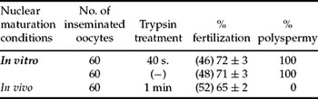
The results show (Table 1) that trypsin treatment before insemination does not cause polyspermy since the same treatment applied to coelomic oocytes does not alter monospermic fertilization. Moreover, these observations indicate that the vitelline envelope from denuded full-grown ovarian oocytes matured in vitro can be penetrated by sperm and that trypsin treatment before insemination is not necessary for fertilization.
Effect of trypsin treatment on the fertilization of denuded oocytes matured in vitro by progesterone

Groups of 20 denuded full-grown ovarian oocytes matured in vitro by progesterone (1 μg/ml), with or without trypsin treatment before insemination, and coelomic oocytes matured in vivo (controls) with trypsin treatment, were inseminated with homologous sperm (106 sperm/ml). The inseminated oocytes were cultured in 10% AR for 2 h and fixed for cytological examination. Polyspermy was defined as more than two pronuclei in the animal hemisphere of the egg. Values are the mean ±SEM of three experiments (20 oocytes per experiment). Each experiment was performed on a different animal.
Ability of the vitelline envelope to establish a slow block to sperm entry
To determine the ability of the vitelline envelope to establish a slow block to sperm entry and the time required, denuded full-grown ovarian oocytes matured in vitro by progesterone, with or without trypsin treatment, were activated by electric shock and inseminated 30, 60 or 120 s after activation. The inseminated oocytes were cultured in 10% AR for 2 h and fixed for cytological examination.
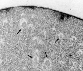
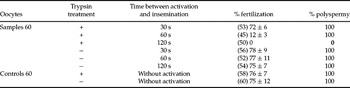
The results indicate (Table 2) that trypsin treatment of the ovarian envelope before activation produces modifications in the properties of the envelope, since oocytes inseminated 120 s after activation cannot be penetrated by sperm. In contrast, in the oocytes without trypsin treatment inseminated 120 s after activation, numerous male pronuclei could be observed in the animal hemisphere (Fig. 1). These results demonstrate that the ovarian envelope without trypsin treatment before activation is unable to block sperm entry.
Polyspermy in Bufo arenarum denuded oocytes matured in vitro by progesterone. Numerous male pronuclei (arrows) can be observed in the animal hemisphere of an egg fixed 90 min after insemination.

The time courses of slow block to polyspermy in denuded oocytes matured in vitro by progesterone

Groups of 20 denuded full-grown ovarian oocytes matured in vitro by progesterone (1 μg/ml), with or without trypsin treatment before activation, were inseminated with homologous sperm at different times after activation. The inseminated oocytes were cultured in 10% AR for 2 h and fixed for cytological examination. Polyspermy was defined as more than two pronuclei in the animal hemisphere of the egg. Values are the mean ± SEM of three experiments (20 oocytes per experiment). Each experiment was performed on a different animal.
Effect of cortical granule products on the ability of the vitelline envelope to establish a slow block to sperm entry
In Bufo arenarum, cortical granule products from the ovoposited oocytes (tCGP and dCGP) cause ultrastructural transformations in the vitelline envelope that block fertilization (Gómez, Reference Gómez1992).
In order to determine whether the vCGP can block fertilization, that is to say, whether it can disturb the vitelline envelope to prevent sperm entry, denuded full-grown ovarian oocytes matured in vitro by progesterone were trypsin-treated and inseminated with previous vCGP treatment for 30 min. The inseminated oocytes were cultured in 10% AR for 2 h and fixed for cytological examination
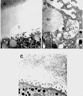
A female pronucleus with 100% polyspermy (Table 3) could be observed in about 30% of the inseminated oocytes. In 70% of the oocytes there was inhibition of sperm entry. Cytological examination of these oocytes revealed characteristic signs of activation, such as the pigment path of female nuclei; a female pronucleus could also be observed. Ultrastructural analyses indicated striking changes in the contents of the cortical granules identical to those observed in oocytes activated by sperm or electric shock (Fig. 2).
Biological activity of vCGP on Bufo arenarum denuded oocytes matured in vitro by progesterone

All the oocytes were treated with trypsin and inseminated with homologous sperm. The inseminated oocytes were cultured in 10% AR for 2 h and fixed for cytological examination. Polyspermy was evaluated for more than two pronuclei in the animal hemisphere of the egg. Values are the mean ± SEM of three experiments (20 oocytes per experiment). Each experiment was performed on a different animal.
Cortex of the animal hemisphere of an oocyte incubated with vCGP. (A), (B) Cortical granule with membrane-like material 30 min after incubation. ×4800; ×12 000. (C) Cortex of the animal hemisphere without cortical granules. ×7000.

Sperm binding assay
Sperm binding was determined using vitelline envelopes isolated from denuded full-grown ovarian oocytes matured in vitro by progesterone with or without trypsin treatment and from denuded full-grown ovarian oocytes matured in vitro by progesterone with or without trypsin treatment, activated by electric shock.
The vitelline envelopes isolated from each group were incubated in 2 ml of 10% AR containing the diffusible factor (1:1). Then, 20 μl of sperm suspension (final concentration 2 × 10⁶) was added and 10 min later the envelopes were placed in 10% AR and washed repeatedly until examination with an optical microscope. The vitelline envelopes isolated from the activated oocytes were incubated with sperm 2 min after activation.
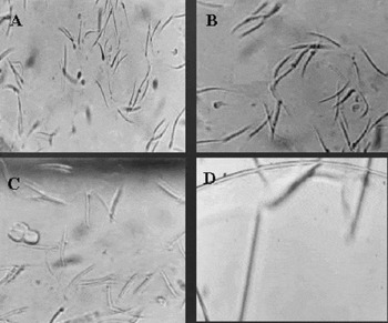
The examination of the vitelline envelopes isolated from the oocytes with or without trypsin treatment showed a high density of bound sperm on the envelope surface (Fig. 3). In the vitelline envelopes isolated from the oocytes with trypsin treatment before activation no bound sperm could be seen on the envelope surface (Fig. 3). In contrast, the vitelline envelope isolated from the oocytes without trypsin treatment before activation showed a high density of bound sperm (Fig. 3).
Vitelline envelope isolated from Bufo arenarum denuded oocytes matured in vitro by progesterone. (A) Vitelline envelope isolated from an oocyte with trypsin treatment, incubated with sperm for 15 min, showing high density of bound sperm. ×1000. (B) Vitelline envelope isolated from an oocyte without trypsin treatment, incubated with sperm for 15 min, showing a high density of bound sperm. ×1000. (C) Vitelline envelope isolated from an activated oocyte without trypsin treatment incubated 2 min after activation with sperm for 15 min, showing a high density of bound sperm. ×1000. (D) Vitelline envelope isolated from an oocyte with trypsin treatment and activated, incubated 2 min after activation with sperm for 15 min. It does not show bound sperm. ×1000.

The results indicate a loss of the sperm binding capacity in the vitelline envelopes isolated from the oocytes with trypsin treatment before activation. The protease action of trypsin might be involved in the loss of sperm binding and therefore in the block to polyspermy.
Interaction between vCGP denuded matured in vitro oocytes, perivitelline space and vitelline envelopes
In anuran eggs, after fertilization or activation, the contents of the cortical granules are initially expelled into the perivitelline space. We know that the material diffuses through the perivitelline space and through the matrix of the vitelline envelope itself. In order to study the contribution of the cortical granule material to the block to polyspermy we examined the space by scanning electron microscopy in oocytes matured in vitro and artificially activated and in fertilized oocytes.
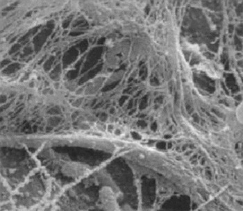
Groups of 20 oocytes matured in vitro were activated, inseminated 10 s after activation, and fixed 10 min later. In order to observe the perivitelline space, vitelline envelopes were manually pulled off. Fig. 4 shows sperm trapped by nets, which would be involved in the block to polyspermy.
Perivitelline space of Bufo arenarum denuded oocytes matured in vitro by progesterone, activated, inseminated 10 s after activation and fixed 10 min after insemination. The images shown sperm trapped by nets. ×8600.

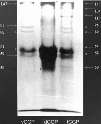
Electrophoretic profiles obtained by SDS-PAGE of vCGP, dCGP and tCGP were compared (Fig. 5). Gels were documented with a Gel-Doc 1000 system (Bio-Rad Lab) and analysed using Molecular Analyst software for image analysis systems.
SDS-PAGE of cortical granule products from Bufo arenarum oocytes. vCGP, from denuded oocytes matured in vitro by progesterone; dCGP, from ovoposited oocytes with vitelline envelope; tCGP, from ovoposited oocytes without vitelline envelope.

In the vCGP (oocytes matured in vitro) and tCGP (oviposited oocytes without vitelline envelopes) two bands were detected at 90 and 96 kDa. These bands were not found in dCGP (oviposited oocytes with vitelline envelopes). The results show that vitelline envelopes without trypsin treatment cannot retain either gp90 or gp96.
Electrophoretic analysis
Solubilized vitelline envelope components isolated from oocytes matured in vitro were analysed by SDS-PAGE to determine their macromolecular composition.The vitelline envelopes were isolated under different experimental conditions:
1. from oocytes matured in vitro;
2. From oocytes matured in vitro, with trypsin treatment
3. from oocytes matured in vitro, activated by electric shock;
4. from oocytes matured in vitro, with trypsin treatment and activated by electric shock.
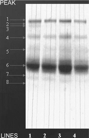
Electrophoretic analyses revealed small differences (Fig. 6).
SDS-PAGE of vitelline envelopes from Bufo arenarum denuded oocytes matured in vitro by progesterone. Line 1, proteins from vitelline envelopes of denuded oocytes matured in vitro; line 2, proteins from vitelline envelopes of denuded oocytes matured in vitro with trypsin treatment; line 3, proteins from vitelline envelopes of denuded oocytes matured in vitro and activated; line 4, proteins from vitelline envelopes of denuded oocytes matured in vitro with trypsin treatment and activated.

When the Bufo arenarum vitelline envelopes of denuded full-grown ovarian oocytes matured in vitro by progesterone were analysed, we found the conversion of gp40 (oocytes matured in vitro, without trypsin treatment) to gp38 (ascribable to trypsin activity or to CGP activity) and the conversion of gp70 to gp68 (ascribable to trypsin activity plus CGP activity).
Discussion
Denuded full-grown oocytes of the amphibian Bufo arenarum matured in vitro by progesterone are polyspermic (Oterino et al., Reference Oterino, Sánchez Toranzo, Zelarayán and Bühler1997), although cortical granule exocytosis seems to occur following a normal chronological sequence (Oterino et al., Reference Oterino, Sánchez Toranzo, Zelarayán, Valz-Gianinet and Bühler2001).
The slow block, which is dependent on cortical granule exocytosis, involves physico-chemical transformations of the vitelline envelope also found in ovoposited oocytes (Gómez, Reference Gómez1992). In B. arenarum, as the egg traverses the oviduct, the vitelline envelope is altered by a trypsin-like protease secreted by the oviduct (Cabada et al., Reference Cabada, Mariano and Raisman1978; Miceli et al., Reference Miceli, Fernández, Raisman and Barbieri1978a, Reference Miceli, Fernández and Del Pinob; Katagiri et al., Reference Katagiri, Iwao and Yoshizaki1982; Miceli & Fernández, Reference Miceli and Fernández1982; Takamune et al., Reference Takamune, Yoshizaki and Katagiri1986; Hedrick & Hardy, 1991). The pars recta secretion, termed oviductin, induces an increased susceptibility of the vitelline envelopes to sperm lysins (Miceli et al., Reference Miceli, Fernández, Raisman and Barbieri1978).
Denuded oocytes of B. arenarum, matured in vitro, were inseminated with trypsin treatment, the same as coelomic oocytes, because they are not influenced by oviductal secretions. However, we found that denuded oocytes can be fertilized with or without trypsin treatment, suggesting that this “immature” vitelline envelope is totally sperm-permeable.
However, the insemination experiments in activated oocytes showed that this envelope is unable to block sperm entry during the first 60 s after activation without trypsin treatment. The analysis of SDS-PAGE profiles showed two bands at 90 and 96 kDa in the vCGP (oocytes matured in vitro) and tCGP (oviposited oocytes without vitelline envelopes), but not in the dCGP (oviposited oocytes with vitelline envelopes). The results show that vitelline envelopes without trypsin treatment cannot retain either gp90 or gp96. This suggests that these glycoproteins are involved in the block to polyspermy and that trypsin treatment of matured in vitro oocytes is necessary to enable vitelline envelopes to block polyspermy.
Presumably, one of the functions of the vitelline envelope is to provide binding sites for sperm fertilization. This binding process is very important because if it is blocked fertilization will not take place. In amphibians, the change in the gamete adhesion step at the level of the vitelline envelope can be ascribable to the ultrastructural alteration of the envelope during the passage of the oocyte through the uppermost portion of the oviduct (Grey et al., Reference Grey, Working and Hedrick1977; Katagiri et al., Reference Katagiri, Iwao and Yoshizaki1982; Mariano et al., Reference Mariano, Gómez de and Pisanó1984; Gerton & Hedrick, Reference Gerton and Hedrick1986; Takamune & Katagiri, Reference Takamune and Katagiri1987; Hardy & Hedrick, Reference Hardy and Hedrick1992). Sperm can bind and penetrate the ovoposited oocyte but not the coelomic oocyte (Omata & Katagiri, Reference Omata and Katagiri1996).
The loss of the binding capacity after fertilization or activation may be due to the interaction between products released by the cortical granules and the vitelline envelope during oocyte activation. This process could hinder or modify the sites of interaction of the vitelline envelope with the sperm (Cabada et al., Reference Cabada, Mariano and Gómez1987; Linsay & Hedrick, 1989; Yamasaki & Katagiri, Reference Yamasaki and Katagiri1991). The loss of the binding capacity in vitelline envelopes isolated from B. arenarum oocytes matured in vitro with trypsin treatment and activated by electric shock suggests that previous trypsin treatment is a necessary step for sperm block to occur. When in vitro matured oocytes were incubated with vCGP, vitelline envelopes with trypsin treatment were able to block sperm entry; these oocytes also exhibited the characteristic signs of activation. These results support the idea that B. arenarum oocytes can be activated by external stimuli and suggest the presence of unknown oocyte surface receptors linked to the activation machinery in response to fertilization.
Several groups have reported biochemical modifications in the vitelline envelope of amphibians. Hardy & Hedrick (Reference Hardy and Hedrick1992) have described the conversion of gp43 (glycoprotein component of the coelomic oocyte) to gp41 (in the uterine oocyte) caused by a serine active site protease, oviductin, with trypsin-like specificity. Oviductin can also convert gp43 to gp41 in isolated vitelline envelopes (Lindsay et al., Reference Lindsay, Wieduwilt and Hedrick1999).
Electrophoretic profiles obtained by SDS-PAGE of solubilized vitelline envelopes from oocytes matured in vitro revealed slight differences like those found in Xenopus laevis (Hedrick & Nishihara, Reference Hedrick and Nishihara1991) and B. arenarum (Barisone et al., Reference Barisone, Albertali, Sánchez and Cabada2003). These differences are especially marked in the conversion of gp42 (glycoprotein component of the coelomic oocyte) to gp41 (in the uterine oocyte). This modification has been implicated in the acrosome reaction. In B. arenarum we observed the conversion of gp40 (matured in vitro oocytes, without trypsin treatment) to gp38 (ascribable to trypsin activity or CGP activity) and the conversion of gp70 to gp68 (ascribable to trypsin activity plus CGP activity). Taking into account that only the vitelline envelopes of in vitro matured oocytes, with trypsin treatment and activated, can block sperm entry, we may suggest that the conversion of gp70 to gp68 is related to the changes associated with sperm binding. All these observations indicate that the slow block, which is dependent on cortical granule exocytosis and physico-chemical transformations of the vitelline envelope, is effectively established in in vitro matured oocytes, suggesting that failure in the prevention of polyspermy would be related to the fast block to polyspermy that is established before cortical granule breakdown. This hypothesis remains to be investigated in future studies.