INTRODUCTION
Large and comprehensive inventories of the deep-sea fauna in the South Atlantic–Weddell Sea system are generating a torrent of new data, an important part of which relates to the discovery of numerous new species (Janussen et al., Reference Janussen, Tabachnick and Tendal2004; Plotkin & Janussen, Reference Plotkin and Janussen2007, Reference Plotkin and Janussen2008; Janussen & Reiswig, Reference Janussen and Reiswig2009; Vieira et al., Reference Vieira, Cosme and Hajdu2010; Göcke & Janussen, Reference Göcke and Janussen2011, Reference Göcke and Janussen2013b; Lopes et al., Reference Lopes, Bravo and Hajdu2011; Rapp et al., Reference Rapp, Janussen and Tendal2011, Reference Rapp, Göcke, Tendal and Janussen2013). The Demospongiae have been comparatively less studied, so we have published detailed descriptions of the demosponges sampled during the SYSTCO I expedition in Göcke & Janussen (Reference Göcke and Janussen2013b). Here we conclude the study of these sponges by focusing on the little known family Phellodermidae Van Soest & Hajdu, Reference Van Soest, Hajdu, Hooper and van Soest2002.
Phellodermidae is currently understood (Van Soest & Hajdu, Reference Van Soest, Hajdu, Hooper and van Soest2002) as belonging in the Myxillina Hajdu, Van Soest & Hooper, Reference Hajdu, van Soest, Hooper, van Soest, van Kempen and Braekman1994, and characterized by the possession of monactinal megascleres and arcuate chelae. The family comprises two genera, namely Echinostylinos Topsent, 1927 and Phelloderma Ridley & Dendy, Reference Ridley and Dendy1886. Both are nearly exclusively deep-water dwelling, with the notable exception of E. reticulatus sensu Bergquist & Fromont (Reference Bergquist and Fromont1988), reported from 55–110 m depth.
In the present study we re-describe the holotype of Phelloderma radiatum Ridley & Dendy, Reference Ridley and Dendy1886, and correct some flaws in the original description. We describe a new Phelloderma species, remarkable for its possession of abundant oxychaetes (onychaete-like microxeas) and propose the transfer of Esperiopsis flava Lévi, Reference Lévi and Crosnier1993 to Phelloderma. Additionally, we contrast the myxilline affinities of Phellodermidae implied in the current classification, to an alternative scenario of mycaline affinity. The latter is suggested by the great similary exhibited by the robust arcuate isochelae and polytylote mycalostyles of Phelloderma spp. and those of Abyssocladia Lévi, 1964.
MATERIALS AND METHODS
The sponge described herein as the holotype of a new species was collected during the ANT XXIV/2 SYSTCO I expedition with RV ‘Polarstern’ during the winter 2007–2008. The Antarctic deep-sea bentho-pelagic SYSTem COupling program is a continuation and follow-up of the successful ANtarctic benthic DEEP-sea biodiversity cruises (ANDEEP I and II in 2001–2002 and ANDEEP III in 2005). SYSTCO I took place from 28 November 2007 to 4 February 2008. The sponge specimen described herein was collected during SYSTCO I on 3 January 2008 from Station no. 039/11 by a giant box corer at a depth of 2151.5 m. For a full report on SYSTCO I demosponges and a map of their occurrences see Göcke & Janussen (Reference Göcke and Janussen2013a, Reference Göcke and Janussenb). The specimen was photographed immediately upon collection, and fixed in 96% ethanol soon after. Skeletal architecture was studied on histological sections by light microscopy. These were prepared following the technique of Vacelet (Reference Vacelet2006), and dissociated spicules following the technique of Boury-Esnault & Rützler (Reference Boury-Esnault and Rützler1997). The measurements of spicules were made using a calibrated stage micrometer, and the numbers of measured spicules are indicated specifically for each case. Spicule dimensions are given as minimum–maximum, with indication of standard deviation, including maximum length and thickness for the megascleres. All sponges from the ANDEEP and SYSTCO expeditions are deposited in the Senckenberg Naturmuseum, Frankfurt am Main (SMF). These are available online via the electronic Senckenberg collections management database (SESAM). Furthermore, we re-examined the holotype of Phelloderma radiatum, which was loaned from the Natural History Museum, London (BMNH), and some specimens of the same species reported from the Antarctic by Hentschel (Reference Hentschel1914), which were loaned from the Zoological Museum of Berlin (ZMB).
RESULTS
SYSTEMATICS
Order POECILOSCLERIDA Topsent, 1928
Suborder MYXILLINA Hajdu, Van Soest & Hooper, Reference Hajdu, van Soest, Hooper, van Soest, van Kempen and Braekman1994
Family Phellodermidae Van Soest & Hajdu, Reference Van Soest, Hajdu, Hooper and van Soest2002
Phelloderma Ridley & Dendy, Reference Ridley and Dendy1886
EMENDED DIAGNOSIS
Phellodermidae with a single category of frequently polytylote (subtylo)styles radially organized, and stout arcuate isochelae or arcuate cleistochelae, to which oxychaetes may be added.
TYPE SPECIES
Phelloderma radiatum Ridley & Dendy, Reference Ridley and Dendy1886: 347 (by monotypy).
REMARKS
The finding that Echinostylinos may also have only a single category of megascleres (Cosme & Hajdu, Reference Cosme, Hajdu, Lavrado and Brasil2010; possibly also Mycalopsis lingua Koltun, 1970), the finding of oxychaetes in a species of Phelloderma (see below), and the transfer of Abyssocladia Lévi, 1964, from the synonymy of Phelloderma, to the Cladorhizidae, proposed by Vacelet (Reference Vacelet2006), all demanded a redefinition of Phelloderma. The main distinguishing aspect now to differentiate between genera of Phellodermidae is the basically radial architecture and encrusting growth of Phelloderma vs a more erect architecture in Echinostylinos, which can exhibit axially condensed or somewhat plumo-reticulated skeletal patterns.
Esperiopsis flava Lévi, Reference Lévi and Crosnier1993, from New Caledonia, is best assigned to Phelloderma on account of its neatly arcuate chelae of cleistochelae morphology, and single category of polytylote styles in a radial-like arrangement [‘styles très nombreux, tous orientés, ascendants’, Lévi (Reference Lévi and Crosnier1993)].
Phelloderma radiatum Ridley & Dendy, Reference Ridley and Dendy1886
(Figures 1–3; Table 1)
Phelloderma radiatum Ridley & Dendy, Reference Ridley and Dendy1886: 347–348, Reference Ridley and Dendy1887: 113–114, pl. 23, figure 8. Hentschel, Reference Hentschel1914: 69–70.
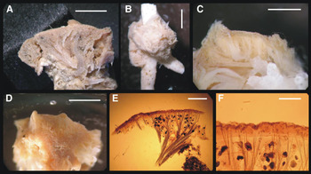
Phelloderma radiatum Ridley & Dendy, Reference Ridley and Dendy1886: (A) holotype (BMNH 1887.5.2.167), specimen laterally sliced: (B) ZMB Por 4826 specimen 77, habitus, sponge is growing on bryozoan: (C) ZMB Por 4826 specimen 502, close-up of laterally sliced part: (D) ZMB Por 4826 specimen 502, dermal surface: (E, F) holotype (BMNH 1887.5.2.167), skeletal sections. Scale bars: A, 5 mm; B, 2.5 mm; C, 2 mm; D, 2.5 mm; E, 1 mm; F, 500 µm.

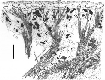
Phelloderma radiatum Ridley & Dendy, Reference Ridley and Dendy1886, holotype (BMNH 1887.5.2.167). Skeletal architecture. Scale bar: 500 µm.

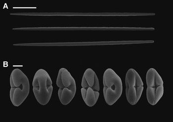
Phelloderma radiatum Ridley & Dendy, Reference Ridley and Dendy1886, holotype (BMNH 1887.5.2.167): (A) Polytylote styles: (B) Arcuate cleistochelae. Scale bars: A, 100 µm; B, 10 µm.

Comparative spicular micrometric data on all Phelloderma spp. Values given in µm.

TYPE MATERIAL
Holotype: BMNH 1887.5.2.167 Challenger expedition Station 320, 14 February 1876 (37°17′S– 53°52′W, off the mouth of the Rio de la Plata), 1097.28 m depth.
COMPARATIVE MATERIAL EXAMINED
ZMB Por 4826, Deutsche Südpolar Expedition, Gauss-Station (66°2′9″S–89°38′E), 385 m, 16 April, 25 June, 22 November 1902, three specimens.
DIAGNOSIS
Subglobular, encrusting sponge with radially arranged skeleton of plumose tracts, and characteristic polytylote styles and arcuate cleistochelae.
DESCRIPTION
Subglobular sponge (Figure 1A), originally encrusting over Sporadopora (Cnidaria, Hydrozoa). Surface is smooth, slightly hispid, ectosome is dense. Few oscula present, situated on short papillae (Figure 1A, D). Ectosome without specialized skeleton, except an accumulation of chelae. Below the ectosome, a weakly developed second layer of tissue can be found, overlying the choanosome (Figures 1, 2). A high amount of foreign material, predominantly sand grains, is regularly distributed within the tissue. Texture in ethanol is spongy, chewy. Colour in ethanol is brown–beige. Within the tissue, some embryos of ~400 µm in diameter are present.
Skeleton. Radial arranged plumose spicular tracts (Figures 1E, F, 2), branching towards the surface, with styles spreading as bouquets in the subectosomal region. Microscleres free within the tissue, predominantly in the ectosome, often on its surface.
Spicules (Figure 3; Table 1). Main spicules are straight polytylote styles (Figure 3A), rarely moderately curved, with the pointed end often blunted. Sizes variable, 490–600 × 10–18 µm. Microscleres are arcuate isochelae with three broad alae on each side (Figure 3B). The middle alae often grow very closely to each other, in most cases they even seem to coalesce. This phenomenon is rather rare among lateral alae. Specimens with all alae being free are also present, in these cases, the ends of the alae are more rounded and alae are less broad. Chelae are 36–44 µm long with terminations 17–23 µm high.
DISTRIBUTION
The Patagonian coast off the mouth of Rio de la Plata; Antarctica, off Wilhelm II Land.
REMARKS
In their original description, Ridley & Dendy (Reference Ridley and Dendy1886, Reference Ridley and Dendy1887) reported a distinct ‘cork-like cortex lying immediately below the dermal membrane, about 0.24 mm thick’. This cortical structure was even used as a character for determination by Hentschel (Reference Hentschel1914) in his report of five Antarctic specimens, which is the only further report of the species. In our re-examination of the holotype, as well as of Hentschel's specimens, we could not see such a prominent structure, we only detected a thin layer of specialized tissue immediately beneath the ectosome, which can hardly be considered a diagnostic character for the species. The term ‘corky’ is not very precise to describe the texture of the sponge. As it appears to us, this structure which we may identify as the ‘cork-like cortex’ does not have a very special consistency strongly differing from common ectosomal structures present in other sponge species.
Ridley & Dendy (Reference Ridley and Dendy1887) gave a single value for the size of each spicule type, which are probably the maximal sizes. In Table 1 we document the actual size variation and mean values for the spicules, which in most cases are often distinctly below the maximal values given by Ridley & Dendy (Reference Ridley and Dendy1887). In the case of the styles, we were not able to find their maximum, as the largest styles we found in new preparations from the holotype were 600 µm long only, despite originally reported to reach 650 µm. Hentschel (Reference Hentschel1914) reported similar size variations from his Antarctic specimens. His sponges are similar to the holotype in spicular morphology, both in dimensions and shape, as well as in their general body plan. The only distinct difference from the holotype is their high fragility, which might be due to their thin growth. As the general shape of the sponges is better preserved than that of the holotype (due to preparations), we chose to include some pictures of their anatomy in Figure 1B–D.
Phelloderma oxychaetoides sp. nov.
(Figures 4–6; Table 1)
Phelloderma oxychaetoides sp. nov., holotype (SMF 11726): (A) specimen attached to substrate. The smaller sponge is a Tentorium semisuberites (SMF 11727): (B) cross-section of the specimen showing neat radial architecture with divergent megasclere tracts running toward the surface, intermingled to abundant embryos: (C) abundance of oxychaetes in the ectosomal and subectosomal regions: (D) embryo bearing a dense mass of arcuate isochelae in its central portion. Scale bars: A, 10 mm; B, 1 mm; C, D, 100 μm.

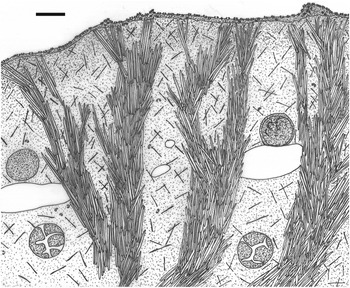
Phelloderma oxychaetoides sp. nov., holotype (SMF 11726, A–D). Skeletal architecture. Scale bar: 500 µm.

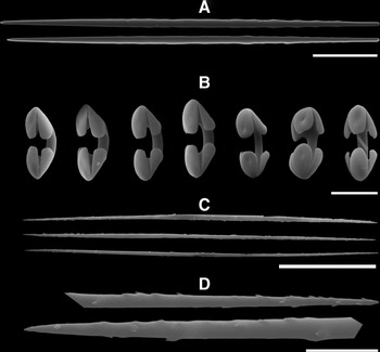
Phelloderma oxychaetoides sp. nov., holotype (SMF 11726, A–D): (A) polytylote mycalostyles: (B) arcuate isochelae: (C) oxychaete microscleres: (D) detail of extremities of the oxychaetes. Scale bars: A, 100 μm; B, 20 μm; C, 30 μm; D, 3 μm.

TYPE MATERIAL
Holotype: SMF 11726 SYSTCO I Station 39-11 (64°28.80′S–02°52.52′E; Weddell Sea, Antarctic), 2151.5 m depth, coll. RV ‘Polarstern’, 3 January 2008.
DIAGNOSIS
The only species of Phelloderma with abundant oxychaetes.
DESCRIPTION
The specimen is a round encrusting sponge with a diameter of 19 mm and a maximal thickness of 3 mm overgrowing a basalt stone (Figure 4A). The sponge thins out towards the periphery. Its color in life was light yellowish, almost white. Preserved in ethanol, it is white. The specimen was growing close to a small Tentorium semisuberites (SMF 11727).
Skeleton (Figures 4B–D, 5). Unspecialized ectosome, traversed by the terminations of choanosomal tracts of megascleres in vicinity of oscula (Figure 4B); otherwise smooth, slightly wavy surface. Ectosome is thin, with distinct aggregation of chelae. Choanosome with radially disposed tracts of styles (ca 100–250 μm across, Figure 4B), only slightly divergent when approaching the ectosome. Isochelae are common, and oxychaetes abundant (Figure 4C) in all parts of the sponge. The holotype bears abundant embryos (~290 μm in diameter), one of which possesses a dense central mass of seemingly disorganized isochelae of the same dimensions as those spread in the mesohyl of the adult sponge (Figure 4D).
Spicules (Figure 6; Table 1). Megascleres: styles (Figure 6A) varying little in dimensions, smooth, fusiform, occasionally slightly curved, frequently slightly polytylote, tapering gradually to moderately sharp ends, 580–710 × 14–17 µm. Microscleres: stout, deeply-curved arcuate isochelae, 27–38 µm (Figure 6B); oxychaetes (onychaete-like microxea, Figure 6C, D), iso-terminated (oxeote), with microspines often unevenly dispersed and all pointing in the same direction, 118–168 μm.
DISTRIBUTION
Provisional Weddell Sea endemic, from 2151.5 m depth.
ETYMOLOGY
The species is named for the occurrence of the microsclere type oxychaeta, which is so far unique for the genus.
REMARKS
The dubious ‘cork-like cortex’ of genotypic species P. radiatum reported by Ridley & Dendy (Reference Ridley and Dendy1887), and subsequently used as a diagnostic character by Hentschel (Reference Hentschel1914), turned out to be a rather vague feature in our re-examination of the species’ holotype (see above). So, the absence of such a structure in the new species cannot be considered an impediment for assigning it to Phelloderma. Assignment to this genus is confirmed by strong similarities between our new species and the genotypical P. radiatum, such as the general body plan and organization of the skeleton, as well as the characteristic polytylote styles with slightly blunted sharp ends and arcuate isochelae.
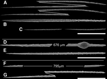
In spite of the overwhelming abundance of oxychaetes found in the new species, we first thought these were onychaetes, and a suspicion on their possible allochthonous origin did not completely fade away until a detailed SEM study of the onychaetes of Tedania (Tedaniopsis) tantula (Kirkpatrick, 1907) collected in the same area was conducted (Figure 7). All the onychaetes studied showed aniso-terminations and occurred in two or three distinct size classes, the larger of which being much longer than the oxychaetes in P. oxychaetoides sp. nov. One of the onychaete categories was obviously fusiform (Figure 7B) and another had a conspicuous subterminal tyle (Figure 7D), that rendered both further distinct from the oxychaetes. We considered it safe thus to assume the latter as proper for the new Phelloderma species described herein. Rather similar oxychaetes to those in P. oxychaetoides sp. nov. are also present in Celtodoryx Perez, Perrin, Carteron, Vacelet & Boury-Esnault, Reference Perez, Perrin, Carteron, Vacelet and Boury-Esnault2006, which furthermore has arcuate isochelae not too different from the ones in our new species. Nonetheless, the genus as defined by Perez et al. (Reference Perez, Perrin, Carteron, Vacelet and Boury-Esnault2006) shows several distinct differences from the new sponge, which clearly underline it does not belong there. Celtodoryx possesses a distinctly plumoreticulate skeleton and tylotes with terminations bearing distinct spines. Our new sponge clearly has a skeleton made of short plumose fibres and polytylote styles as the sole megascleres, both characters being in good accordance with the current diagnosis for Phelloderma (see above). Furthermore, Celtodoryx is only known from the China Yellow Sea and the North Sea (as an invader species, see Henkel & Janussen, Reference Henkel and Janussen2011), whereas Phelloderma has been known already long ago to be an Antarctic genus (Hentschel, Reference Hentschel1914).
Diverse onychaetes for comparison: Tedania (Tedania) rhoi Sim & Lee, 1998 (A–C, Er Tuo, Dalian, Yellow Sea, SMF 10808): (A) onychaetes–I: (B) onychaete–II: (C) onychaetes–III. Tedania cf. tantula Kirkpatrick, 1907 (D–E, ANDEEP III, Station 74–7, SMF 11757): (D) gigantic onychaete–I: (E) onychaete–II. Tedania cf. tantula (F–G, ANDEEP III, Station 74–7, SMF 11756): (F) gigantic onychaete–I: (G) onychaete–II. Scale bars: 30 μm.

After Vacelet's (Reference Vacelet2006) transfer of Abyssocladia to the Cladorhizidae (Mycalina), Phelloderma remained poorly represented by only two species. The new species described here clearly differs from P. radiatum and P. polypoides, mainly by the abundant oxychaetes found in the new species, but also by its smooth dermal surface and the very low size variability of styles and chelae (Table 1).
IDENTIFICATION KEY FOR PHELLODERMA SPP.
1. Oxychaetes present in abundance………P. oxychaetoides sp. nov.
Oxychaetes absent………2
2. Styles polytylote, thicker than 10 μm………3
Styles smooth, thinner than 10 μmP. polypoides
3. Arcuate cleistochelae smaller than 35 μm………P. flava
Arcuate cleistochelae larger than 40 μm………P. radiatum
DISCUSSION
The acceptance of arcuate chelae within the Mycalina, once restricted to a few anisochelae of disputable ontogeny occurring in some Asbestopluma Topsent, 1901 (Cladorhizidae), is now supported by the inclusion of several species of Abyssocladia in this suborder (Vacelet, Reference Vacelet2006; Ise & Vacelet, Reference Ise and Vacelet2010; Kelly & Vacelet, Reference Kelly and Vacelet2011; Lopes et al., Reference Lopes, Bravo and Hajdu2011; Lopes & Hajdu, Reference Lopes and Hajdu2014). This move demands a reconsideration of the proposed transformation series for isochelae, viz. palmate–arcuate–anchorate (sensu Hajdu et al., Reference Hajdu, van Soest, Hooper, van Soest, van Kempen and Braekman1994), as is similarly done by Hajdu et al. (Reference Hajdu, de Paula, Redmond, Cosme, Collins and Lobo-Hajdu2013), who recognized the anchorate morphotype to be the most basal chelae, from which the other types derive. It is also important to note that recognition of the validity of Abyssocladia as proposed by Vacelet (Reference Vacelet2006), and followed by many authors is one thing. Another is its assignment to the Cladorhizidae, which was not settled on firm ground until recently, as alternative, equally parsimonious evolutionary morphological scenarios could be easily built.
The arguments underlying Vacelet's (Reference Vacelet2006) proposition assembled the stipitate morphology, widespread possession of sigmancistras and likely carnivorous feeding habit, as suggestive of greater proximity to Cladorhizidae than to Phelloderma. These might all be symplesiomorphic traits, or homoplasy, as they occur in distinct families within the Mycalina or elsewhere in the Demospongiae. Sigmancistras had already been recognized as a likely symplesiomorphy by Vacelet (Reference Vacelet2006), for their occurrence in both Cladorhizidae and Guitarridae, and are thus as good a character to propose affinity to the former family, as they are to propose to the latter. Stipitate, or nearly so, representatives of Esperiopsis Carter, 1882 (e.g. E. symmetrica Ridley & Dendy, Reference Ridley and Dendy1886, E. desmophora Hooper & Lévi, 1989, E. bathyalis Lopes & Hajdu, 2004, E. koltuni Ereskovsky & Willenz, 2007—Esperiopsidae, Mycalina), also (likely) carnivorous, were suggested as possibly best assigned to Abyssocladia by Lopes et al., (Reference Lopes, Bravo and Hajdu2011). This leaves the possible parallelism of poriferan carnivory resting on Euchelipluma alone, if Cercicladia Rios, Kelly & Vacelet, Reference Ríos, Kelly and Vacelet2011 truly belongs in Cladorhizidae. Of course, the possibility cannot be entirely ruled out that Euchelipluma actually belongs in the Cladorhizidae, as placochelae-like microscleres have already been reported for Asbestopluma, i.e. the anisoplacochelae of A. anisoplacochela Kelly & Vacelet, Reference Kelly and Vacelet2011.
Additional morphological traits in Abyssocladia were given high relevance in proposing its phellodermid affinity in the Systema Porifera (Van Soest & Hajdu, Reference Van Soest, Hajdu, Hooper and van Soest2002)—the combined occurrence of mycalostyles and a particular kind of stout arcuate isochelae, called abyssochelae. In the present classificatory scheme (Vacelet, Reference Vacelet2006; Van Soest et al., Reference Van Soest, Boury-Esnault, Hooper, Rützler, de Voogd, Alvarez de Glasby, Hajdu, Pisera, Manconi, Schoenberg, Janussen, Tabachnick, Klautau, Picton, Kelly, Vacelet, Dohrmann, Cristina Díaz and Cárdenas2014), nevertheless, Phelloderma spp. and Abyssocladia spp. are classified in two distinct suborders, an option which is further corroborated by the present finding of oxychaetes in P. oxychaetoides sp.nov., as well as by molecular evidence brought up by Vargas et al. (Reference Vargas, Erpenbeck, Göcke, Hall, Hooper, Janussen and Wörheide2012). These authors observed on the basis of two molecular markers (28S rDNA and COI) that Abyssocladia is best classified within the Cladorhizidae, which come out as the sister group of Mycale within the Mycalina, whereas Phelloderma appears to be well nested within the Myxillina.
Undoubtedly, Van Soest & Hajdu (Reference Van Soest, Hajdu, Hooper and van Soest2002) were led by the frequently combined occurrence of auxiliary monactinal megascleres and arcuate chelae in Phelloderma, Echinostylinos, and alleged synonyms, to propose their myxilline affinity. But an alternative proposal might be considered, drawn from the observation that both genera actually possess species devoid of any auxiliary megascleres (e.g. Phelloderma spp.; Echinostylinos sp. in Cosme & Hajdu, Reference Cosme, Hajdu, Lavrado and Brasil2010); and at the same time recognizing that auxiliary megascleres were also known from mycalines, as is the case with many cladorhizids (e.g. Asbestopluma—A. agglutinans Vacelet, Reference Vacelet2006) and a few mycalids (e.g. Mycale—M. arenaria Hajdu & Desqueyroux-Faúndez, Reference Hajdu and Desqueyroux-Faúndez1994; Phlyctaenopora—P. bitorquis Topsent, 1904). In summary, auxiliary megascleres may represent affinity to either the Myxillina or the Mycalina, but the oxychaetes reported here for Phelloderma are a strong back-up for the first option.
This morphological conundrum is mixed up further by the current state of affairs regarding the accepted distribution of oxychaetes and chelae transformation series. The first of these, hitherto restricted to Chaetodoryx Topsent, 1927 and Celtodoryx Perez, Perrin, Carteron, Vacelet & Boury-Esnault, Reference Perez, Perrin, Carteron, Vacelet and Boury-Esnault2006 (both Coelosphaeridae, Myxillina), while the latter, besides its neat observation within the Myxillina, also occurs as a somewhat undervalued evolutionary parallel achievement within the Cladorhizidae.
The crucial question, not only for the classification of Phelloderma but also for the phylogenetic system of the whole Poecilosclerida, is the value of the interpretation of microsclere micromorphology as being selectively neutral, as suggested by several specialists (e.g. Dendy, Reference Dendy1921; Hajdu & Desqueyroux-Faúndez, Reference Hajdu and Desqueyroux-Faúndez1994; Hajdu et al., Reference Hajdu, van Soest, Hooper, van Soest, van Kempen and Braekman1994; Hajdu & Van Soest, Reference Hajdu and van Soest1996). As morphological and some molecular data have already brought some insight into the relationships within the Poecilosclerida, especially within the Mycalina (Hajdu et al., Reference Hajdu, de Paula, Redmond, Cosme, Collins and Lobo-Hajdu2013; Redmond et al., Reference Redmond, Morrow, Thacker, Diaz, Boury-Esnault, Cardenas, Hajdu, Lobo-Hajdu, Picton, Pomponi, Kayal and Collins2013), still further studies will be necessary for a better understanding of the complex issues of systematics and phylogeny within the highly diverse order Poecilosclerida, especially concerning the Cladorhizidae, as well as other carnivorous sponges currently classified within the poecilosclerid families Esperiopsidae and Guitarridae,
ACKNOWLEDGEMENTS
Lena Kirchhof (Senckenberg Frankfurt) is thanked for preparing drawings of sponge skeletons and for assistance in preparing the SEM-pictures of P. radiatum; Heike Szmutka (Senckenberg Frankfurt) is thanked for spicule preparation of P. radiatum holotype. We want to give thanks to Emma Sherlock (British Museum of Natural History) for lending us the holotype of P. radiatum, and to Carsten Lüter (Museum für Naturkunde Berlin) for lending us Hentschel's specimens of P. radiatum. We thank also our colleagues Sergio Vargas and Gert Wörheide (Munich University) for important cooperation on the molecular phylogeny of the Poecilosclerida, Alexander Plotkin (St Petersburg University) for SEM photos, and Wolfgang Dröse (Göttingen University) for histological sections.
FINANCIAL SUPPORT
We thank the CeDAMar Taxonomist Exchange Program for a grant which enabled the visit by E. Hajdu in May 2010 to Senckenberg for collaborative work on ANDEEP/SYSTCO Demospongiae. E.H. further thanks the National Counsel of Technological and Scientific Development (CNPq), as well as Carlos Chagas Filho Research Support Foundation of Rio de Janeiro State (FAPERJ) for grants and/or fellowships that enabled completion of this work. Thanks are due to the Deutsche Forschungsgemeinschaft for financing projects by D.J. on Antarctic Porifera (JA 1063/14-1,2, JA-1063/17-1). This is ANDEEP-SYSTCO publication no. 152.