Arteriovenous malformations, commonly located in the brain, liver, and limbs, are well recognized as producing congestive cardiac failure in fetuses and newborns.Reference McMahon1 Arteriovenous malformations within the thorax, but outside the pulmonary capillary bed, however, are extremely rare. We report our experience with an arteriovenous fistula unusually located between the left subclavian artery and the left brachiocephalic vein. The fistula was already known to be causing cardiac failure during fetal life, and has been effectively treated by surgery postnatally.
Case report
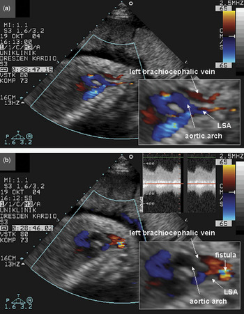
A large arteriovenous malformation extending from the left subclavian artery to the left brachiocephalic vein was identified during fetal echocardiography (Fig. 1) at 26 weeks of gestation. A follow-up echocardiogram at 28 weeks of gestation showed signs of congestive cardiac failure, with rightward deviation of the cardiac long axis, slight dilation of the pulmonary trunk, enlargement of the right atrium and ventricle, and a moderate pericardial effusion. The fetus was treated successfully by administration of digoxin to the mother.
Fetal echocardiography at 28 weeks of gestation shows (a) the aortic arch in blue, with the origin of the left subclavian artery, and the left brachiocephalic vein in red. Turbulent flow is seen across the arteriovenous fistula between the vessels (b). The spectral Doppler (b) shows the peak velocity of flow inside the fistula to be 4 metres per second. LSA – left subclavian artery.

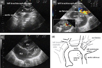
The male infant, weighing 3,320 grams, was born at 38 weeks of gestation with Apgar scores of 9, 9, and 9. Because of clinical and radiological signs of wet lungs, we commenced continuous positive airway pressure ventilation. The clinical examination showed signs of obstruction to the return of venous blood from the head and neck, with slight oedema of the upper body and distension of the cervical veins. There was no evidence of cyanosis, but the infant showed mild intercostal and epigastric retractions, periodic respiration, intermittent apnoea, and slight hepatomegaly. The peripheral pulses were slightly decreased. A loud continuous murmur, graded 4 from 6, was audible across the upper left chest, heard maximally in the left infraclavicular region, with diffuse radiation to the right side and the back. Chest radiography revealed cardiomegaly due to right atrial and right ventricular distension. Postnatal echocardiography confirmed the presence of an arteriovenous malformation between the left subclavian artery and the left brachiocephalic vein (Fig. 2). Doppler interrogation showed a maximum velocity of 4.0 metres per second. The right atrium and ventricle were markedly increased in their size (Fig. 2c), and the pressure in the right ventricle, as measured on the basis of an increased gradient of tricuspid regurgitation, and also by the right-to-left shunt across the oval foramen and the arterial duct, was increased. The contractility of both ventricles, however, was normal. In addition, the pulmonary trunk was elongated, and ectasia was noted in the ascending aorta, the aortic arch, and the proximal part of the left subclavian artery (Fig. 2a, b). There was no evidence of coarctation. Cerebral ultrasound showed no anomalies, apart from decreased enddiastolic flow due to the arteriovenous malformation.
Transthoracic echocardiography on the first day of life from the suprasternal notch (a) shows the dilated left brachiocephalic vein, and an enlarged left subclavian artery rising from the aortic arch. Colour Doppler imaging in the same view (b) shows the arteriovenous fistula draining into the left brachiocephalic vein. The modified four chamber view (c) demonstrates enlargment of the right atrium and right ventricle. In panel d, we show a schematic drawing of the pathological anatomy. Abbreviations : LSA – left subclavian artery, RA – right atrium, RV – right ventricle, LA – left atrium, LV – left ventricle, av- arteriovenous.

The day after birth, the fistula was resected. During the surgical procedure, coarctation was noted at the junction of the aortic isthmus with the arterial duct. The coarctation lesion was resected, and the aortic pathways reconstituted by end-to-end anastomosis and resection of the arterial duct. The floor of the oval fossa was also partially resected, the hole created being closed by a pericardial patch containing a 5 millimetre fenestration. The postoperative course was complicated by capillary leak syndrome and bilateral pleural effusions, confirmed as being due to chylothorax. With administration of diuretics and a diet free of fatty acids, the symptoms disappeared. Treatment with digoxin and spironolactone was continued for 12 weeks after surgery. The boy was discharged home 4 weeks after surgery. He is now 4 years old, and doing well. Follow-up evaluations have shown no abnormalities of cardiac function, with normal flow observed across the aortic isthmus. The fenestration in the pericardial patch remains open, albeit now measuring 3 millimeters in diameter, and the right atrium, right ventricle, pulmonary trunk, ascending aorta, the proximal part of the left subclavian artery, the superior caval vein, and the left brachiocephalic vein all remain slightly enlarged.
Discussion
Our patient is of interest for several reasons. First, the location of the fistula is extremely rare. Second, signs of cardiac failure were already found at 28 weeks of gestation, this being to the best of our knowledge, the first description of such an intrathoracic arteriovenous malformation diagnosed during fetal life.
Congenital arteriovenous malformations are a rare cause of congestive cardiac failure in newborns. A review of the literature suggests that, except for intracranial and intrahepatic fistulas, the onset of failure is usually not in the immediate newborn period, but rather later on in life. Arteriovenous fistulas may arise anywhere because of the embryonal development of the vascular bed,Reference Gomes and Bernatz2 but the most common sites are in the head, abdomen, pelvis, and limbs. There are several reports of intrathoracic arteriovenous fistulas, but they usually involve the intercostal or internal mammary arteries. There is one report of an arteriovenous fistula between the aorta, or one of its principal branches, and a principal vein.Reference Soler, Mehta and Garcia3
Reviewing the literature, we found 14 reports of congenital arteriovenous malformations between the brachiocephalic arteries and systemic veins. Of these, only 6 were detected in the neonatal period,Reference Milstein and Juris4–Reference Dogan, Yilmaz and Ozkutlu9 all presenting during the first days of life because of congestive cardiac failure. Although not specific, echocardiographic findings of cardiomegaly, deviation to the right of the cardiac long axis as seen on the four-chamber view, and/or signs of congestive cardiac failure, without any other cardiac abnormalities, should lead to the exclusion of an arteriovenous malformation permitting high flows. Irrespective of the site of such malformations, if they are haemodynamically significant during intrauterine life, then the increased venous return to the right heart, and increased flow in the pulmonary trunk, may account for the increased size of the right cardiac chambers.Reference Sapire, Lobe and Swischuk5
As also suggested by other authors, congestive cardiac failure may not manifest acutely, but instead develop to compensate for the increase in blood volume, and the shift of blood from the normal circulation into the malformation. Both volume and pressure overload may contribute to the failure.Reference Holman10 Our patient is one of the very few thus far reported to have an arteriovenous fistula involving the great veins and arteries in the mediastinum. As already indicated, as we can tell, he is the only one to be diagnosed prenatally. In his case, the early detection and treatment of his lesion produced a very satisfactory result.