Introduction
CHD is a leading contributor to paediatric morbidity and mortality, affecting nearly 9 per 1000 live births. Among the acyanotic CHDs, post-tricuspid left-to-right shunt lesions contribute a significant proportion of CHDs and are amenable to repair by surgical and or interventional techniques. Reference Saxena1, Reference Kannan 2
Moderate to large post-tricuspid left-to-right shunt lesions, including ventricular septal defect, patent ductus arteriosus, and aortopulmonary window, result in chronically increased pulmonary blood flow and sustained left atrial volume overload. The shunted blood returning from the pulmonary circulation imposes a progressive haemodynamic burden on the left atrium, leading to atrial dilation and, if untreated, pulmonary vascular remodelling and pulmonary arterial hypertension. The degree of this volume load is closely related to the pulmonary-to-systemic flow ratio (Qp/Qs), which serves as an objective marker of shunt severity and correlates with the extent of left atrial enlargement.Reference Díaz, Zócalo and Bia 3 , Reference Hancock, Deal, Mirvis, Okin, Kligfield and Gettes 4
Left atrial enlargement thus represents a clinically relevant surrogate marker of disease severity and evolving diastolic dysfunction in children with post-tricuspid shunts, making its early recognition critical for timely intervention.Reference Blume, Mcleod and Barnes 5 , Reference Taggart, Cetta, O’Leary, Seward and Eidem 6
The quantification of an increased left-to-right shunt can be roughly understood by the clinical presentation and symptomatology but needs true predictive estimates for precise management decisions. Although routine chest X-rays may help, the clinching information of shunt estimates is provided by echocardiography. Echocardiographic evaluation of left atrial volume and the left atrial–to–aortic root ratio provides reliable and reproducible measures of atrial volume overload, with important implications for clinical decision-making and longitudinal follow-up.
Although echocardiography remains the gold standard for detecting left atrial enlargement, its availability is often limited in resource-constrained settings due to high costs, limited expertise, and regional disparities in access to paediatric cardiology services.Reference Guo, Huang, Bai and Hsieh 7 While patients with known post-tricuspid shunts should ideally be followed up by paediatric cardiologists on a regular basis, in many low-and middle-income settings such specialist access is intermittent at best. In this context, electrocardiography—being widely available and inexpensive—offers potential as an adjunctive screening tool. Reference Alingandula, Murthy P.S.V. and Komma8– Reference Truong, Charipar and Ptaszek 10 The practical translation of such criteria would likely be through automated algorithmic interpretation built into modern electrocardiography machines, enabling primary care physicians to identify children who warrant urgent specialist referral or expedited echocardiographic evaluation, rather than expecting manual application of complex electrocardiography indices at the point of care.
Several electrocardiography-based parameters, including the Macruz index (P wave/PR segment ratio), bifid P wave morphology (P mitrale), and the Morris index (negative P-terminal force in lead V1), have been studied in this context.Reference Macruz, Perloff and Case 11 –Reference Hopkins and Barrett16 However, most available literature focuses on adult populations or small paediatric cohorts, with variable and often suboptimal diagnostic accuracy. Reference Lee, Appleton and Lester17– Reference Waggoner, Adyanthaya, Quinones and Alexander 21
This study introduces a novel approach to electrocardiography-based detection of left atrial enlargement by modifying the placement of lead V1 from the standard 4th right intercostal space to the 2nd intercostal space.Reference Lacalzada-Almeida, Izquierdo-Gómez and Laynez-Cerdeña 22 This repositioning may enhance the sensitivity of detecting subtle changes in atrial depolarisation by increasing the prominence of the terminal negative component of the P wave.Reference White, Gormley, Jimenez, Evans and Clark 23 Specifically, we assessed the area subtended by the negative deflection of the biphasic P wave in this modified position—a novel electrocardiographic criterion not widely studied in paediatric populations.
Thus, this study was envisaged to evaluate the diagnostic accuracy of conventional electrocardiography parameters and compare it with the novel method of lead placement, i.e., the V1-at-2nd intercostal space criterion in detecting echocardiographically confirmed left atrial enlargement in children with post-tricuspid shunts.
Methodology
This cross-sectional observational study was conducted at the Division of Paediatric Cardiology at a tertiary care hospital in North India from April 2023 to June 2024. Ethical clearance was obtained from the institutional ethics committee. Children aged 1 month to 18 years with echocardiographically diagnosed post-tricuspid left-to-right shunt lesions (ventricular septal defect, patent ductus arteriosus, or aortopulmonary window) were enrolled after obtaining informed consent. Exclusion criteria included chest wall deformities, non-sinus rhythm, unanalysable P waves, poor echocardiographic windows, or complex/cyanotic CHDs.
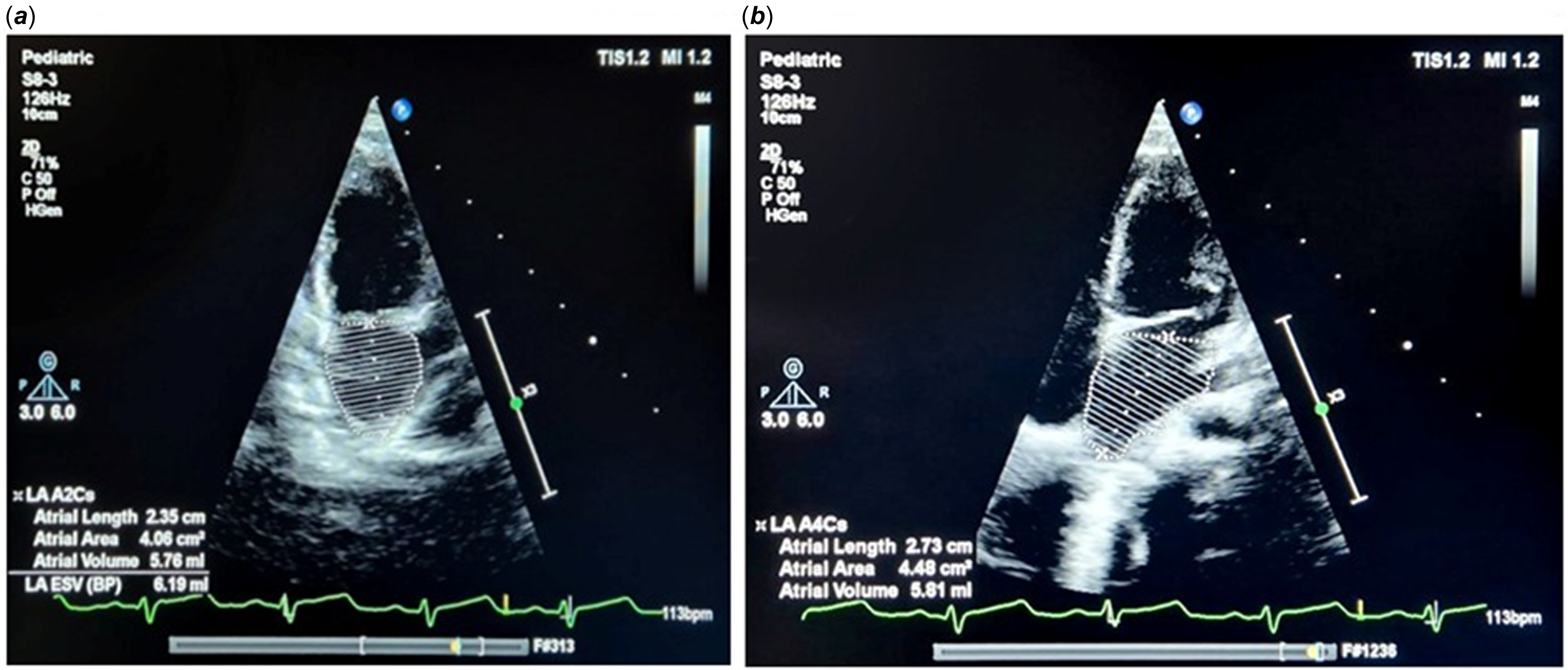
Echocardiography was performed using a Philips EPIQ 7 system with a 5–8 MHz probe. Left atrial volume was measured using the biplane area-length method from apical four-chamber views at end-systole. (Figure 1) Left atrial volume >2 Z-scores (Boston Children’s Hospital norms) was defined as left atrial enlargement.Reference Lopez, Saurers and Barker 24 –Reference Colan26
Echocardiogram depicting calculation of left atrial volume (a) by the 2D biplane area-length method (apical two-chamber view). (b) by the area-length method (apical four-chamber view).

Figure 1. Long description
Panel A: An echocardiogram image in the apical two-chamber view. The image shows a cross-sectional view of the heart with measurements labeled as LA A 2 C s, Atrial Length 2.35 centimeters, Atrial Area 4.09 square centimeters, and Atrial Volume 5.78 milliliters. The left atrial volume is calculated using the 2D biplane area-length method. Panel B: An echocardiogram image in the apical four-chamber view. The image shows a cross-sectional view of the heart with measurements labeled as LA A 4 C s, Atrial Length 2.73 centimeters, Atrial Area 4.48 square centimeters, and Atrial Volume 5.81 milliliters. The left atrial volume is calculated using the area-length method.
A trained technician performed the electrocardiography under the supervision of a qualified researcher. The interpretation of the electrocardiography was conducted by a researcher distinct from the one analysing the echocardiographic data, ensuring that both investigators were independently blinded to the findings of the alternate modality.
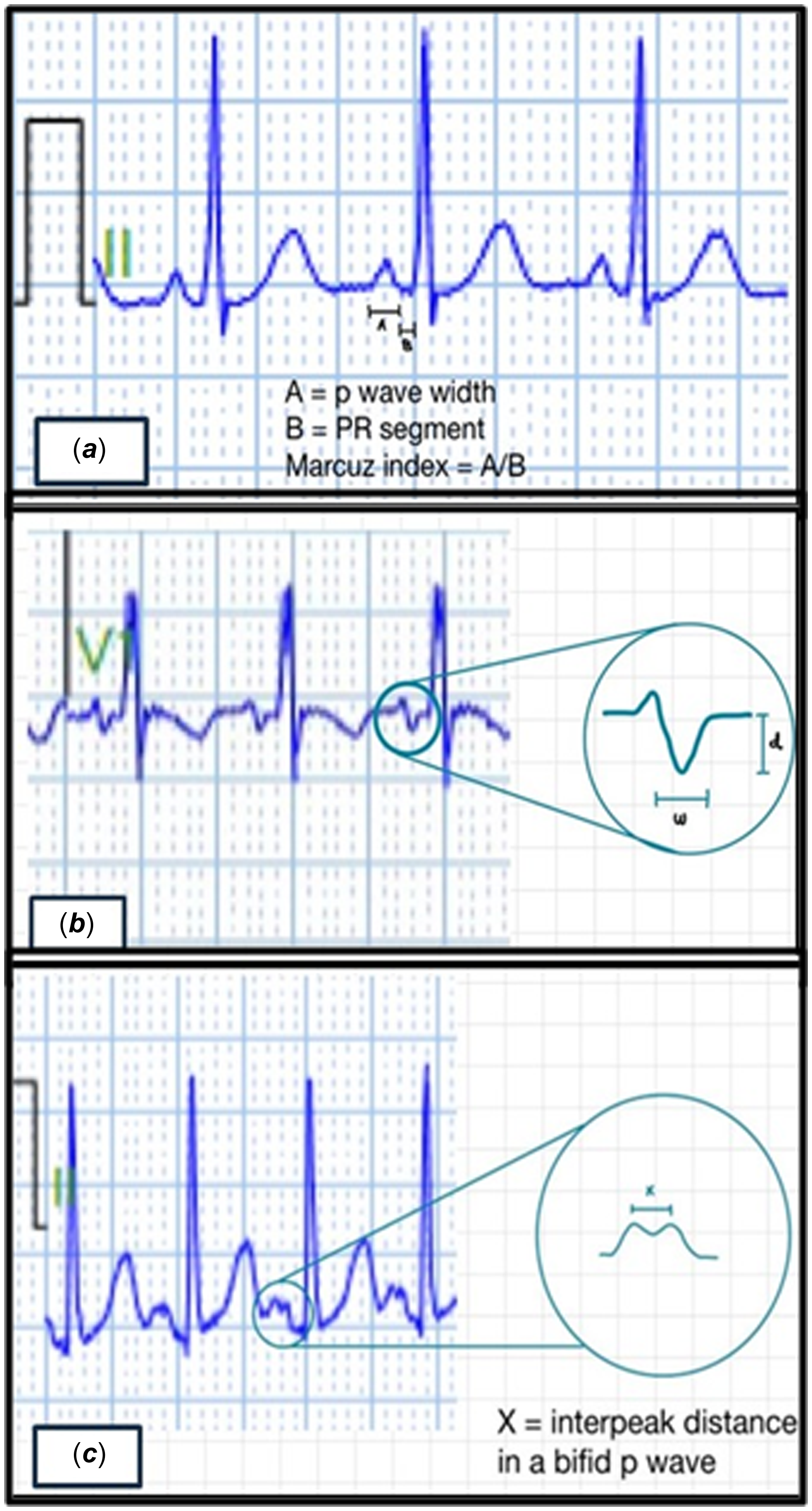
Standard 12-lead electrocardiographs were recorded using the Biocare electrocardiography-1210 system. The following parameters were analysed: (See Figures 2 and 3)
Depicting the calculation of various indices for left atrial enlargement (a) Macruz Index: the ratio of P wave width and PR segment. (b) Morris index: the product of the width (w) and depth (d) of the P wave’s negative component.

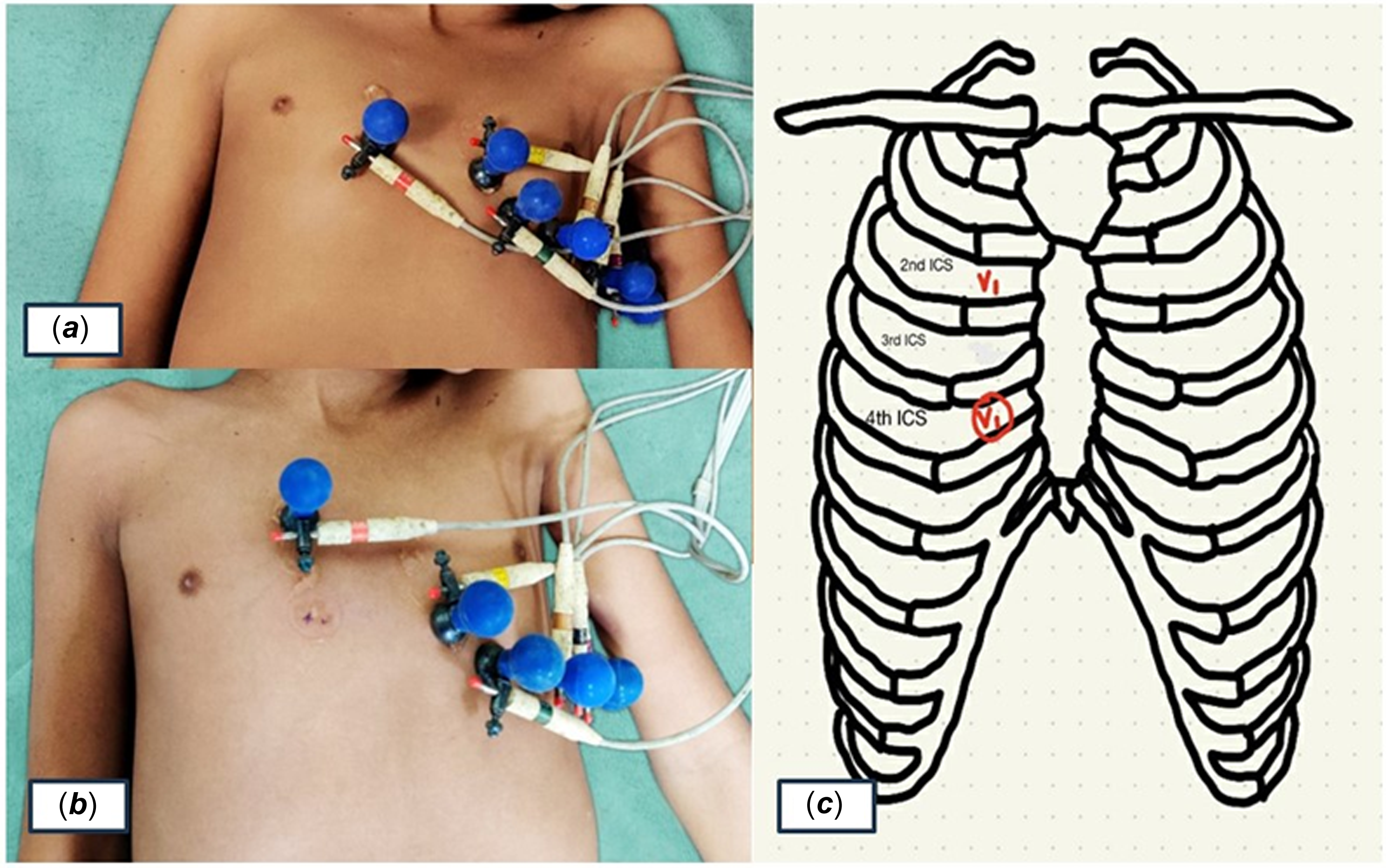
Image depicting lead position while recording ECG in a child: (A) conventional position of V1 at the right 4th intercostal space, (B) V1 placed at the right second intercostal space, and (C) cartoon depicting V1 at two different comparative positions.

-
• Morris Index: Area of terminal negative P wave in lead V1 (≥40 mm·ms) at the standard 4th intercostal space. Reference Morris, Estes, Whalen, Thompson and Mcintosh13,Reference O’Connor, McDaniel and Brady27
-
• Modified V1 Criterion (Novel Method): Area of terminal negative P wave ≥151.2 mm·ms in lead V1 placed at the 2nd right intercostal space.
-
• Macruz Index: P wave/PR segment ratio >1.6 in lead II. Reference Macruz, Perloff and Case11, Reference SZoe 28
-
• Bifid P wave: Interpeak distance ≥40 ms in lead II (P mitrale).
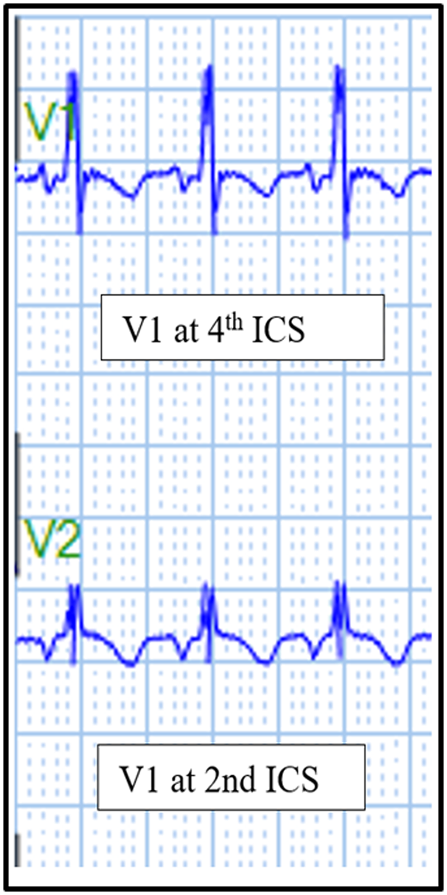
The novel V1-at-2nd intercostal space parameter was introduced to investigate whether repositioning enhances the detection of left atrial enlargement by better visualising atrial depolarisation. Electrocardiography results were compared against echocardiographic findings to classify true/false positives and negatives. Diagnostic performance was assessed using sensitivity, specificity, predictive values, and accuracy. Figure 4 depicts an instance of electrocardiography obtained from V1 positioned and right second and right fourth intercostal space in a patient with a ventricular septal defect.
Electrocardiogram depicting a scenario where the P wave’s terminal negative deflection becomes more prominent as the V1 electrode is positioned higher up (from the 4th intercostal space to the 2nd intercostal space).

Statistical analysis
The optimal cutoff for the novel novel P-terminal force in V1 at the 2nd intercostal space was determined using receiver operating characteristic curve analysis, with the threshold selected at the point maximising the Youden index (sensitivity + specificity – 1). To assess the internal validity of this receiver operating characteristic-derived threshold, a stratified 10-fold cross-validation was performed: the dataset was randomly partitioned into 10 equal subsets, with the model trained on 9 folds and tested on the remaining fold in each iteration. The mean area under the curve (AUC), sensitivity, and specificity across all folds (with 95% confidence intervals) are reported. Comparison of AUC values between parameters was performed using the DeLong test. Odds ratios with 95% confidence intervals were calculated using binary logistic regression to quantify the strength of association between each electrocardiography parameter and echocardiographically confirmed left atrial enlargement.
Results
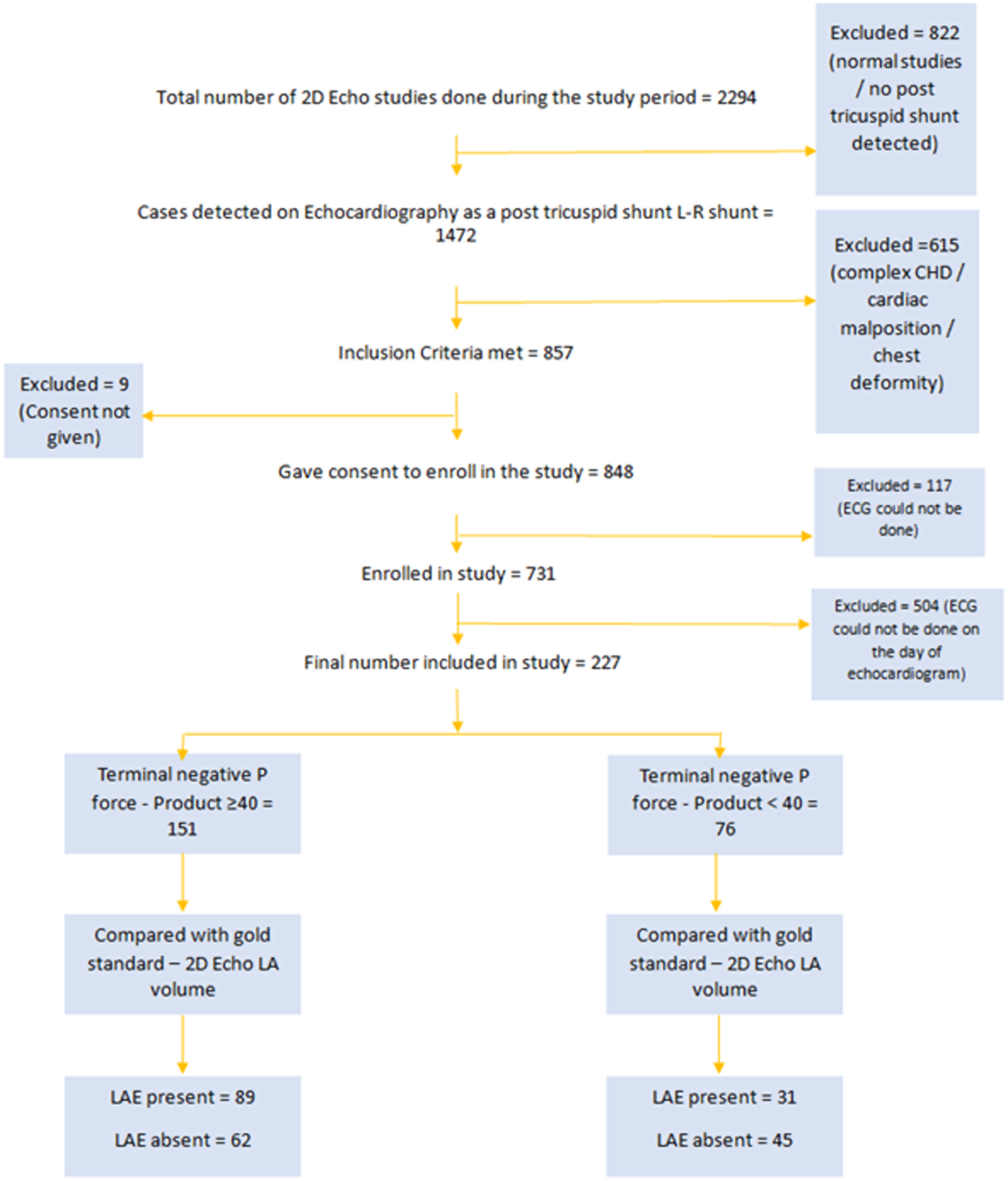
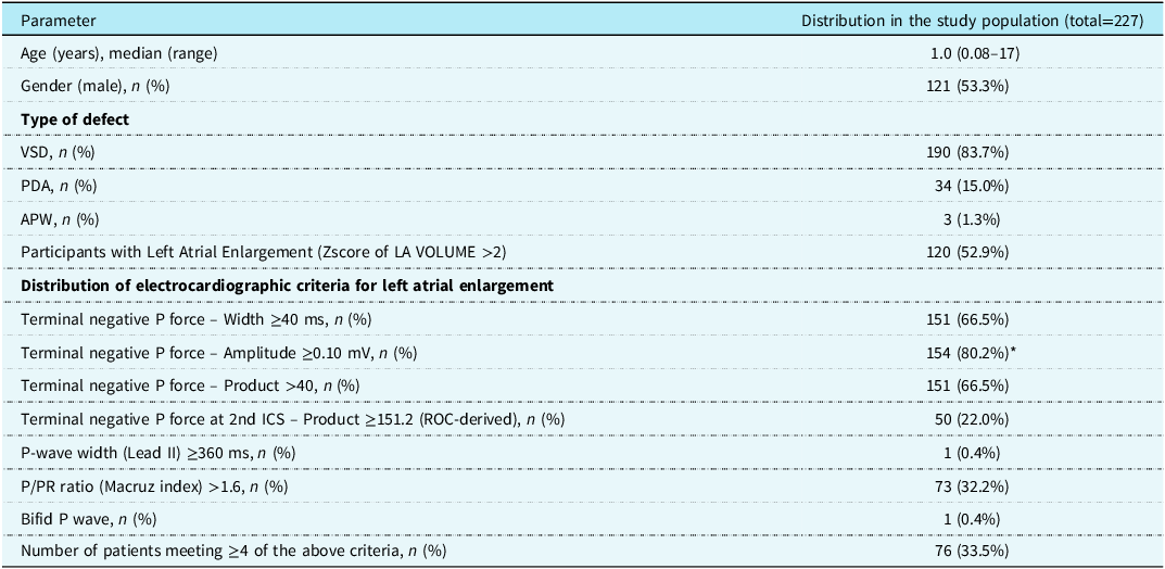
Out of 2294 echocardiograms performed during the study period, 227 children aged 1 month to 18 years with post-tricuspid shunt lesions (ventricular septal defect, patent ductus arteriosus, and aortopulmonary window) were included. The mean age was 3.13 ± 3.99 years; 53.3% were male. The majority of children (n = 104; 45.8%) were infants. Only 11 (4.8%) children were above the age of 12 years. Ventricular septal defect was the most common lesion (83.7%), followed by patent ductus arteriosus (15%) and aortopulmonary window (1.3%). Left atrial enlargement, defined as a left atrial volume >2 Z-scores on 2D echocardiography, was present in 52.9% (120/227) of participants. Table 1 depicts the baseline characteristics of the study population. Figure 5 depicts the study flow diagram.
Flow diagram summarising patient recruitment and key findings.

Demographics and distribution of electrocardiographic criteria for left atrial enlargement in the study population

* Percentage calculated among participants with available amplitude measurements (n = 192).
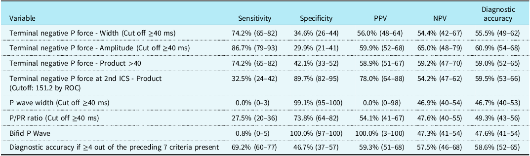
Among the traditional electrocardiography parameters, novel P-terminal force in V1 at the 4th intercostal space (cutoff ≥40 mm·ms) demonstrated a sensitivity of 74.2%, specificity of 42.1%, and diagnostic accuracy of 59.0% (p = 0.010). The Macruz index and bifid P wave showed lower sensitivity at 27.5% and 0.8%, respectively (Table 2).
Performance of study parameters for predicting left atrial enlargement. (The standard criteria taken for comparison is the 2D echo-derived LA volume Z score >2)
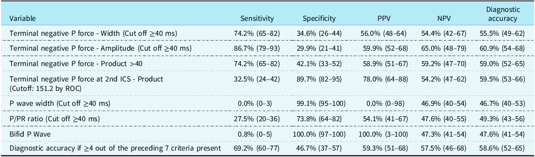
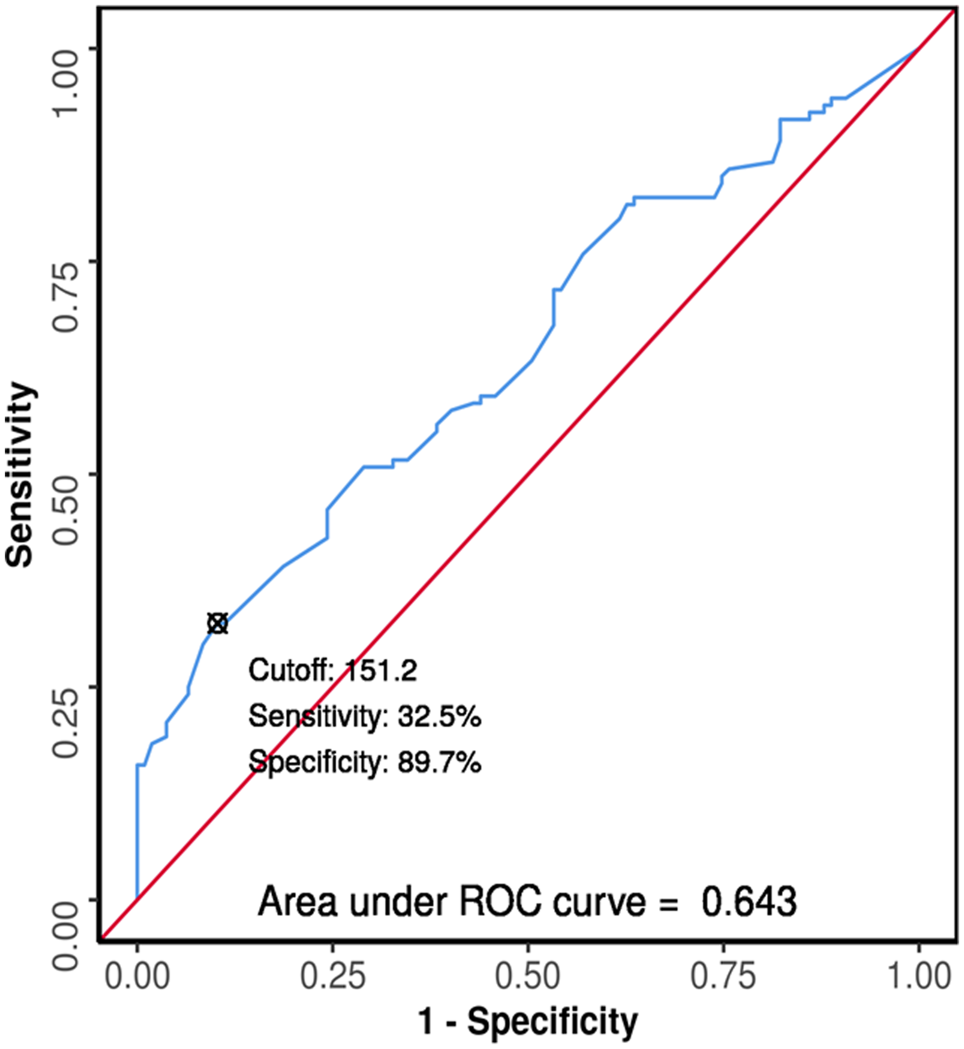
The novel parameter, novel P-terminal force in V1 measured at the 2nd right intercostal space, incorporated calculating the area of the terminal negative component of the P wave at a threshold>151.2 mm·ms, demonstrated a specificity, sensitivity, and an overall diagnostic accuracy of 89.7%, 32.5%, and 59.5% (p < 0.001), respectively (Table 2). The receiver operating characteristic analysis showed an AUC of 0.643, significantly outperforming several conventional markers like the P/PR ratio (AUC = 0.529, p = 0.032). (Figure 6) With an odds ratio of 4.20 and a positive predictive value of 78%, the novel method was among the most specific electrocardiography-based predictors of left atrial enlargement.
ROC curve analysis showing diagnostic performance of ECG: terminal negative P force at 2nd ICS—product in predicting LAE (n = 227).

Internal validation of the novel cutoff
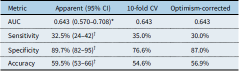
To address potential optimism in the receiver operating characteristic-derived threshold (≥151.2 mm·ms) for the novel novel P-terminal force in V1-at-2nd intercostal space criterion, internal validation was performed by 10-fold stratified cross-validation, in which the optimal cutoff was re-derived within each training fold and applied to the held-out fold, and by bootstrap optimism correction (B = 1000 iterations, Harrell). The cross-validated AUC matched the apparent value (0.643), with a bootstrap 95% confidence interval of 0.570–0.708, confirming statistically significant though modest discrimination. As expected for a data-derived threshold, the cutoff-dependent metrics showed mild optimism: optimism-corrected sensitivity, specificity, and accuracy were 30.0%, 87.0%, and 56.9%, respectively (Table 3). The Youden-optimal cutoff ranged from 118.4 to 151.2 mm·ms across folds (median 145.9), indicating that 151.2 mm·ms represents one valid threshold within a narrow plausible range. Prospective external validation in an independent paediatric cohort remains necessary before clinical adoption.
Internal validation of the novel NPTFV1-at-2nd-ICS criterion (cutoff ≥151.2 mm·ms) for the prediction of LAE (n = 227)

*Bootstrap percentile 95% CI (B = 2000).
†Wilson 95% CI as reported in Table 2.
Discussion
Large post-tricuspid shunts result in progressive LA volume overload. Detecting left atrial enlargement among children with post-tricuspid shunt lesions using electrocardiography could be a novel, cost-effective, widely available, technically less cumbersome, easily applicable surrogate diagnostic modality for shunt assessment if associated with reasonable sensitivity and specificity. Traditional electrocardiography indices have shown inconsistent performance in paediatric settings, partly due to variability in thoracic anatomy and lead positioning. Reference Kahn, Scheuer, Wachtel, Grishman and Donoso12,Reference Birkbeck, Wilson, Hall and Meyers29
Our study showed that among all parameters, novel P-terminal force in V1 amplitude and product demonstrated the highest diagnostic performance towards prediction of left atrial enlargement in standard lead positioning. However, their specificity remained modest.
Early paediatric investigations, including those by Reynolds and Levin, suggested an association between abnormal PTFV1 and atrial overload; however, reliance on radiographic or angiographic surrogates limited diagnostic precision. Reference Reynolds30, Reference Levin, Frand and Baltaxe 31 Subsequent paediatric investigations refined this understanding. Maok and Krongrad (1984) demonstrated pronounced lesion-specific variability in electrocardiography performance, reporting moderate sensitivity in ventricular septal defect (50–54%) but strikingly poor sensitivity in patent ductus arteriosus (8%), attributed to the shorter duration and reversibility of atrial dilation. These observations closely mirror the present study, which similarly identified inferior diagnostic performance of PTFV1 in patent ductus arteriosus compared with ventricular septal defect.Reference Maok and Krongrad 14
The most robust contemporary paediatric analysis by Ng et al. (2020), encompassing 492 children, evaluated electrocardiography criteria against echocardiographic diameter-based measures and demonstrated consistently low-to-moderate sensitivity (20.8–55.0%) despite high specificity (73.6–96.1%) for PTFV1 and related indices. While methodologically rigorous and adequately powered, the reliance on LA diameter and left atrial–to–aortic root ratio rather than volumetric assessment may have limited detection of early or asymmetric atrial remodelling.Reference Ng, Ahmad, Budhram, He, Balakrishnan and Mondal 19
In contrast, the present study uniquely employed indexed left atrial volume derived from the 2D biplane area-length method as the reference standard, a parameter shown to better correlate with cardiovascular morbidity. In an unselected paediatric cohort with post-tricuspid left-to-right shunts, classical PTFV1 thresholds yielded high sensitivity but poor specificity, whereas receiver operating characteristic-derived higher cutoffs improved specificity at the expense of sensitivity. This diagnostic trade-off aligns with age-related variability reported by Batra et al. (2018), who demonstrated superior electrocardiography performance in younger populations.
The novel criterion of measuring novel P-terminal force in V1 at the 2nd intercostal space introduced a key advancement. By repositioning the V1 lead, the atrial activity’s negative terminal component became more pronounced, leading to improved specificity (89.7%). This method matched the performance of traditional indices in sensitivity-adjusted receiver operating characteristic comparisons and offered a significantly better positive likelihood ratio compared to the P/PR ratio and bifid P wave.
The moderate effect size (point-biserial correlation = 0.28) and statistically significant association (p < 0.001) indicate that this novel parameter could serve as a specific screening tool for left atrial enlargement, especially valuable where echocardiography is not routinely available.
On comparative evaluation of performance of conventional terminal negative P wave amplitude (had the highest AUROC0.657), with the novel 2nd intercostal space product marker (AUROC = 0.643), the later performed equally well (p = 0.898, DeLong’s test), and with better specificity, reinforcing its value in limiting false positives in routine screening.
In subgroup analysis, the diagnostic accuracy of the novel marker in children with ventricular septal defects remained moderate (accuracy: 62.1%, specificity: 72.2%) using a tailored receiver operating characteristic-derived cutoff (118.4 mm·ms), suggesting good adaptability in common CHD subsets.
It is important to acknowledge that the overall diagnostic accuracy of all electrocardiography-based parameters evaluated in this study, including both traditional and novel criteria, ranged from 47% to 61%, indicating that electrocardiography alone cannot reliably substitute for echocardiographic assessment of left atrial enlargement in children with post-tricuspid shunts. This finding is consistent with prior literature demonstrating the inherent limitations of surface electrocardiography in detecting volumetric atrial changes, particularly in the paediatric population where age-related thoracic and cardiac size variability introduces additional confounding. Nevertheless, the high specificity (89.7%) and positive predictive value (78%) of the novel 2nd intercostal space parameter suggest potential utility as a ‘rule-in’ tool: a positive result may warrant prioritised echocardiographic evaluation, while a negative result should not be used to exclude left atrial enlargement.
Limitations
In the absence of Indian norms, Boston Children’s Hospital norms were employed to define left atrial enlargement, which could have affected the outcome for most of our children lagging behind in comparison to their growth parameters. Its cross-sectional design precludes longitudinal assessment of changes over time. The moderate sample size may limit the generalisability of findings. Additionally, conducting electrocardiography studies in younger children poses challenges, as paediatric thoracic anatomy varies with age; thus, uniform placement of precordial leads at the second intercostal space (intercostal space-2) may be suboptimal in older children. Finally, inter-observer variability in electrocardiography interpretation was not assessed.
The receiver operating characteristic-derived cutoff for the novel 2nd intercostal space parameter was established and internally validated (10-fold cross-validation) within the same cohort. The absence of an independent external validation cohort limits the generalisability of this threshold. Prospective validation in an unrelated, heterogeneous paediatric population is essential before this criterion can be recommended for routine clinical use.
Conclusion
In this cohort of children with post-tricuspid left-to-right shunts, measuring the terminal negative P wave force in V1 at the 2nd right intercostal space demonstrated high specificity (89.7%) for detecting echocardiographically confirmed left atrial enlargement, though at the cost of limited sensitivity (32.5%). While none of the electrocardiography parameters evaluated can replace echocardiographic assessment, this novel criterion may have a role as an adjunctive ‘rule-in’ tool for prioritising referrals in settings where echocardiography is not readily available. Prospective validation in an independent cohort is needed before clinical adoption.
Author contributions
The study was conceptualised by DK and DDB. RAP and DDB were the primary researchers involved in data collection and analysis. AM and SS contributed to patient management. The first draft was written by RAP and critically reviewed by DDB, SS, and DK. All authors agree to the final submitted draft.
Financial support
No funding received.
Competing interests
None.
Ethical standard
Obtained from the institutional ethics committee.