Introduction
Sinus venosus atrial septal defect constitutes approximately five to six percent of atrial septal defectsReference Vicenty-Rivera, Molina-Lopez, Diaz-Rodriguez and Molinary-Jimenez 1 and is frequently associated with partial anomalous pulmonary venous drainage.Reference El-Andari, Moolla and John 2 In contrast, partial anomalous pulmonary venous drainage occurs in approximately ten to fifteen percent of patients with secundum atrial septal defect.Reference Shimajiri, Harada, Kinoshita and Mikami 3 Anatomical variation and nonspecific symptoms may delay diagnosis. Although echocardiography is the first-line imaging modality, it may not fully delineate venous anatomy.Reference Oe, Honda, Yamano and Kawasaki 4 Anomalous hepatic venous drainage to the coronary sinus has been reported only in a small number of cases.Reference Karolczak, Mądry and Zacharska-Kokot 5 Although arterial ductal connections are common congenital findings, the presence of two such connections is rare. This report describes a rare coexistence of sinus venosus atrial septal defect, partial anomalous pulmonary venous drainage, anomalous hepatic venous drainage, two arterial ductal connections, and cor triatriatum in an infant, emphasising diagnostic and surgical considerations.
Case presentation
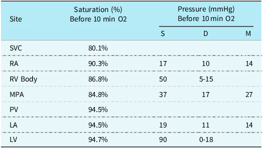
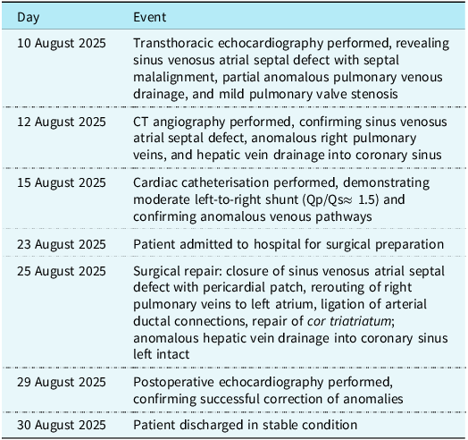
An eleven-month-old female infant weighing approximately eight kilograms presented with dyspnoea, poor weight gain, sleep disturbance, and recurrent respiratory infections. Physical examination revealed a grade three systolic murmur at the upper left sternal border. Chest radiography showed normal cardiac size with pulmonary vascular congestion. Transthoracic echocardiography demonstrated malalignment of the interatrial septum, partial anomalous pulmonary venous drainage, and mild pulmonary valve stenosis. Due to suboptimal visualisation of the superior atrial septum, CT angiography was performed and confirmed sinus venosus atrial septal defect with a malaligned interatrial septum (Figure 1). Three right pulmonary veins drained into the right atrium, and hypoplastic left pulmonary veins drained into the left atrium. In addition, a systemic vein arising below the diaphragm draining into the coronary sinus was visualised on multi-planar reconstructions (Figure 2) and highlighted on three-dimensional reconstructions (Figure 3). Cardiac catheterisation demonstrated a left-to-right shunt with a pulmonary-to-systemic flow ratio (Qp/Qs) of approximately 1.5 and mildly elevated mean pulmonary artery pressure of approximately 27 mmHg, which decreased with oxygen administration (Table 1, Supplementary Video 1). Surgical correction consisted of median sternotomy and cardiopulmonary bypass. Unexpected intraoperative findings included cor triatriatum and two arterial ductal connections. The atrial septal defect was repaired with a pericardial patch. Left pulmonary veins were augmented and redirected to the left atrium, and right pulmonary veins were rerouted. Hepatic venous drainage was left intact because of minimal flow and lack of hemodynamic significance. Both arterial ductal connections were ligated. The patient was successfully weaned from bypass and discharged in stable condition. The key diagnostic, therapeutic, and follow-up events are summarised in Table 2.
Multiplanar reconstruction (MPR) CT image in sagittal oblique view demonstrating the superior caval vein, inferior caval vein, and a sinus venosus atrial septal defect with a malaligned interatrial septum. (SCV = Superior Caval Vein; ICV = Inferior Caval Vein; SV-ASD = Sinus Venosus Atrial Septal Defect).

Multiplanar reconstruction (MPR) CT images demonstrating complex venous anatomy. ( a ) Coronal oblique view. ( b ) Sagittal oblique view showing the course of the anomalous hepatic vein. ( c ) Axial oblique view. Arrowheads delineate the anomalous hepatic vein draining into the coronary sinus and its connection to the right atrium.

Multiplanar CT evaluation of the systemic venous anomaly. ( a ) Coronal oblique view showing the malaligned interatrial septum and hepatic vein. ( b ) Sagittal oblique view tracing the anomalous venous course towards the coronary sinus. ( c ) Axial oblique four-chamber view demonstrating the right atrium, right ventricle, left ventricle, and coronary sinus. (IAS = Interatrial Septum; HV = Hepatic Vein; RA = Right Atrium; RV = Right Ventricle; LV = Left Ventricle; CS = Coronary Sinus).

Haemodynamic and oxygen saturation data before and after 10 minutes of supplemental oxygen. S: systolic, D: diastolic, M: mean

SVC = superior caval vein; RA = right atrium; RV = right ventricle; MPA = main pulmonary artery; LA = left atrium; LV = left ventricle.
Clinical timeline of the patient, summarising key diagnostic, therapeutic, and follow-up events

Discussion
The embryological development of the sinus venosus, pulmonary veins, and systemic venous pathways is closely interconnected. Disruption of normal sinus venosus incorporation into the right atrium together with abnormal pulmonary venous incorporation into the left atrium may explain the coexistence of a sinus venosus atrial septal defect and partial anomalous pulmonary venous drainage. Persistence of embryonic systemic venous channels may account for anomalous hepatic venous drainage to the coronary sinus. Cor triatriatum, resulting from incomplete incorporation of the common pulmonary vein into the left atrium, further supports a shared disturbance in pulmonary venous development. In addition, abnormal remodelling of the sixth aortic arches during fetal life may explain the presence of arterial ductal connections. Overall, these findings suggest a common developmental disturbance rather than isolated anomalies.
The exact genetic or epigenetic basis in this patient remains unknown. However, complex clusters of cardiac anomalies like these have been associated in the literature with mutations in transcription factors such as NKX2.5 or TBX5, which regulate cardiac septation and venous development. Epigenetic mechanisms may also contribute to such malformations, although no specific testing was performed in this case.
Echocardiography is often insufficient for detailed anatomical assessment, whereas CT angiography provides accurate visualisation of venous pathways. Partial anomalous pulmonary venous drainage occurs in approximately one percent of autopsies and typically produces a left-to-right shunt.Reference Ebrahimi, Mandegar and Jafari Fesharaki 6 In contrast, anomalous hepatic venous drainage to the coronary sinus often contributes minimally to shunting and may not require surgical intervention.Reference Karolczak, Mądry and Zacharska-Kokot 5
The anomalous hepatic venous drainage to the coronary sinus was left uncorrected due to its low-flow nature and lack of a significant contribution to pulmonary hypertension, as cardiac catheterisation demonstrated only mildly elevated pulmonary artery pressure. Two arterial ductal connections and cor triatriatum may be missed preoperatively because of small size or atypical imaging features. Thorough intraoperative evaluation is essential in complex congenital heart disease. Although multiple anomalies may suggest a genetic basis, current guidelines do not recommend routine testing in the absence of syndromic features or family history.Reference Veldtman, Bauersfeld and Babu-Narayan 7
Conclusion
This case illustrates a highly unusual combination of congenital cardiac anomalies. Advanced imaging, meticulous preoperative planning, and readiness for unanticipated intraoperative findings are essential for achieving favourable outcomes. Certain defects may not be identifiable preoperatively and may require recognition and management during surgery.
Supplementary material
The supplementary material for this article can be found at https://doi.org/10.1017/S1047951126111378.
Acknowledgements
The authors thank the patient’s parents for his participation and cooperation. We also express our gratitude to Emam Reza Hospital for their support.
Financial support
This research received no specific grant from any funding agency.
Competing interests
The authors report no conflicts of interest.
Ethical standard
Written informed consent for publication was obtained from the patient’s legal guardians. No identifiable patient information is included.
AI disclosure
AI tools were used for language editing only.