Introduction
A specific pattern of scent glands – dorsal abdominal (hereinafter just DAGs) in larval instars and metathoracic (hereinafter just MTGs) in adults – is considered autapomorphic for the order Heteroptera within the Insecta (e.g. Wheeler et al., Reference Wheeler, Schuh and Bang1993; Schuh & Slater, Reference Schuh and Slater1995; Weirauch, Reference Weirauch2006a,b). The existence of scent glands is also one of the features that clearly differentiates Heteroptera from other Hemiptera taxa (Schaefer, Reference Schaefer1972).
Dufour (Reference Dufour1833) first mentioned the scent glands, MTGs, in adult Heteroptera; Künckel d'Herculais (Reference Künckel d'Herculais1866) first observed the presence of DAGs in their larvae. The heteropteran larval DAGs developed as serially homologous structures medially on the abdominal dorsum. They probably occurred originally between all the abdominal terga in the ancestor. More recently, the larvae possess mostly two to three DAGs, either paired or unpaired (Dupuis, Reference Dupuis1947; Cobben, Reference Cobben1978; Staddon, Reference Staddon1979; Schuh & Slater, Reference Schuh and Slater1995).
The general basic function of the glands' secretion is defensive (Remold, Reference Remold1962, Reference Remold1963; Staddon, Reference Staddon1979). Therefore, DAGs were originally considered exclusively larval structures, functionless in adults, since their MTGs have taken over that role. Verhoeff (Reference Verhoeff1893) was the first to suggest a possible occurrence of DAGs, namely their external openings (ostioles), in the adults. Later, Gulde (Reference Gulde1902) and Henrici (Reference Henrici1939) studied reservoirs of DAGs and the histological structure of glands alone in several heteropterans adults. Eventually, Dupuis (Reference Dupuis1947) recognized even sexual dimorphism of the adult DAGs persistent in predaceous Asopinae (Pentatomidae). Recently, many cases of the DAGs' persistence in adults of various Heteroptera taxa have become known (e.g. Cobben, Reference Cobben1978; Aldrich, Reference Aldrich1988; Davidová-Vilímová & Podoubský, Reference Davidová-Vilímová and Podoubský1999; Staddon, Reference Staddon1999; Weirauch, Reference Weirauch2006a).
The basic histological structure of the Heteroptera scent glands corresponds to that of insect epidermal glands generally (Staddon, Reference Staddon1979; Noirot & Quennedey, Reference Noirot and Quennedey1991; Quennedey, Reference Quennedey and Harrison1998). The glands are based on the cuticular structures and differentiated epithelial cells, and their units function as glandular (secretory) structures. The reservoirs of glands are lined with a cuticle (known as the ‘intima’) continuous with the cuticle of the body wall. The reservoir, bearing numerous cuticular efferent (conducting) ductules of the glandular units, opens externally by the ostiole (Staddon, Reference Staddon1979; Noirot & Quennedey, Reference Noirot and Quennedey1991; Quennedey, Reference Quennedey and Harrison1998).
Most often, only the externally visible structures associated with DAGs have been studied through the entire ontogeny (e.g. Putshkov & Putshkova, Reference Putshkov and Putshkova1956; Putshkov, Reference Putshkov1961), and these also have been used for diagnostic purposes. Mostly, the internal structures have been studied only for the situation of DAGs in 5th larval instars in comparison with adults, e.g. Percy et al. (Reference Percy, MacDonald and Weatherston1980) – Apateticus bracteatus (Fitch, 1856), Pentatomidae, Farine (Reference Farine1987, Reference Farine1988) – Dysdercus cingulatus (Fabricius, 1755), Pyrrhocoridae, Lucchi & Solinas (Reference Lucchi and Solinas1990) and Lucchi (Reference Lucchi1993) – Nezara viridula (Linnaeus, 1758), Pentatomidae, Cassier et al. (Reference Cassier, Nagnan, Llosa, Andre and Guillaumin1994) – Lincus spurcus (Rolston, 1989), Pentatomidae, Davidová-Vilímová & Podoubský (Reference Davidová-Vilímová and Podoubský1999) – Pentatomoidea, Weirauch (Reference Weirauch2006a) – Reduviidae. Only three studies have concerned the entire or nearly the entire ontogeny of DAGs. Oetting & Yonke (Reference Oetting and Yonke1978) studied all instars of three alydids: Alydus eurinus (Say, 1825), Alydus pilosulus (Herrich-Schaeffer, 1848) and Megalotomus quinquesoinosus (Say, 1825), and Staddon (Reference Staddon1995) described changes of DAGs during the entire ontogeny of Oncopeltus fasciatus (Dallas, 1852), Lygaeidae. In all these species, DAGs are functionless in the adults. Davidová-Vilímová (Reference Davidová-Vilímová2006) studied states of DAGs reservoirs from 2nd larval instar to adult of Aradus betulae (Linnaeus, 1758), Aradidae, in which DAGs can be functional in the adults.
The present paper deals with a study of DAG cuticular structures in the full ontogenetic development of the model species Elasmucha ferrugata (Fabricius, 1787). The species belongs to the family Acanthosomatidae (Pentatomoidea), members of which commonly provide parental care, and the adult persistence of DAGs has been recognized in at least all Central European genera (e.g. Evans et al., Reference Evans, Staddon, Games, McCaffery and Wilson1990). Especially concerning the adult persistent DAG/DAGs, it remains unresolved which structure or phenomenon can prove functionality of the gland in the dried material of Heteroptera for cases where we cannot study histological structures. Therefore, we also endeavour to answer the question, which cuticular structures by their existence signal that the DAGs are truly functional.
Material and methods
Material studied
All larval instars and adults of the acanthosomatid species studied, Elasmucha ferrugata (Fabricius, 1787), were collected in the Czech Republic from the following localities: central Bohemia, the Zbiroh environment (map grid 6148 of faunistic grid mapping system: Pruner & Míka, Reference Pruner and Míka1996), during 2008–2009; southern Bohemia, the Křižanov environment (6552, 6652), during 2008–2009; and southern Bohemia, the Oslov environment (6551, 6651), during 1992–1994. The specimens were collected on the following host plants: Ericaceae: Vaccinium myrtillus (Linnaeus, 1753); Grossulariaceae: Ribes rubrum (Linnaeus, 1753) and Ribes nigrum (Linnaeus, 1753). The material of the 1st larval instar originated from egg batches deposited by females reared in the laboratory under common garden conditions. The material is deposited in the collection of Charles University, Faculty of Science, Department of Zoology.
Methods
Fixation of material
Larvae from the 3rd to 5th instars and adults were fixed in 75% or 96% ethanol. The specimens of 1st and 2nd larval instars were fixed with 2.5% solution of glutaraldehyde in 0.1M phosphate buffer (PBS) at pH 7.4 for minimally 24 h, then transferred into PBS solution and dehydrated in a graded series of ethanol (30%, 45%, 60%, 75%, 96%, each for optimally 30 min), then into 99.8% ethanol as the final fixation.
Dissection
In addition to standard equipment (e.g. fine forceps and scissors), a hypodermic needle was used in dissection. Head and thorax were removed, as were soft tissues from the abdomen. The abdominal dorsum, including DAGs reservoirs, was cleared in hot 10% KOH solution (time period depending on the size of the sample), washed in PBS, then dehydrated in 30%, 45%, 60%, 75%, 96% and 99.8% ethanol for study by scanning electron microscopy (SEM). For light microscopy study, dehydration was only to 75% ethanol.
Light microscopy
The dorsum was stained with Chlorazol Black E, fixed on a slide into glycerine and studied using an Olympus BX40 compound microscope with Quick Photo Micro 2.3 and Helicon Focus software.
Ductule counts
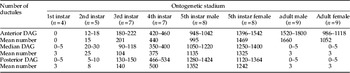
The swollen opening of a glandular unit's cuticular ductule is recognizable on the reservoir intima as a sclerotized dark ring. The ductules were counted from microphotographs of the reservoir in the glycerine slides. A square grid of defined size was placed onto the photograph in Quick Photo Micro 2.3 software. The ductules were counted in one square and related to the reservoir as a whole (table 1).
Number of ductules opening into DAG reservoirs of Elasmucha ferrugata.

Scanning electron microscopy
The dissected and macerated samples were cleaned in an ultrasonic bath (1st and 2nd instars in 75% ethanol, all other stadia in 99.8% ethanol, for different time periods depending on their levels of sclerotization, with each individual sample being monitored continually during cleaning), dried by the critical point method, and then coated with gold. Microphotographs were prepared using SEM JEOL 6380 LV.
Terminology
The basic classification and terminology suggested for cuticular structures of the MTGs (Kment & Vilímová Reference Kment and Vilímová2010a,b) were modified herein for the DAGs.
Internal parts of DAGs
Cuticular reservoir surrounded by glandular units, unicellular and multicellular, the latter always with two canal cells. The following cuticular structures are distinguishable (e.g. Staddon, Reference Staddon1979; Farine, Reference Farine1987, Reference Farine1988; Lucchi & Solinas, Reference Lucchi and Solinas1990; Lucchi, Reference Lucchi1993): receiving canal, sacculus and conducting canal=ductule with swollen opening into reservoir. Valve is a cuticular projection of the reservoir posterior wall, closing the ostiole of the reservoir.
Scent efferent system
Only external parts of the system are developed in DAGs. Internal parts, represented in particular by a vestibule, are developed only in MTGs. Ostiole is an opening of gland reservoir, slot-shaped or semicircular-shaped. Evaporatorium (evaporative area) is in Heteroptera, according to the definition of Nichols & Schuh (Reference Nichols and Schuh1989), “part of cuticle associated with the orifice [ostiole], modified for assistance in rapid evaporation of scent gland products, provided with a specialized microsculpture…” A cuticular process in the shape of an elongated ear located laterally to an ostiole and toward the apex rising above the surrounding surface is called an ear-shaped structure (ear). Cuticular ridge is formed by a transverse, concave, structure raised anteromedially and medially to the ostiole.
All external structures are located on a dorsal medial sclerite distinguishable by distinct pigmentation and sclerotization. Several patterns of cuticle close to the ostiole are differentiated: wrinkled cuticle (wrinkles) running radially toward ostiole of anterior DAG; network-shaped cuticle=irregular net of ridges forming polygon sculpture (=cells), with fine, waved trabeculae on their bottoms, cells connected by elevated ridges (this type of cuticle is located around ostioles of median and posterior glands); loaf-like sculpture is a newly described and named type of microsculpture of specific pattern resembling scale-like sculpture, but its processes are shorter and more swollen (this type of cuticle is located around the ostiole of the anterior gland).
Results
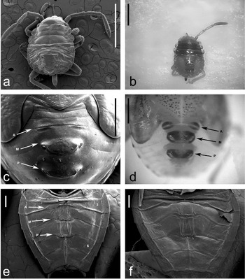
Three DAGs, anterior, median and posterior, are developed in Elasmucha ferrugata (fig. 1a–f). The reservoirs of DAGs are always located anteriorly from ostioles, beneath terga 3, 4 or 5. All the reservoirs have a distinct orange colour. The ostioles lie in intersegmental positions on distinct dark, sclerotized medial sclerites in all larval instars, where they are entirely exposed and located far from wing pads, if these are developed (fig. 1c, d). Abdominal dorsum of adults is slightly sclerotized, including cuticles surrounding ostioles (fig. 1e, f). The entire dorsum is completely covered by wings.
Dorsal view of Elasmucha ferrugata body. 2nd instar (a) under SEM, (b) under optical microscope. 5th instar female (c) under SEM, (d) under optical microscope. Adult under SEM: (e) male, (f) female. A, area of anterior DAG; M, area of median DAG; P, area of posterior DAG; 3, 3rd abdominal segment; 6, 6th abdominal segment. Scale bar: 1 mm.

Anterior DAG
Anterior DAG is paired, located in the 3rd abdominal segment, with paired ostioles between terga 3 and 4 (fig. 1a–f).
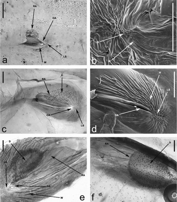
Anterior DAG has paired reservoirs in all larval instars and adults. The reservoir has a wide basal part, an intermediate narrowed part and an apical globular part, shifted slightly laterally from centre of the base width (fig. 2a, e). Cuticular intima of the reservoir is completely developed already in the 1st instar (fig. 2a), the reservoir occurring in an identical characteristic shape in all ontogenetic stadia including adults; only its sclerotization and size increase, either copying the size of paired medial sclerites in larval instars (fig. 2a, c, e) or larger than these in adults (fig. 3a).
Anterior DAG of Elasmucha ferrugata larvae. (a) left part of DAG of 1st instar, ventral view, under optical microscope; (b) dorsal view, under SEM. (c) left part of DAG of 3rd instar, ventral view, under optical microscope; (d) right part, dorsal view, under SEM. (e) DAG of 5th instar, ventral view, under optical microscope, right part of female; (f) left part of male. D, ductules; LS, loaf-like microsculpture; OA, ostiole of anterior DAG; RA, reservoir of anterior DAG; W, wrinkled cuticle. Scale bar: 50 μm.

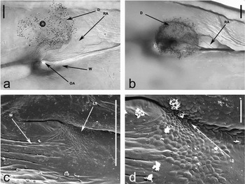
Right part of anterior DAG of Elasmucha ferrugata adults. Ventral view, under optical microscope, (a) female, (b) male. Dorsal view, under SEM, (c) female, (d) male. D, ductules; LS, loaf-like microsculpture; OA, ostiole of anterior DAG; RA, reservoir of anterior DAG; W, wrinkled cuticle. Scale bars: (a–c) 100 μm; (d) 10 μm.

The swollen openings of cuticular ductules of glandular units into the reservoir, if developed, were recognized only in the apical part of the reservoir (fig. 2c, e). No sclerotized openings of ductules were recognized in the reservoir of the anterior DAG of the 1st instar (fig. 2a, table 1). Ductules occur from 2nd instar, with a maximum of 18, and an average of 15 ductules. Their number increases to an average 201 ductules in 3rd instar (fig. 2c), 440 in 4th instar, 995 in 5th male instar, and 1469 in 5th female instar (fig. 2e, f). In adults, there are more ductules in males (averaging 1660) than in females (averaging 1052) (fig. 3a, b, table 1).
The reservoir opens by a transverse, slot-like ostiole, which is slightly roofed by posterior margin of tergum 3 in the dorsal view (figs 2d and 3c). The ostiole lies laterally on medial sclerite; each of these paired sclerites is drop-shaped with a wider side directed laterally (figs 1d and 2c). The sclerite is highly sclerotized and distinctly pigmented, gradually more so during ontogeny to 5th instar; it is slightly sclerotized in 1st instar, gradually more so from 2nd to 4th and 5th instars (fig. 2d). Its sclerotization is reduced to faint in adults of both sexes (fig. 3a, c).
The area of loaf-like microsculptured cuticle is in close contact with the lateral margin of the ostiole in all larval instars and adults (figs 2d and 3c, d). The size of particular loafs is constant, but their numbers increase during ontogeny, resulting in gradually larger area. The surface with loaf-like microsculpture roughly increases (related to medial sclerite) from 1st instar to adult female, being most recognizable under a light microscope (figs 2c and 3a, b), with the exception of ecdysis from 1st to 2nd instar, when it decreases.
The area with the wrinkled cuticle is conspicuous, located radially around the ostiole. Wrinkles continue from the area with the loaf-like microsculpture in the shape of a ring, more conspicuously medially along the ostiole (fig. 2b, d, e), with the longest wrinkles pointed medially and posteriorly in larval instars (fig. 2b, d). In adults, the wrinkles do not touch the loafs; they begin a short distance from the loaf-like sculptured area and are roughly transverse, posteriorly to ostiole, i.e. parallel with it (fig. 3a, c). The area with the wrinkled cuticle is rather larger than the loaf-like sculptured cuticle in all stadia, occupying at least one third of medial sclerite.
Median DAG
Median DAG is unpaired, located in the abdominal segment 4, with paired ostioles between terga 4 and 5 (fig. 1a–f).
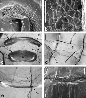
The reservoir is unpaired, sac-shaped, transversally elongated in all larval instars and adults (fig. 4c, e). The relative size of the reservoir, deduced from the condition of its intima, is similar in adults as it is in 5th instar; the reservoir is not distinctly reduced. Ductules of glandular units are regularly located on the whole surface of the reservoir intima. The number of their openings is quite low in 1st instar, maximally five, mostly wholly undeveloped (fig. 5a). The number slowly increases in 2nd instar (fig. 5c), up to 25 on average, and more in subsequent instars: 104 on average in 3rd instar, 375 in 4th instar and very high numbers occur in 5th instar (1135 in males and even 1325 in females) (fig. 4c). Maximally just five openings of ductules were recognized in whole intimas of adult reservoirs in both sexes, which is still relatively large (fig. 4d, e).
Median DAG of Elasmucha ferrugata. Detail of external structures associated with right ostiole of 4th instar, (a) dorsolateral view, under SEM; (b) left part of evaporatorium, dorsal view. Medial sclerite of 5th instar male, (c) ventral view, under optical microscope. Medial sclerite of adult, ventral view, under optical microscope, (d) female (head in the left direction), (e) male; (f) female, dorsal view, under SEM. CE, cell of evaporatorium, E, evaporatorium; ES, ear-shaped structure; MSAR, dorsal medial sclerite of right part of anterior DAG; MSM, dorsal medial sclerite of median DAG; OM, ostiole of median DAG; R, ridges of evaporatorium; RA, reservoir of anterior DAG; RM, reservoir of median DAG; RP, reservoir of posterior DAG; T, trabeculae of evaporatorium. Scale bars: (a, b) 20 μm; (c–f) 200 μm.

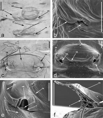
Median DAG of Elasmucha ferrugata larvae. 1st instar, (a) ventral view, under optical microscope; (b) external structures associated with left ostiole, dorsal view, under SEM. Medial sclerite of 2nd instar, (c) ventral view, under optical microscope; (d) dorsal view, under SEM. Detail of external structures of 3rd instar, (e) under SEM, left ostiole, dorsal view; (f) ear-shaped structure, anterolateral view. CP, cuticular process; CR, cuticular ridge; D, ductules; E, evaporatorium; ES, ear-shaped structure; LP, loaf-like processes; OM, ostiole of median DAG; OP, ostiole of posterior DAG; RM, reservoir of median DAG; RP, reservoir of posterior DAG; V, valve. Scale bar: 50 μm.

Medial sclerite is unpaired, transversally elongated, roughly elliptical (figs 4c and 5c, d), slightly sclerotized in 1st instar (fig. 5a), progressively more sclerotized and pigmented from 2nd to 5th instars. Its sclerotization is strongly reduced in adults; the area of adult medial sclerite does not differ from the surrounding sclerotization (fig. 4d, e).
Paired ostioles are located sublaterally on the medial sclerite. They are slot-shaped in 1st instar, without valves and directed posterodorsally (fig. 5b), of semicircular shape with a straight posterior border directed dorsally in 2nd to 5th instar (fig. 5d, e). The functional ostiole of median DAG, which should be open through the cuticle, was not found in adults. Only an area of thin, pale cuticle is in the location of the larval ostiole (fig. 4e).
A semicircular cuticular valve can tightly close the ostiole. The valve is developed from 2nd instar (fig. 5d). The valve is posterolaterally continual with a posterior reservoir wall, its anterior margin is tilted inside the reservoir if the ostiole is open (fig. 5e). Loaf-like processes are developed on the posterolateral area of the valve surface; this sculpture is very similar to the loaf-like sculpture associated with the ostiole of the anterior DAG, but the processes are lower and with broader apices (fig. 5e). The slot-shaped ostiole of 1st instar has an undeveloped valve. Inside the ostiole, loaf-like processes can be seen in the posterior wall of the reservoir, close to the ostiole (fig. 5b). In adults, the similar area with slightly indicated loaf-like processes occurs in the location of the larval ostiole.
A wide, U-shaped cuticular ridge is developed from 2nd to 5th instars, where it is most conspicuous (fig. 5d); it is not developed in 1st instar and is completely reduced in adults (fig. 4d). The ridge runs medially between the ostioles, its lateral arms are bent anteriorly from the ostioles but does not reach them.
Distinct cuticular structure is developed laterally from ostiole. The thickened cuticular process is developed in 1st instar (fig. 5b), the ear-shaped structure is developed in an identical position from 2nd to 5th instars (fig. 5d, e). No external cuticular structure is associated with the ostiole of median DAG in adults (fig. 4f). The cuticular process in 1st instar is a flattened, apically rounded process located laterally from the ostiole, surpassing the intersegmental line posteriorly. The base of the ear-shaped structure lies laterally from the ostiole. The body of the structure runs anterolaterally, with the flattened dorsal part bearing a shallow groove (fig. 5d, e). The apex of the structure is distinctly elevated above the surrounding area (fig. 5e, f). The cuticle of the dorsal and anteromedial surface of the ear-shaped structure is smooth (fig. 5f), that of the posterolateral surface is covered by a network microsculpture (figs 4a and 5f) continuing from the evaporatorium, which is located posteriorly and posterolaterally from the ostiole (fig. 4a).
The cuticle of evaporatorium is modified into the network microsculpture (figs 4b and 5e). Elevated ridges delimit cells of irregular shapes. Surfaces of cells are depressed and can bear fine ridges (fig. 4b). The entire pattern resembles the structure of a bee honeycomb. The size of the cells is constant, their number increases during the ontogeny from 2nd to 5th instars (figs 4a and 5d).
The network microsculpture was not recognized in the 1st instar (fig. 5b); only a very slight indication of it was found in one specimen. The cuticle is only slightly wrinkled around the ostiole in 1st instar. The evaporatorium with network microsculpture is developed from 2nd to 5th instar. That microsculpture covers a continually larger, wide, semicircular area posteriorly from the ostiole, posterolaterally from the ear-shaped structure and on the posterolateral surface of that structure (figs 4a and 5d, e). A low number of cells with only slightly wrinkled surfaces of the cells between the ridges occur in 2nd instar (fig. 5d). Higher numbers of cells were recognized in 3rd instar; they expand mostly on the posterolateral surface of the ear-shaped structure (fig. 5e, f). The ridges are higher and the cell surfaces conspicuously more wrinkled from 3rd to 5th instars (fig. 4a, b), thus resembling a pattern of MTGs' mushroom structure. The surface of the evaporatorium is slightly smaller in females than in males in 5th instar. Neither the evaporatorium nor its remnant was recognized in adults (fig. 4d–f).
Posterior DAG
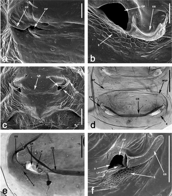
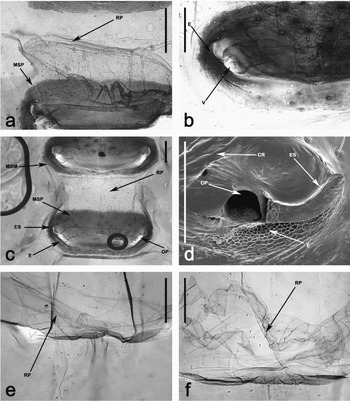
Posterior DAG is unpaired, located in the abdominal segment 5, with paired ostioles between terga 5 and 6. All cuticular structures associated with posterior DAGs are either very similar or identical to those of median DAG (fig. 6a–c, e and fig. 7a, b, d). Therefore, only states of structures that differ are described below.
The medial sclerite is relatively smaller than that of the medial DAG (fig. 1a–f).
Ductules of glandular units are regularly located on the entire reservoir intima surface. Maximally five ductules were recognized in 1st instar, but mostly they are not developed at all. The number slowly increases in 2nd instar, up to eight on average (fig. 6d) and is quite higher in subsequent larval instars, up to 140 on average in 3rd instar, 500 on average in 4th instar, 1352 on average in 5th instar males (fig. 7c) and 1242 on average in 5th instar females. Maximally, only five ductules were recognized in adults of both sexes (fig. 7e, f).
Posterior DAG of Elasmucha ferrugata larvae. Detail of external structures, under SEM, (a) left side of 1st instar, dorsal view; (b) right side of 2nd instar, dorsolateral view. Medial sclerite of 2nd instar, (c) dorsal view, under SEM; (d) ventral view, under optical microscope. External structures associated with right ostiole of 4th instar, (e) ventral view, under optical microscope; (f) dorsal view, under SEM. CP, cuticular process; CR, cuticular ridge; E, evaporatorium; ES, ear-shaped structure; MSP, dorsal medial sclerite of posterior DAG; LP, loaf-like processes; OP, ostiole of posterior DAG; RP, reservoir of posterior DAG; V, valve. Scale bars: (a, b) 20 μm; (c–f) 100 μm.

Posterior DAG of Elasmucha ferrugata. Medial sclerite of 4th instar, (a, b) ventral view, under optical microscope. 5th instar male, (c) reservoir, ventral view, under optical microscope; (d) detail of external structures associated with right ostiole, dorsal view, under SEM. Medial sclerite of adult, ventral view, under optical microscope, (e) male, (f) female. CR, cuticular ridge; E, evaporatorium; ES, ear-shaped structure; MSM, dorsal medial sclerite of median DAG; MSP, dorsal medial sclerite of posterior DAG; OP, ostiole of posterior DAG; RP, reservoir of posterior DAG; V, valve. Scale bars: (a–c, e, f), 200 μm; (d) 100 μm.

The cuticular valve closing the ostiole is developed from the 2nd instar, but its surface is smooth without loaf-like processes in this instar; the processes are developed from 3rd instar (fig. 6f). There were found only a small number of slightly indicated loaf-like processes in the location of the larval ostiole in adults, and only slightly wrinkled cuticle around the ostiole.
Discussion
Chemical communication is a general phenomenon within the insects. The Heteroptera produce repellent secretions (allomones) from different scent epidermal glands. The general basic function of the glands' secretions is defensive against predators (e.g. Remold, Reference Remold1963; Staddon, Reference Staddon1979; Aldrich Reference Aldrich1988, Reference Aldrich, Carde and Bell1995). Mostly two to three, maximally four, DAGs are developed in larvae of the extant Heteroptera, with the secretions formed by specific mixtures of aromatic volatile compounds, mostly alkenals and alkanes, e.g. n-tridecane (Staddon, Reference Staddon1979; Aldrich, Reference Aldrich1988, Reference Aldrich, Carde and Bell1995).
The DAGs pattern of anterior DAG paired, median and posterior unpaired, all with paired ostioles, occurring also in Elasmucha ferrugata, is without doubt a common feature of Pentatomoidea larvae (e.g. Cobben, Reference Cobben1978; Staddon, Reference Staddon1979; Percy et al., Reference Percy, MacDonald and Weatherston1980; Lucchi & Solinas, Reference Lucchi and Solinas1990; Cassier et al., Reference Cassier, Nagnan, Llosa, Andre and Guillaumin1994; Davidová-Vilímová & Podoubský, Reference Davidová-Vilímová and Podoubský1999). The condition of DAGs recognized in E. ferrugata is identical with published data concerning the Acanthosomatidae (Evans et al., Reference Evans, Staddon, Games, McCaffery and Wilson1990; Staddon, Reference Staddon1990; Davidová-Vilímová & Podoubský, Reference Davidová-Vilímová and Podoubský1999), except that Evans et al. (Reference Evans, Staddon, Games, McCaffery and Wilson1990) state that the anterior DAG have no functionality in the 5th instar. In contrast, we confirm that that gland is without a doubt functional in both sexes.
The DAGs, originally supposed to be exclusive larval structures, often persist as the functional glands in the adults. We confirm that the anterior DAG is functional in the E. ferrugata adult. The same condition was found in E. grisea by Evans et al. (Reference Evans, Staddon, Games, McCaffery and Wilson1990) and Staddon (Reference Staddon1990). All the cuticular structures of adult DAGs are homologous with those of the 5th instar. They present no different structures developed de novo only in adults. The persistence of the anterior DAG seems to be a common character of Acanthosomatidae adults, corroborated in all European members. The different numbers of ductules represent the first case of sexual dimorphism of adult anterior DAG observed in Acanthosomatidae, with males having about 1.6 times more ductules than females. We found maximally five ductules both in the median and posterior DAGs of E. ferrugata adults. Even if a small volume of secretion could be produced, however, the glands are evidently not functional because their ostioles are closed.
Until now, it has not been shown which cuticular structures, if any at all, could prove that a gland was functional in dried, old material. Verhoeff (Reference Verhoeff1893) had first mentioned the existence of ostioles (so-called pores) of DAGs in adults of several Heteroptera and supposed their persistent function. However, ostioles, or the scars of ostioles, are often distinct in heteropteran adults in studies using only optical stereomicroscope (e.g. in Cydnidae: Lis & Hohol-Kilinkiewicz, Reference Lis and Hohol-Kilinkiewicz2002) and do not refer to fully functional glands. The occurrence of a reservoir, or the cuticular intima of a reservoir, also has been used often as proof of functional DAGs in adults (e.g. Podoubský, Reference Podoubský1997; Davidová-Vilímová & Podoubský, Reference Davidová-Vilímová and Podoubský1999). We have shown in the present study, however, that the reservoir intima is present in adults even without proper functional glandular units and open ostioles. Therefore, it is clear that the occurrence of only the cuticular intima of the gland reservoir does not prove a functional gland.
Presumably, higher numbers of ductules, representing proper gland units, imply functional glands. Hamilton et al. (Reference Hamilton, Gough, Staddon and Games1985), Staddon et al. (Reference Staddon, Thorne and Knight1987) and Staddon (Reference Staddon1990, Reference Staddon1995, Reference Staddon1999) confirmed functionality of DAGs in several heteropteran taxa by the presence of ductules. Weirauch (Reference Weirauch2006a), similarly in Reduviidae, used ductules as well as their swollen medial parts, saccules.
Histological studies and/or chemical analyses of the gland reservoirs' content are undoubtedly the best methods for proving true functionality of the scent glands, but these methods may be used only for fresh material. There exist several histological studies documenting the presence of functional glandular units themselves (e.g. Henrici, Reference Henrici1939; Roth, Reference Roth1961; Aldrich et al., Reference Aldrich, Yonke and Oetting1972; Oetting & Yonke, Reference Oetting and Yonke1978; Percy et al., Reference Percy, MacDonald and Weatherston1980; Farine, Reference Farine1987, Reference Farine1988; Lucchi & Solinas, Reference Lucchi and Solinas1990; Lucchi, Reference Lucchi1993; Cassier et al., Reference Cassier, Nagnan, Llosa, Andre and Guillaumin1994). The location of glandular units is known in several pentatomoidean species, Apateticus bracteatus (Pentatomidae: Percy et al., Reference Percy, MacDonald and Weatherston1980), Scaptocoris divergens (Cydnidae: Roth, Reference Roth1961), Nezara viridula (Pentatomidae: Lucchi & Solinas, Reference Lucchi and Solinas1990; Lucchi, Reference Lucchi1993) and Elasmucha ferrugata (Acanthosomatidae: present paper). The pattern as to the arrangement and also number of the glandular units seems to be roughly unified in higher taxa, e.g. Pentatomoidea. Much more data is needed before drawing any general conclusions, however, as only sparse information has been published to date.
The number of ductules, as the indicator of gland functionality, increases during E. ferrugata's ontogenetic development from the 1st instar to adults in the anterior DAG and from the 1st to 5th instars in median and posterior DAGs. We found no ductules only in the anterior DAG of the 1st instar. A peculiar situation concerns the number of ductules in the anterior gland in the 5th instar, compared with the adult of both sexes. The number is in larvae conspicuously higher in females than in males. In adults, however, the ductules are more numerous in males than in females. We have no explanation for that curious situation; why is the highest number of ductules and thus activity of the anterior gland the greatest in females in the 5th instar? Generally, the increasing number of ductules during ontogeny corresponds with the situation described in Oncopeltus fasciatus by Staddon (Reference Staddon1995) and in the alydids described by Aldrich et al. (Reference Aldrich, Yonke and Oetting1972).
In E. ferrugata, we recognized that the anterior DAG is quite different from the identical median plus posterior DAGs, with the exception in the 1st instar; the characters of median plus posterior DAGs are simpler than in the older instars. The different structure of the glands is defined more probably by topography in the abdomen than by order (first gland developed, second, etc.). The Pentatomoidea anterior DAG, i.e. between terga 3 and 4, is thus quite different from the identical median and posterior glands between terga 4 and 5, and 5 and 6, respectively, while the Alydidae have two identical glands, median and posterior (e.g. Aldrich et al., Reference Aldrich, Yonke and Oetting1972).
We can find no general pattern across all Heteroptera as to which larval DAG persist into the adult stage. Unfortunately, we have only scattered information. In Pyrrhocoridae, of three larval unpaired DAGs, two glands, mostly anterior and median, persist in adults (e.g. Farine, Reference Farine1987, Reference Farine1988). In Rhopalidae, of two larval unpaired DAGs, either both can persist or only the posterior one (summary in Davidová-Vilímová et al., Reference Davidová-Vilímová, Nejedlá and Schaefer2000). In Reduviidae, larvae have three unpaired glands, while in adults various possible combinations of persistent glands exist (Weirauch, Reference Weirauch2006a). In Pentatomoidea, of three larval DAGs, the anterior one that is morphologically and histologically different from the other two is the most conspicuous as persisting into adults (also Staddon, Reference Staddon1999). Probably, the different external morphological modifications associated with glands reflect quite different composition and thus also secretory function of anterior versus median and posterior DAGs. That could also explain why only the anterior DAG is persistent in adults, as it can have a different function already in the larvae.
The functional adult DAGs produce pheromones. The glands are often sexually dimorphic, either morphologically or/and physiologically, most conspicuously in predaceous Asopinae (Pentatomidae) (e.g. Staddon, Reference Staddon1979; Aldrich, Reference Aldrich, Carde and Bell1995). The volatile compounds, including particularly aliphatic, aromatic terpenoid constituents, form the secretion, for example, of pentatomids (Aldrich et al., Reference Aldrich, Rosi and Bin1995). E-2 hexenal is mostly the essential compound as the attractant (Staddon, Reference Staddon1979). It was shown that heteropteran pheromones have been exploited as kairomones by a wide spectrum of parasitoids, e.g. Tachinidae (Diptera) and Scelionidae (Hymenoptera). That phenomenon has been often used to manage the behavior of insect pests and their natural enemies (Aldrich, Reference Aldrich1988, Reference Aldrich, Carde and Bell1995; Aldrich et al., Reference Aldrich, Rosi and Bin1995, Reference Aldrich, Khrimian, Zhang and Shearer2006, Reference Aldrich, Khrimian and Camp2007). Only Evans et al. (Reference Evans, Staddon, Games, McCaffery and Wilson1990) analyzed chemicals from acanthosomatid scent glands. The DAG secretion of adults is a species-specific mixture composed of the monoterpens (also Aldrich et al., Reference Aldrich, Rosi and Bin1995). The analysis of compounds of adult DAG secretion was not aimed at in the present study.
The quite different condition of the external cuticular structures associated with the median and posterior DAGs in the 1st instar of E. ferrugata, in contrast to older instars, corresponds with the thesis that these glands are not functional in the 1st instar, which is under the care of the female (see below).
The occurrence of an ear-shaped structure, developed in association with the ostioles of median and posterior DAGs, can be considered as characterizing an advanced state. Lucchi (Reference Lucchi1993) recognized that the muscle opening the valve is inserted internally on the ear-shaped structure in N. viridula. In addition to the aforementioned action, the ear-shaped structure could function also in evaporation of the secretion. Its position and shape are similar to the peritreme of the MTGs, of the type spout (Kment & Vilímová, Reference Kment and Vilímová2010b); it also can direct the secretion toward an intruder. Moreover, that suggestion can be supported by the fact that part of the ear-shaped structure's surface is covered by a microsculpture identical with the evaporatorium with which it is contiguous.
The physical properties of the evaporatorium cuticle, i.e. permeability to secretion, were studied by Remold (Reference Remold1962, Reference Remold1963). Different types of microsculpture can form the evaporatorium. We found three types in E. ferrugata, loaf-like, wrinkled and network-shaped, the last probably the more preferable pattern for evaporation of secretion. In Lygaeoidea, for example, there exist many more types and their combinations (Xue & Bu, Reference Xue and Bu2007, Reference Xue and Bu2008). Lygaeoidea are often assumed to be a sister group of Pentatomoidea, having a common ancestor (e.g. based on MTGs: Schaefer, 1972). However, their DAGs scent system evolved quite differently. Lygaeoidea live mostly on or in a substrate, and the activity of gland secretion can be more effective by simple evaporation. Pentatomoidea live more often higher in vegetation, including on shrubs and trees; their secretion can be used more effectively by spraying and directing it in space against an intruder by means of the ear-shaped structure.
The typical loaf-like sculpture exists in connection with the ostiole of the anterior DAG, both larval and adult. The most important event of secretion evaporation probably occurs on the surface with the loaf-like microsculpture, as the wrinkles, continuous with that area, direct the remaining secretion farther from the ostiole, where it evaporates. The area with loaf-like cuticle is conspicuously larger in adults than in the 5th instar, thus corresponding with the higher number of glandular units. The loaf-like processes, representing probably only a slight modification of loaf-like cuticle, are developed particularly on part of the valve surface and on the connected part of reservoir wall, which corroborates the hypothesis that the valve originated from the wall of the gland reservoir (e.g. Lucchi & Solinas, Reference Lucchi and Solinas1990). We can deduce that the rest of the secretion evaporates on the surface with the loaf-like processes of the valve and reservoir wall and does not flow back to the reservoir. Biophysical studies should help to resolve exact behaviour of the scent secretion in contact with the microsculpture of the evaporatorium.
The Acanthosomatidae are generally known to show maternal care. The female could use secretion of the persistent anterior DAG to chemically defend not only herself but also her larvae. The E. ferrugata female safeguards larvae of the 1st and 2nd instars (Hanelová, Reference Hanelová2005). We determined that the anterior DAG possesses no multicellular units in the 1st instar and only a very low number of these in the 2nd instar. Median and posterior glands possess very few ductules in the 1st instar and few in the 2nd instar. Evidently, DAGs are not functional in the 1st instar and have at most only slight activity in the 2nd instar. The defence of young larvae is performed mainly by the female. The secretion of her DAG probably combines several functions, e.g. olfactory repellents and attractants and an alarm pheromone effect in larvae as in E. grisea (Maschwitz & Gutmann, Reference Maschwitz and Gutmann1979; Evans et al., Reference Evans, Staddon, Games, McCaffery and Wilson1990; Staddon, Reference Staddon1990; Aldrich et al., Reference Aldrich, Rosi and Bin1995). Presumably, the DAG secretion of females providing maternal care, as Acanthosomatidae do, can also have an alarm effect for young larvae that is in addition to the repellent effect on an intruder. The following behaviour of E. ferrugata and E. grisea females defending egg batches has been commonly observed (Podoubský, Reference Podoubský1997; J. Vilímová, unpublished data). The female sitting over the egg batch spreads both pairs of wings and uses them to make a fanning motion towards the impulse of danger. The identical behaviour is known in tingids with maternal care (summarized in Tallamy & Wood, Reference Tallamy and Wood1986), which is explained by the female optically enlarging her body size in this way and intimidating an aggressor. It also is possible that the female of E. ferrugata spreads her wings to facilitate evaporation of the DAG's secretion and directs it by fanning it towards an intruder.
However, the higher number of ductules in the anterior DAG occurs in males rather than in females. Such sexual dimorphism is known, e.g. in Pentatomidae: Asopinae, and is explained by the secretion of a sexual pheromone by the male. The same function in the Acanthosomatidae has been supposed by Aldrich et al. (Reference Aldrich, Rosi and Bin1995). We need more data in order to explain the situation in E. ferrugata adults and the behaviour of the females.
Generally, it is supposed that heteropteran DAGs are functional from the 1st instar (e.g. Schuh & Slater, Reference Schuh and Slater1995). More generally, however, the DAGs are perhaps not functional in the 1st instar of the Heteroptera. Oetting & Yonke (Reference Oetting and Yonke1978) found no secretion in DAGs of the 1st instar for the alydid species they studied, nor was the evaporatorium found to be developed. The short lifespan of the 1st instar, about three days during which it does not feed, can explain this phenomenon. In species with maternal care, like E. ferrugata, it can continue until the older instars.
From the external cuticular structures, only well-developed evaporatorium (with different types of microsculpture) can very probably corroborate that the gland is functional. The best proof, however, is the existence of cuticular structures of glandular units, e.g. of ductules. These cuticular structures are moulted during each ecdysis, including that from the 5th instar to adult, and all glandular units are developed as structures de novo from matrix (epithelial) cells. They never degenerate. If they are not developed, it is because matrix cells have lost the capability of forming new gland cells (Henrici, Reference Henrici1939; Staddon, Reference Staddon1995). The occurrence of ductules in the adult reservoir intima undoubtedly proves functionality of the gland, as these glandular units were developed only during the metamorphosis from 5th instar to adult.
Our results show that it is practicable and even necessary to combine studies using both optical and electron microscopy in order to recognize the exact condition of scent glands in dried material (see also Weirauch, Reference Weirauch2006a). What is often reported to be an ‘open ostiole’ from observation by optical microscope is revealed by SEM to be only a scar of the ostiole.
The external cuticular modifications associated with scent glands, namely the evaporatorium, can be successfully used as a diagnostic character at different levels, but it bears no phylogenetic information as to functionality of the gland, which can be sometimes only vestigial.
Acknowledgements
We appreciate the helpful comments on the manuscript from Petr Kment (National Museum, Prague) and Pavel Štys (Charles University, Prague). We thank Jan Šobotník (Institute of Organic Chemistry and Biochemistry Academy of Sciences of the Czech Republic, Prague), Vlasta Pachtová (Charles University, Prague) and Jeffrey R. Aldrich (USDA-ARS, Beltsville) for help with literature and Jan Šobotník for methodical help to KK. We are greatly indebted to Miroslav Hyliš for his kind assistance during work with SEM and to Petr Janšta (both Charles University, Prague) for technical help with the manuscript and to Gale Allen Kirking (English Editorial Services, Brno) for the correction of English. The study was supported by the grant of the Grant Agency of Charles University 100309/2009 and the grant of the Ministry of Education of the Czech Republic MSM 0021620828.