Introduction
Over the last 15 years, semicircular canal dehiscence has become a well-established cause of sound- and pressure-induced vestibular symptoms. It was first described by Minor et al. in 1998, who reported that sound- and pressure-induced vertigo, oscillopsia, or disequilibrium were correlated with the absence of bone overlying the affected semicircular canal on computed tomography scans.Reference Minor, Solomon, Zinreich and Zee 1 Semicircular canal dehiscence causes an abnormal communication between the inner ear and middle cranial fossa, creating a third window effect that leads to abnormal stimulation of the vestibular system.Reference Yew, Zarinkhou, Spasic, Trang, Gopen and Yang 2
Patients are particularly sensitive to bone-transmitted sound and changes in intracranial or middle-ear pressure.Reference da Cunha, de Melo and Lima 3 , Reference Amoodi, Makki, McNeil and Bance 4 Patients may suffer from vertigo, disequilibrium, pulsatile tinnitus, autophony and conductive hearing loss.Reference Minor, Solomon, Zinreich and Zee 1 , Reference Brantberg, Bergenius, Mendel, Witt, Tribukait and Ygge 5 In a subset of patients, conductive hearing loss is the only manifestation, with no vestibular symptoms.Reference Mikulec, McKenna, Ramsey, Rosowski, Herrmann and Rauch 6 Treatment typically involves the avoidance of provoking stimuli, and, in severe cases, surgical plugging or resurfacing of the semicircular canal.Reference Mikulec, Poe and McKenna 7 , Reference Martin, Neal, Monacci and Eisenman 8
The incidence of semicircular canal dehiscence has been reported as 0.7 per cent, based on cadaveric temporal bone studies.Reference Carey, Minor and Nager 9 Semicircular canal dehiscence typically manifests in adulthood; small case series have indicated that the average age of diagnosis is approximately 40 years.Reference Minor 10
To date, there are no large data sets describing demographics such as age, laterality or variation of incidence by gender. Furthermore, laterality of semicircular canal dehiscence has not been well studied. The authors have observed an anecdotally higher incidence of semicircular canal dehiscence in women and involving the left semicircular canal. We present a two-institution case review, combined with a literature review of semicircular canal dehiscence cases, to determine if there is laterality or gender predilection in semicircular canal dehiscence.
Materials and methods
Systematic literature search
The literature search, conducted using PubMed archives, included articles published between 2005 and 2015. The search term used was ‘superior semicircular canal dehiscence’. Abstracts were reviewed for relevance to the goals of our study. We excluded articles that: described the wrong canal (posterior or lateral); did not report patient gender or side of dehiscence; were considered not applicable to the study based on the authors’ judgement; and/or presented duplicate data.
Multi-institutional chart review
We performed a retrospective chart review of all identified semicircular canal dehiscence cases treated at the Massachusetts Eye and Ear Infirmary Department of Otolaryngology between 2000 and 2014 and at the Saint Louis University Department of Otolaryngology between 2005 and 2015. All patients who met the clinical and radiographic criteria for the diagnosis of semicircular canal dehiscence were included in the chart review. The chart review was performed to extract the following data: age, gender and laterality. Data collection for the Massachusetts Eye and Ear Infirmary group did not include age data. Institutional review board approval was obtained at both institutions.
Statistical analysis
Statistical analysis was performed using IBM® SPSS® software, version 22.0.0.0. Gender and laterality preponderance were each analysed using the chi-square goodness of fit test based on an assumption of equal distribution (e.g. an equivalent number of male and female patients). The association between gender and laterality was evaluated using the chi-square test. The difference in age at presentation by gender was tested using the independent samples t-test. The association between laterality and age was assessed using a one-way analysis of variance.
Results
The initial literature search identified 291 articles. Seventy-one articles were excluded because they were not English language or human subject studies. An additional 188 articles were excluded because they were not applicable to the present study, had duplicate data, or did not report gender or laterality. A total of 32 studies remained and were included in the final analysis.Reference Amoodi, Makki, McNeil and Bance 4 , Reference Manzari, Modugno, Brandolini and Pirodda 11 – Reference Lim, Friedland, Boeddinghaus, Thompson, Rodrigues and Atlas 41
The sample size range in the 32 included studies was between 2 and 45. The total sample size extracted from these studies was 387. There were 276 patients with semicircular canal dehiscence identified in the Massachusetts Eye and Ear Infirmary group and 19 patients in the Saint Louis University group. The total number of patients was 682.
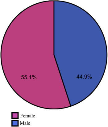
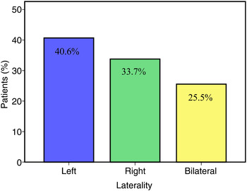
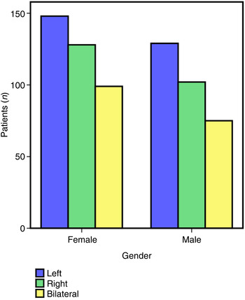
The age range of patients who presented with semicircular canal dehiscence was 5–88 years, with a mean age of 49.75 years (standard deviation = 15.33). Of the 682 patients, 376 (55.1 per cent) were female and 306 (44.9 per cent) were male. Semicircular canal dehiscence was associated with a statistically significant female predominance (chi-square = 7.185, p = 0.007), with a female-to-male ratio of 1.2 to 1 (Figure 1). Among the 682 patients, 277 (40.6 per cent) had left-sided semicircular canal dehiscence, 230 (33.7 per cent) had right-sided semicircular canal dehiscence and 174 (25.5 per cent) had bilateral semicircular canal dehiscence (chi-square = 23.457, p < 0.001) (Figure 2). The left versus right ratio was 1.2 to 1. The unilateral to bilateral ratio was 2.9 to 1. The analysis performed for laterality based on gender revealed no statistically significant association (chi-square = 1.383, p = 0.710) (Figure 3). There was also no statistically significant association between gender and age (t = 0.098, p = 0.922) or laterality and age (F = 2.034, p = 0.132).
Occurrence of semicircular canal dehiscence by gender. Semicircular canal dehiscence has a statistically significant female predominance (chi-square = 7.185, p = 0.007).

Occurrence of semicircular canal dehiscence by laterality. Of 682 patients, 277 had left-sided semicircular canal dehiscence, 230 had right-sided semicircular canal dehiscence and 174 had bilateral semicircular canal dehiscence (chi-square = 23.457, p < 0.001).

Laterality of semicircular canal dehiscence based on gender. There was no statistically significant association between gender and laterality (chi-square = 1.383, p = 0.710).

Discussion
Prior to the discovery of semicircular canal dehiscence, sound-induced vertigo or nystagmus, also known as the Tullio phenomenon, was most commonly associated with Ménière's disease, perilymphatic fistula and congenital deafness.Reference Fox, Balkany and Arenberg 42 – Reference Kwee 44 Semicircular canal dehiscence has recently been established as the major cause of these symptoms. Semicircular canal dehiscence is a relatively new clinical entity, and our knowledge of its incidence, aetiology and pathophysiology remains incomplete. Although the incidence of semicircular canal dehiscence has been reported prior to the current study, based on cadaver and imaging studies, there have been little organised data regarding laterality or gender predilection of the disease.
Previous research has suggested that approximately one-third of cases are bilateral.Reference Banerjee, Whyte and Atlas 45 Computed tomography studies have further demonstrated thinning of the contralateral semicircular canal in unilateral cases. These findings have suggested an underlying congenital cause for semicircular canal dehiscence syndrome.Reference Yew, Zarinkhou, Spasic, Trang, Gopen and Yang 2 A genetic component has also been proposed, based on anecdotal evidence of semicircular canal dehiscence in siblings.Reference Mikulec, McKenna, Ramsey, Rosowski, Herrmann and Rauch 6 Given that the onset of symptoms typically occurs in adulthood and can be initiated by precipitating events, such as direct trauma or activities that induce intracranial or middle-ear pressure changes, the cause of semicircular canal dehiscence is likely to be multifactorial.Reference Carey, Minor and Nager 9 , Reference Minor 10 A predisposing developmental anatomical abnormality, coupled with a secondary inciting event (i.e. trauma or age-related bony erosion), is the likely cause. Our study reveals that bilateral semicircular canal dehiscence occurs in 26 per cent of patients, similar to prior reports (33 per cent). This finding continues to support the concept of a congenital predisposition.
The current study reports a preponderance of left-sided disease in unilateral semicircular canal dehiscence. This may be the result of gross morphological size differences between the right and left cerebral hemispheres, specifically the temporal and parietal lobes. Theoretically, an increase in size of the gyri overlying the arcuate eminence could cause such increases in intracranial pressure and possibly bony tegmen erosion in the area of dehiscence. Alternatively, a difference in brain size during development could lead to a thinner bony tegmen on the side with a larger cerebral hemisphere. A number of morphological asymmetries of the brain have been described, most notably involving an increase in size of the planum temporale and the pars opercularis on the left related to left hemispheric dominance for language function.Reference Goldberg, Roediger, Kucukboyaci, Carlson, Devinsky and Kuzniecky 46 In a study by Goldberg et al., volumetric magnetic resonance imaging analysis revealed that the transverse temporal gyri, lateral superior temporal gyrus, planum temporale and superior parietal gyrus were all larger in the left hemisphere when compared to the right.Reference Goldberg, Roediger, Kucukboyaci, Carlson, Devinsky and Kuzniecky 46 A similar study by Pujol et al. revealed the parietal and temporal regions were consistently larger in the left hemisphere (left temporal volume of 68.8 ml vs 59 ml on the right, p < 0.001; left parietal volume of 36.7 ml vs 26.7 ml on the right, p < 0.001).Reference Pujol, López-Sala, Deus, Cardoner, Sebastián-Gallés and Conesa 47 These morphological asymmetries could be the cause of a thinner bony left-sided tegmen, resulting in a higher rate of left-sided semicircular canal dehiscence syndrome cases. Gender analysis performed in these volumetric brain studies did not reveal a significant gender effect on the overall size of temporal or parietal lobes.
The current study indicates that the female-to-male ratio of semicircular canal dehiscence is 1.2 to 1. If hemispheric size differences cannot account for this disparity, how can we explain a female predilection for semicircular canal dehiscence? It may simply be the result of gender differences in healthcare utilisation. Studies have revealed that women use more health services and incur larger healthcare expenditures than men. The 2013 Health Care Cost and Utilization Report details a higher rate of out-patient visits by women when compared to men. 48 The greatest disparity was between the ages of 26 and 54 years, similar to the age range during which semicircular canal dehiscence is diagnosed. In a study by Bertakis et al., women had a significantly higher mean number of primary care visits and a significantly higher mean number of diagnostic tests ordered.Reference Bertakis, Azari, Helms, Callahan and Robbins 49 Perhaps, given the higher number of visits, and a lower physical and mental health status reported by women, primary care physicians were more likely to request specialist referral or diagnostic testing.Reference Bertakis, Azari, Helms, Callahan and Robbins 49 Conceivably, if women are presenting for medical care more frequently and obtaining more diagnostic testing, this could explain the higher rate of semicircular canal dehiscence syndrome among women, particularly as diagnosis is made primarily by high-resolution computed tomography scanning and vestibular-evoked myogenic potential testing.
-
• Semicircular canal dehiscence syndrome is a well-established cause of sound- and pressure-induced vestibular symptoms
-
• The disease is thought to be caused by a predisposing developmental anatomical abnormality coupled with a secondary inciting event
-
• Semicircular canal dehiscence syndrome is most commonly left-sided and exhibits a female predominance
-
• Left-sided and female preponderance may be the result of cerebral hemisphere asymmetries and healthcare-seeking propensities of women, respectively
Semicircular canal dehiscence syndrome is a relatively new clinical entity, and knowledge of its incidence, aetiology and pathophysiology continues to expand. It is likely that the disease is caused by a combination of congenital and environmental factors. Semicircular canal dehiscence syndrome is most commonly left-sided and exhibits a female predominance. This may be secondary to morphological cerebral hemisphere asymmetries in both sexes coupled with a predilection of women to seek more medical care than men.