Introduction
The increasing number of aged people in the population means that diseases that may affect our lungs are increasingly manifest in older patients and so a question commonly arises as to what extent any observed changes in the lungs’ performance are due to natural ageing processes and how much might be due to a disease process. To answer this question one first needs a full understanding of how the lungs age and then one must consider how this affects their performance. With this knowledge, lung function tests can then be best used to determine the effects of disease processes.
This article will firstly address what is understood about the normal ageing of the lungs and how this affects their function. Then the second part will look at how lung function can be compared against known normal values for an aged population to make deductions in individual patients about whether their function is abnormal or not, and if it is abnormal, how abnormal that is.
Normal lung ageing
What causes the lungs to age?
The normal process of ageing in humans is often evident, even to lay people, from changes in a subject's skin, posture or exercise performance. Any changes that occur within our lungs with age are not so evident. The precise way in which cellular function declines with age is a cause for continuing debate. With each cell division the telomere length shortens and ultimately further cell division is not possible, and this has been linked to ageing and mortality.Reference Kimura, Hjelmborg and Gardner1 How much this effect leads to loss of important cellular function in the lungs is not fully known. The regenerative potential of the lungs is limited so the requirement for cell turnover is minimal. Therefore this mechanism of ageing is perhaps not very important for the lungs.
Intrinsic ageing can arise from metabolic changes as a consequence of oxidative stress within cells. Our alveolar surface (100 square metres) is the second largest contact we make with our environment, with our gut surface area being 300 square metres. We largely choose what we eat and so our gut is exposed to selected substances, but for our lungs we do not have much choice about the constituents of the air we breathe (except for tobacco smoke) and the air can contain many noxious substances (particulates from car exhaust, open fires) and gases (ozone, sulphur dioxide) that can lead to lung damage.Reference Bernard, Samet, Grambsch, Ebi and Romieu2 Inhaled irritants can set up an inflammatory reaction drawing leukocytes to the lungs,Reference Stenfors, Nordenhäll and Salvi3 which then release potentially damaging enzymes, including elastase, which can affect the lung parenchyma.Reference Stockley4 Thus the normal properties of the lungs could be damaged by constant attrition from inflammatory processes set up from inhaling noxious substances. Subjects with better anti-oxidant defence mechanisms might then be better protected from this constant attrition.
A further argument towards the beneficial effects of protection against these inflammatory changes arose from studies in rats exposed to cigarette smoke that showed a protective effect from statins, which have anti-inflammatory and anti-oxidative properties in addition to their lipid-lowering effects, in preventing the smoke-related accelerated lung decline.Reference Lee, Lee and Kim5 This has been confirmed in human studies with the normative ageing study6 showing that the use of statins significantly reduced the normal decline over time in FEV1 and a recent large study in current and former smokers also showed a protective effect from statinsReference Keddissi, Younis and Chbeir7 with regard to lung function.
What effect does ageing have on the lungs?
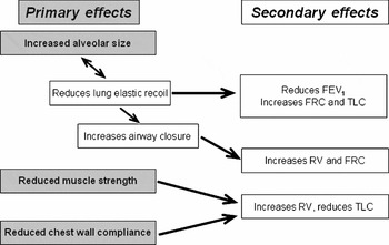
Figure 1 summarizes all the primary changes with age that affect lung function, together with the secondary effects these have, and these will be discussed in turn. The lungs have a central function in achieving satisfactory ventilation so that gas exchange can occur to oxygenate the blood. Both the ventilatory function and gas exchange appear to be reduced with natural ageing.Reference Quanjer, Tammeling, Cotes and Pedersen8 Satisfactory ventilation is achieved by being able to inhale with sufficient speed and depth (that is, volume) to maintain gas exchange. The flow characteristics of our airways depend on the airway cross-sectional area, the airway wall compliance and adequate driving pressure.Reference Dawson and Elliott9 The larger the cross-sectional area, the higher the possible flow that can be achieved. Thus greater flows are possible at higher lung volumes as the airways are pulled to a more open state by increasing lung inflation and outward traction on the airway walls transmitted via the lungs’ tissue elements. In this way peak expiratory flow is maximal only if a full inhalation to total lung capacity is achieved before exhaling and the exhalation is performed with maximum effort. Oedema of the airway walls leads to reduced cross-sectional area without any need for smooth muscle contraction to have occurred.
Outline of the primary effects of ageing on the lungs and the secondary effects these have on the function of the lungs.

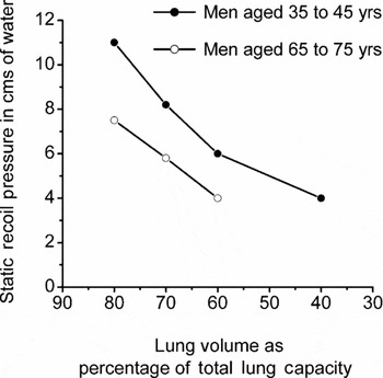
Little is known about age-related changes in airway wall compliance in human lungs. A more compliant, that is floppy, airway leads to a reduced maximum flowReference Dawson and Elliott9 since the airway can collapse more easily. The final determinant of maximum flow is the driving pressure which is very much dependent on the elastic recoil properties of the lungs. It is known that elastic recoil is significantly reduced with ageReference Babb and Rodarte10 (see Figure 2). This loss of recoil with age will also reduce the normal elastic pull on the airways and so the cross-sectional area for a given degree of inflation is less. Conversely, when fibrotic lungs inflate, greater traction than usual is exerted on the airway walls through the lung tissue and so in early interstitial lung disease peak expiratory flow is often much greater than normal.
Static recoil pressure plotted against percentage of total lung capacity for men aged 35–45 years (closed circles) and men aged 65–75 years (open circles). Adapted from Babb et al.Reference Babb and Rodarte10

There is elastin in the lungs and so a first consideration might be that the reduced lung elastic recoil with age would be due to loss of elastin within the lungs, much in the same way that elastin is lost from the skin with age.Reference Callaghan and Wilhelm11 However, pathological specimens of lungs from subjects of different ages who had died of non-respiratory disease showed no change in the ratio of collagen to elastin in the lungs with age,Reference Pierce and Ebert12 but they did find a reduced amount of elastin in the pleura. It is known that the majority of the recoil pressure in normal lungs is not related to elastin or collagen, but due to the effect of surface tension from the air–fluid interface on the surface of the alveoli; this follows the finding that the recoil of the lungs reduces dramatically if they are filled with saline instead of air.Reference Bachofen, Hildebrandt and Bachofen13 In essence the alveoli are like millions of small bubbles and the Laplace lawReference Tenney14 indicates that the surface tension force is much greater for a small bubble compared with a larger one; so smaller alveoli should collapse, pushing their air into larger ones. This does not happen because surfactant lowers the surface tension and so stabilizes the alveoli. The observed loss of recoil with age is in fact mainly due to an increased airspace diameter of the alveoli with age, and this effect has been confirmed by several researchers from studying the pathology of lungs in the elderly using measurements of the distance between airspace walls (called the mean linear intercept, Lm).Reference Gillooly and Lamb15, Reference Verbeken, Cauberghs and Mertens16
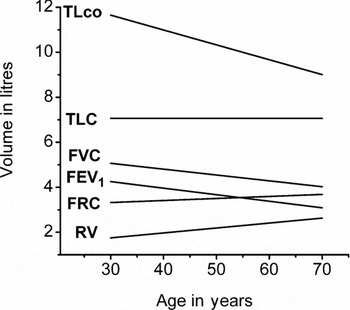
What little is known about changes in surfactant with age suggests there are no significant changes.Reference Rebello, Jobe, Eisele and Ikegami17 Why the airspaces get larger appears to be due to changes in the configuration of elastin in the area around the respiratory bronchiole. From middle age onwards these elastin fibres appear to degenerate and their coiling structure around alveolar ducts is altered, leading to an evenly distributed increase in size in all alveoli with age,Reference Janssens, Pache and Nicod18 with no destruction of alveolar walls themselves. An increase in airspace dimension not only reduces recoil but also leads to a decrease in surface area available for gas transfer relative to the volume of gas inhaled, thus the total lung gas transfer factor (TLco) falls with age (see Figure 3).
Changes with age in total lung capacity (TLC), functional residual capacity (FRC), residual volume (RV), forced vital capacity (FVC) and forced expiratory volume in 1 s (FEV1) all in litres for a man of height 1.77 m as predicted from the ECCS cross-sectional equationsReference Quanjer, Tammeling, Cotes and Pedersen8 together with total lung gas transfer (TLco) in mmol/min/kPa.

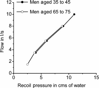
The age-related reduction in maximum achievable flow at a given lung volume could be due to the reduced elastic recoil with age and/or an increase in airway resistance with age. The achieved maximum flow for a given recoil driving pressure is not significantly changed with age,Reference Janssens, Pache and Nicod18 indicating that the airway resistance is not significantly changed with age (see Figure 4). Thus expiratory flow is lower in older subjects due primarily to the loss in recoil driving pressure.
Plot of expiratory flow in l/s against static recoil pressure in young (closed circles) and older men (open circles). Adapted from Verbeken et al.Reference Verbeken, Cauberghs and Mertens16

Measurements of lung elastic recoil require measuring oesophageal pressure (from a swallowed balloon positioned in the lower oesophagus) and recording how this pressure varies with lung volume. This should be done under static conditions, that is, with no air flowing through the airways, to avoid pressure components due to airway resistance. From this pressure versus lung volume curve, one can construct the elastic recoil profile of an individual's lungs (see Figure 2). This curve has been shown to change with age such that the pressure required to inflate the lungs to a certain volume gets less with age. The natural recoil of the lungs, which tends to make them collapse, is thus diminished with age. This has two obvious effects.
Firstly the natural resting position of the breathing system, which is called the functional residual capacity (FRC), is increased. This is the point at the end of gentle tidal exhalation when all the muscles acting on the ventilatory system are relaxed and the natural tendency of the chest wall to spring outwards from this position is exactly balanced by the recoil force of the lungs wanting to collapse. It can be seen that if the natural recoil of the lungs to collapse diminishes with age, then the FRC resting position will rise if the chest wall characteristics at this point of inflation are unchanged. Thus with age, the chest wall gradually moves outwards to a more expanded position as FRC rises. Many subjects will know from experience that their chest measurement tends to increase with age, requiring a larger jacket size when one is 60 years old compared with 30 years old.
Secondly the recoil pressure is the major part of driving pressure for expiratory flow so exhaled flow is more limited with age. This leads to a lower forced expiratory volume in 1 second (FEV1). FVC is less susceptible to change from a lower driving pressure from elastic recoil and so is relatively preserved compared with FEV1. In Figure 3 there is an almost parallel drop in FVC and FEV1 with age, but FEV1 reaches zero sooner (see Figure 3), so the ratio of FEV1 to FVC falls with age and so fixed cut-off levels of FEV1/FVC to define airflow obstruction will falsely identify many older subjects as having airflow obstruction which they do not have.Reference Swanney, Ruppel and Enright19
Inflammation in the airways may lead to airway wall inflammation and structural remodelling of the airways. This may lead to increased airway wall thickness, reduced airway diameter and so an increased airway resistance. Loss of elastic recoil driving pressure and increased airway resistance will diminish the maximum flow possible and lead to premature airway closure during exhalation. The point at which airways start to close is called closing volume and this volume is known to occur at a higher lung volume in older subjectsReference Anthonisen, Danson, Robertson and Ross20 and can occur during tidal breathing in the elderly.Reference Leblanc, Ruff and Milic-Emili21 These changes will also lead to a fall in FVC, but this is less marked than the fall in FEV1, so the FEV1 to FVC ratio falls with age.
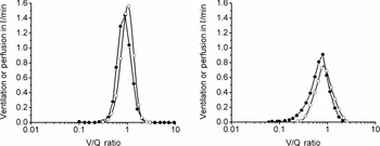
The purpose of ventilating the lungs is to achieve adequate gas exchange for the subject's metabolic needs. Gas exchange in older subjects is not as good as in younger subjects and so the resting arterial blood oxygen tends to fall up to about the age of 70,Reference Cerveri, Zoia and Fanfulla22 and then does not seem to drop further.Reference Hardie, Vollmer, Buist, Ellingsen and Mørkve23 This arises because matching of ventilation to perfusion becomes less good with age. In young subjects in the upright position the distribution of ventilation (V’A) and perfusion (Q’) is relatively narrow, but in older subjects the standard deviation of the distribution of both V’A and Q’ are much larger.Reference Wagner, Laravuso, Uhl and West24 This can be seen in Figure 5 where in the younger subject the amount of blood flow to low V’A/Q’ areas (closed circles) is small. In the older subject on the right, the amount of perfusion to areas of lower and higher V’A/Q’ areas is larger. This means in older subjects a larger proportion of lung units have a very high V’A/Q’, that is wasted ventilation, and a larger proportion of lung units have a low V’A/Q’, that is poorly ventilated but perfused units, when compared with younger subjects. It is these latter low V’A/Q’ units that contribute poorly oxygenated blood to that leaving the lungs. This lowers the average arterial PaO2 and causes an increased alveolar arterial oxygen difference. These areas of low V’A/Q’ occur as a result of the collapse of airways during tidal breathing (see above on closing volume) so some lung units no longer contribute to gas exchange, but are still perfused. In terms of usual lung function tests this effect is evident in the measurement of arterial gases.
Plots of ventilation (open circles) and perfusion (closed circles) against V/Q ratios for the distribution of V/Q in the lungs of a young subject on the left and an older subject on the right. Adapted from Wagner et al.Reference Wagner, Laravuso, Uhl and West24

Muscle strength declines with age and is a predictor of future disability.Reference Rantanen, Guralnik and Foley25 The respiratory muscles are also affectedReference Enright, Kronmal and Manolio26 with the diaphragm strength in 70-year-olds being on average between 13 and 25% lower than that in 20- to 30-year-olds, depending on how the strength was being tested.Reference Polkey, Harris and Hughes27, Reference Tolep, Higgins and Muza28 So although the reduced elastic recoil might mean subjects could achieve a higher TLC, because recoil was lower, the lower muscle strength means the TLC remains unchanged (see Figure 3). Furthermore, the compliance of the chest wall changes with age with the chest becoming stiffer.Reference Mittman, Edelman, Norris and Shock29 The chest wall increasingly resists deviation from the natural resting state. Thus with increasing age as muscle strength is reduced, the residual volume (RV) increases because squeezing the chest down further is problematic. Also TLC does not increase as anticipated by the drop in elastic recoil with age because the weaker muscles cannot expand the chest wall further.
Obesity is an increasing problem in western societies and is more prevalent in older age groups.Reference Ogden, Carroll and Curtin30 In young to middle-aged workers excess weight gain has been found to be associated with a greater decline in FEV1, FVC and their ratio.Reference Wang, McCabe, Petsonk, Hankinson and Banks31 Normal ageing should not involve the above observed tendency to weight gain with age, which relates to a personal failure to adjust food intake to a reduced exercise and metabolic demand. Thus changes in lung function due to obesity must be identified and not attributed to ageing per se.
Defining normal lung function in the elderly
Most clinical tests have normal ranges that do not vary significantly with age; for example, many blood tests are like this although renal function decreases with age. Most lung function indices are known to vary with age, sex and height.Reference Quanjer, Tammeling, Cotes and Pedersen8, Reference Hankinson, Odencrantz and Fedan32 The data upon which these findings are made are usually obtained from normal subjects lacking any disease or relevant symptoms and who have never smoked. As people get older it is increasingly difficult to find subjects who fulfil these criteria and so it is difficult to find adequate data from older subjects on which to base prediction equations of normality.
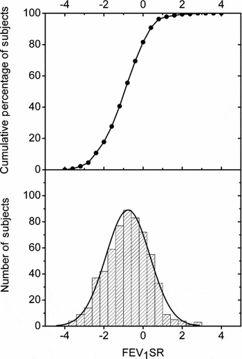
In Europe the commonly applied ECCS equationsReference Quanjer, Tammeling, Cotes and Pedersen8 were derived from subjects only up to 70 years of age and the entry criterion for subject data was to some extent permissive of smoking if the subjects were deemed normal. The use of such equations for older age groups means extrapolating beyond the prediction limits and this may be unsafe. Data have recently been presented on a cohort of 95-year-old subjects from Denmark and found the ECCS equations were reasonably robust for this age group in terms of scatter about the mean, but were not accurate in predicting the mean,Reference Jacobsen, Sigsgaard, Pedersen, Christensen and Miller33 finding the subjects to be about 0.77 of a SD below that predicted (see Figure 6). The same was true when using a set of equations derived from American subjects in the closer age range of 65–85 years.Reference Enright, Adams, Boyle and Sherrill34 This deviation from expected might be accounted for by the known increase in kyphosis in elderly peopleReference Bartynski, Heller and Grahovac35 and so the height coefficients in the prediction equations would not be properly accounting for the size effects on lung function in the elderly. However, these observed differences in Figure 6 could be accounted for by a 6 cm height reduction for women and a 12 cm reduction in men, both of which are unrealistic.
Histogram of FEV1 for 598 subjects in a cohort of 95-year-old subjects when expressed as standardized residuals (FEV1SR) using the ECCS prediction equationReference Quanjer, Tammeling, Cotes and Pedersen8 together with a Gaussian distribution plotted in the lower panel and the cumulative distribution in the upper panel. For a normal population the mean should be zero and 95% of subjects should be between ±1.96. The mean ± SD FEV1SR value was –0.77 ± 1.12.

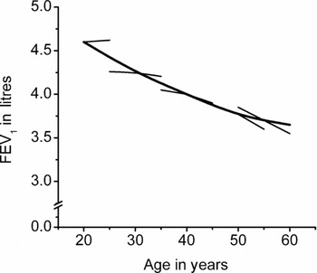
The above highlights the problem of using prediction equations based on cross-sectional studies. Such studies in fact include several cohorts of people who have had different upbringing, nutrition and atmospheric exposures which are used to derive a unifying age coefficient for all ages that is misleading. The magnitude of this cohort effect on FVC has been estimated to be such that for each previous generation going back for over 100 years the FVC was on average about 125 ml smaller than it was for the subsequent generation.Reference Glindmeyer, Diem, Jones and Weill36 The six cities study in the USAReference Ware, Dockery and Louis37 showed that longitudinal decline in lung function for an individual deviates from the accepted single rate of decline as taken from cross-sectional prediction equations. This is shown in Figure 7 where in younger subjects there is no decline but an increase in FEV1, whereas in older subjects the observed longitudinal change is an increasing rate of decline.Reference Van Pelt, Borsboom and Rijken38 This effect was also seen in the seminal paper by Fletcher and Peto on FEV1 decline.Reference Fletcher, Peto, Tinker and Speizer39 It is difficult to find older subjects who have not had disease and who have never smoked or been exposed to deleterious atmospheric pollution, and such subjects may not be representative of normality since they are the few survivors to that age, which in effect selects them as different from other subjects in their birth cohort. Such a healthy survivor effect was evident in the six cities study,Reference Ware, Dockery and Louis37 but only accounted for a very small amount of the variation in lung function observed. More recent work has looked at deriving a single prediction equation to cover all age groupsReference Stanojevic, Wade, Stocks and Hankinson40 and the ERS is currently supporting a task force to finalize such equations for European subjects.
Age dependence of FEV1 cross-sectionally in never smokers as a single continuous line plotted from age 20 to 60 and the longitudinal observed change with age as separate shorter and thinner lines. Adapted from van Pelt et al.Reference Van Pelt, Borsboom and Rijken38

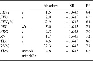
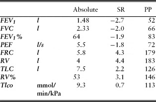
Current best practice is to compare a subject's result with a predicted value derived from a suitable prediction equation using the method of standardized residuals.Reference Quanjer, Tammeling, Cotes and Pedersen8 This converts the deviation from the predicted value (termed the residual) into the number of standard deviations (SD) it is away from predicted. Thus conventional normal ranges for patients suspected of having disease are the 90% confidence limits, which are ± 1.645 SD from predicted. So a result outside these limits can be deemed abnormal and a cause is then sought to explain this deviation. This method can be used for all indices and allows patterns of lung function abnormality to be correctly appreciated. Table 1 shows the lower limit of normal for various lung function indices for an elderly man of short stature expressed as absolute units, standardized residuals (SR) and per cent predicted. It is clear that using per cent of predicted with an 80% value as the cut-off taken to define normality would lead to erroneous conclusions about this subject's lung function, as they are all exactly at the lower limit of normal.
Lung function results for a man aged 70 of small stature at 1.6 m expressed as absolute units, standardized residuals (SR) and as per cent of predicted (PP) using ECCS equationsReference Quanjer, Tammeling, Cotes and Pedersen8

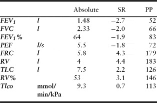
Table 2 shows the results for a middle-aged male patient referred for lung function testing. Just looking at the per cent of predicted results for this man's FEV1, FVC and FEV1 as a percentage of FVC (FEV1%), one might think this was a restrictive defect as the FEV1% might be deemed normal at 83% of predicted with the other two indices appearing to be significantly reduced. In fact the FEV1% is abnormal at –1.9 SR and the static lung volumes confirm the findings of marked airflow limitation (increased RV). The difference between TLC and RV is 3.5 l and this is the slow vital capacity that is much larger than the FVC indicating dynamic air trapping during the forced expiration due to airway collapse. Thus the method of SR is the best for interpreting which results are abnormal and identifying patterns of abnormality (that is, restrictive from obstructive).
Lung function results for a male patient aged 58 of height 1.63 m expressed as absolute units, standardized residuals (SR) and as per cent of predicted (PP) using ECCS equationsReference Quanjer, Tammeling, Cotes and Pedersen8
Assessing degree of lung function deficit in older people
When assessing if an elderly subject has abnormal lung function, the first step is as above to relate the recorded value to a predicted by expressing it in terms of standardized residuals. If this value is outside the normal range then a different method is needed to determine how abnormal this result is. At first sight the SR value, that is the number of SD from predicted, might be a good indicator of severity. Because the actual magnitude of most lung function indices changes with age (Figure 3) and the normal scatter about the predicted is relatively constant irrespective of age, it is possible for a 21-year-old subject, for example, with cystic fibrosis to have an FEV1 that is 8SD below predicted, whereas for a patient aged 70 their FEV1 would have to be less than zero (clearly nonsensical) for the value to be this far from predicted. Thus in older subjects, lung function expressed in SR values can never appear to be as ‘abnormal’ as can occur in younger subjects. However, it cannot be said that older subjects never have such severe disease as younger subjects, so SR values cannot be used sensibly to define the degree of abnormality.
An alternative method for determining the severity of abnormality in a lung function index, such as FEV1, has been by using the method of per cent of predicted. This has been in use since the early 1960s,Reference Bates, Macklem and Christie41 with a rough rule of thumb applied that 80% of predicted was the cut-off for normality. This might be close to correct for some indices in just a few patients, but is incorrect for most indices under most circumstances.Reference Miller and Pedersen42 Whilst this method is no good for determining if a test is abnormal, it is much better for expressing severity than the SR method.Reference Miller and Pedersen42 However, standardizing lung function such as FEV1, by dividing it by height cubedReference Fletcher, Peto, Tinker and Speizer39, Reference Miller and Pedersen42 or by the sex-specific lower limit seen in patients,Reference Miller and Pedersen42 has been shown to better relate FEV1 to subsequent mortality in normal subjectsReference Miller, Pedersen and Dirksen43, Reference Chinn, Gislason and Aspelund44 and in patients with COPD.Reference Miller, Pedersen, Lange and Vestbo45 Thus the best principle for assessing degree of abnormality may be to stop looking at a subject's function in terms of the relation to a predicted value, but instead look at it in terms of where it stands with regard to ‘the bottom line’. The absolute value of the index does not work as well as those that standardize by powers of height, which take some sex and size effect into account, or by the observed sex-specific lower limit.Reference Miller and Pedersen42 Currently these methodologies are only evidenced through data from FEV1 and will need to be explored with other indices. In the meantime, for indices other than FEV1, using per cent of predicted may be the best means for expressing lung function severity.
Even at very advanced age, lung function in terms of FEV1 remains a good predictor of subsequent mortalityReference Jacobsen, Sigsgaard, Pedersen, Christensen and Miller33 and these newer methodologies allow one to make estimates of likely survival and changes in survival if rates of lung function decline can be influenced by therapy or lifestyle changes. Such estimates are still pertinent in an elderly population since decisions about treatment options and likely outcomes, for example for various surgical interventions, may be better determined by using them.
Conclusion
In an increasingly elderly population, patients with lung disease that requires assessment and management will be evermore frequent. Elderly patients deserve just as rigorous criteria for making a diagnosis as younger patients, and so clinicians must strive for the most accurate interpretations of lung function tests to make these diagnoses. It is clear that age per se has a significant effect on lung function. Estimates of FEV1 loss in lifelong non-smokers aged over 50Reference Xu, Weiss, Dockery, Schouten and Rijcken46 suggest that a normal pair of lungs could sustain an individual for perhaps up to 120 years, but clearly other degenerative or disease processes will bring a conclusion to life before then. Adult humans have a considerable reserve of lung function that permits extremes of exercise to be undertaken, which was necessary for our forebears to hunt for food and avoid our predators. As our lungs age, this reserve of lung function gradually diminishes.
This article has outlined how these ageing processes affect lung function and these changes cannot be classified as disease. Identifying the effect of true disease processes on our lungs requires an intelligent use of lung function tests, as well as the use of clinical history taking, examination and imaging. Defining normal limits for lung function in the very old presents some difficulties that can be overcome as reasonable prediction equations pertinent for all ages become available to make this aspect of lung function testing more uniform across patient groups. How to assess the degree of severity of a given lung function deficit is currently undergoing a rethink and assessing how far a test is from the ‘bottom line’ looks to be a more promising way of doing this.
Conflicts of interest
The authors declare no conflicts of interest.