INTRODUCTION
A meta-analysis of 17 randomised trials has shown a reduction in 10-year recurrence and an increase in 15-year survival rates for patients receiving post-surgical breast irradiation for breast cancer. 1 However, normal tissue toxicity can limit the therapeutic ratio and reduce quality of life for these patients,Reference Hurkmans, Borger and van der Horst 2 confirming that the dose delivered to the heart and lung should be minimised.Reference Mackenzie, Fyles, Wang and Chung 3 – Reference Gregoire and Mackie 6 Several studies have reviewed the level of cardiac toxicity and the potential risk to patients having radiotherapy.Reference Paszat, Mackillop and Groome 7 – Reference Bouillon, Haddy and Delaloge 10 The Early Breast Cancer Trialists’ Collaborative Group demonstrated that late cardiac mortality and morbidity usually become evident at 10–15 years following radiotherapy. 11 More recently, Darby et al.Reference Darby, Ewertz and McGale 4 reported a radiation-related increase in major coronary events within 5 years post-radiotherapy. The volume of heart within the radiation field is a key factor that results in radiation-induced perfusion defects in the irradiated myocardium. This may be exacerbated by irradiation of the left anterior descending (LAD) artery, leading to coronary artery stenosis.Reference Mackenzie, Fyles, Wang and Chung 3 The potential detrimental cardiac effect from radiotherapy may also be compounded by the widespread use of anthraxcycline-based chemotherapy and trastuzumab, which are associated with significant cardiac toxicity.Reference Bowles, Wellman and Feigelson 12
Due to the long latent interval to development of late cardiac complications, most studies on cardiac effects of breast radiotherapy are based on data from patients treated in the 1970–90s; since then radiotherapy planning and treatment techniques have changed markedly. However, Taylor et al. suggest that, even with modern treatment delivery techniques, part of the heart regularly receives >20 Gy in left-sided breast radiotherapy. As the risk of a major coronary event increases linearly with dose, with no apparent lowest threshold dose, any irradiation of the heart should be avoided if possible.Reference Darby, Ewertz and McGale 4 , Reference Taylor, Povali and McGale 13
Cardiac shielding using multileaf collimation is routinely used to treat left-sided breast cancer.Reference Landau, Adams and Webb 14 However, due to the position of the tumour bed relative to the heart, cardiac shielding is not always possible without compromising breast tissue coverage. Raj et al. suggest that this is more likely to be an issue for patients with inferiorly located tumours.Reference Raj, Evans and Prosnitz 15
Deep inspiration breath-hold (DIBH) techniques have been shown to help move the chest wall away from the heart and reduce cardiac irradiation.Reference Sixel, Aznar and Ung 16 – Reference Remouchamps, Vicini and Sharpe 18 One of these techniques, Elekta’s Active Breathing CoordinatorTM (ABC) (Elekta, Crawley, UK) is used to deliver radiotherapy for lung cancer.Reference McNair, Brock and Richard 19
This paper describes findings from our initial experience on using the DIBH technique with ABC for the treatment of left-sided breast cancer.
MATERIALS AND METHODS
The study was initiated by the Breast Development Group at our Institution, registered with the Trust audit committee and approved by the Institution’s New Interventional Procedures Committee.
Design
Retrospective observational study.
Aims
The primary aim was to assess the effectiveness of DIBH using ABC in reducing heart dose. Secondary aims included; the effect ABC would have on lung dose, tumour bed dose coverage, treatment reproducibility, patient tolerance and workflow.
Patient selection and sample size
The first 12 consecutive patients referred for adjuvant radiotherapy (40 Gy in 15 fractions) to the left breast or chest wall who would benefit from the ABC technique were included in the study.
At the time of the study participants were identified for the use of ABC, following planning of their tangential breast treatment using a conventional free-breathing (FB) computed tomography (CT) planning scan. Additional dose from a second CT scan, using ABC was justified, in accordance with Ionising Radiation (Medical Exposures) Regulations, 20 when ≥1 cm of heart was included in the treatment field and either breast tissue or tumour bed coverage would be compromised by the use of heart shielding.
CT scanning
In all, 45 minutes slots were scheduled for coaching and acquiring sequential 5 mm slices on a single slice wide bore CT scanner (Philips AcQSim CT Scanner; Philips Medical Systems, Guildford, UK) with the patient in DIBH. Patients were scanned in a supine position on a breast board with both arms raised, identical to the FB scan. Before scanning, patients were coached to breathe normally through the ABC apparatus until they achieved regular respiration and a stable respiratory trace on the lung volume display, determining a breathing baseline. The maximum inspiratory capacity was measured using the ABC spirometer; the breath hold (BH) threshold was set at 75% of the maximum capacity. This was followed by training in assisted respiration suspension, increasing BH times stepwise from the initial 5 seconds to 20 or 25 seconds. Patients were eligible for ABC when able to confidently and repeatedly hold their breath for a minimum of 20 seconds to reduce the number of treatment beam interruptions and, consequently, minimise delays on treatment units. A minimum lung volume of 1 L was required to warrant sufficient chest wall movement and maximise cardiac sparing.
Planning
Tangential beams were simulated using Prosoma virtual software (Oncology Systems Limited, UK) and doses calculated using the Oncentra planning system, version 4.3 (Elekta) and a Collapsed Cone dose computation algorithm.
Tumour bed volumes and tangential field borders were defined by the patient’s respective clinician for both ABC and FB scan. The heart, following previously published delineation guidelines, 21 and ipsilateral lung outlines were delineated for both ABC and FB scans.
Treatment and imaging
Radiotherapy was carried out for each patient using the ABC plan, with the FB plan only to be used if there were any issues encountered with patient set-up/compliance, or mechanical problems with the ABC equipment. Orthogonal megavoltage images were taken to assess the isocentre position at the first three fractions and then once weekly, as per departmental breast imaging protocol.
Analysis
Dose volume histograms (DVHs) were used to analyse the heart dose reduction using the ABC technique—the reduction in heart exposure between FB and ABC was also measured on axial CT images.
The ipsilateral lung dose was recorded and analysed using DVHs to determine the effect ABC technique had on lung volume tolerances.
Radiation doses for both the heart and ipsilateral lung were measured against established critical organ dose constraints. 22 The guidelines recommend that the volume of heart receiving at least 13 Gy (V13 Gy) should be <10% and the volume of the ipsilateral lung receiving at least 18 Gy (V18 Gy) should be <15%. The heart volume receiving at least 5 Gy (V5 Gy) and mean heart dose was also assessed.
Tumour bed dose coverage was measured as the dose received by 98% of the tumour bed volume (D98%), following the International Commission on Radiation Units 83 proposal to use of near minimum (2%) and near maximum (98%) doses when measuring homogeneity within a planning target volume (PTV).Reference Gregoire and Mackie 6 Tumour bed dose was further analysed using DVHs.
Treatment reproducibility was measured on orthogonal megavoltage images, recording the inter-fraction movement in the craniocaudal, lateral and vertical axes.
Two plans, a FB and ABC plan were produced for all patients in this study. It was felt necessary to prepare both plans during the implementation phase as it was not known how well patients would tolerate the technique and also to accommodate for any machine breakdown. Therefore, patient tolerance was measured as the number of eligible patients at CT scanning and those completing their treatment on the ABC plan without reverting back to the FB plan during treatment. BH time, lung threshold volume and patient age were recorded to inform future practice.
Patients were divided into two groups, those with upper quadrant tumours and those with lower quadrant or central tumours to help determine the effectiveness of the ABC technique between the groups. This was based on the premise that cardiac shielding is not always possible without compromising breast tumour bed coverage, and is more likely to be an issue for patients with inferiorly located tumours.Reference Raj, Evans and Prosnitz 15
The treatment time from entering to leaving the treatment room was documented to determine the impact on the day-to-day running of the unit.
RESULTS
In total, 12 patients referred for adjuvant left-sided breast-only radiotherapy, stages 1–3, and treated with the ABC technique between June and December 2012 were assessed. Of these, eight received breast irradiation post-breast conservation (two patients had no tumour bed PTV delineated) and four received chest wall irradiation post-mastectomy. The median age (range) of participants, 49 (34–69) years, was lower than for the wider early breast cancer population. This is due to chance as there were no exclusion criteria on age during patient selection.
Heart dose
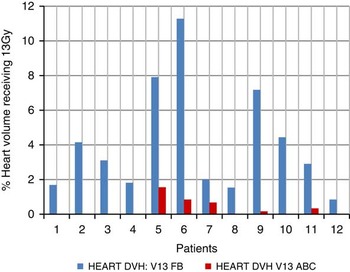
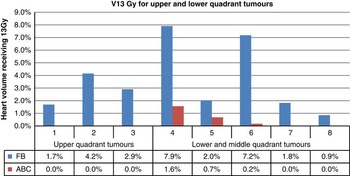
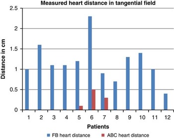
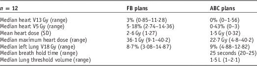
The median (range) heart dose for FB and ABC scans, respectively, was 2·1 Gy (1·2–5·0 Gy) and 1·4 Gy (1·2–2·2 Gy). The heart V13 Gy on FB and ABC plans are demonstrated in Figure 1. The reduction in the percentage of the heart receiving 13 Gy using ABC ranged from 1% (patient 12) to 10% (patient 6). Table 1 summarises the results of the DVH analysis. The median (range) heart volume receiving 13 Gy in FB was 3% (0·85–11·28%) compared with 0% (0–1·56%) for ABC plans. The median (range) maximum heart dose was 36·1 Gy (9·1–40·2) for FB plans compared with 22·7 Gy (4·8–40·2) for DIBH plans. The heart V13 Gy for FB and ABC plans are shown in Figure 2 based on tumour location. With ABC, the chest wall moved away from the heart during inspiration and there was no heart in the treatment field in 9 out of 12 cases. In the remaining three patients 1, 5 and 3 mm of heart remained in the field (Figure 3).
The percentage heart volume receiving 13 Gy or more for 12 patients planned on free-breathing (FB) and Active Breathing Coordinator (ABC) scans. Abbreviation: DVH, dose volume histogram.

The percentage heart volume receiving 13 Gy for upper and lower quadrant tumours (n=8). Abbreviations: FB, free breathing; ABC, Active Breathing Coordinator.

Heart included in the tangential fields as measured on axial CT slice. Abbreviations: FB, free breathing; ABC, Active Breathing Coordinator.

The median heart volume receiving >13 Gy (V13 Gy) and 5 Gy (V5 Gy), mean average dose and median maximum dose to the heart, and the median left lung volume receiving >18 Gy, in the free-breathing (FB) and Active Breathing Coordinator (ABC) plans.

Lung dose
The median left lung V18 Gy for FB and ABC plans, respectively, were 8·7% (3·08–14·87) and 9% (4·88–12·82) (Table 1).
Tumour bed dose
The median (range) dose received by 98% of the tumour bed in FB and ABC, respectively, was 97·3% (92·8–99·47) and 97·5% (96·5–98·6). Improved tumour bed coverage was observed in four out of six (67%) patients who had wide local excisions and defined tumour bed volumes (two tumour bed volumes were not outlined).
Treatment reproducibility
The extent of inter-fraction movement as assessed by orthogonal imaging in the craniocaudal, lateral and vertical direction were, respectively, 2·6 mm (1·6–5·1), 3·4 mm (1·0–6·0) and 1·6 mm (1·0–3·0).
Tolerance
All 12 patients completed treatment on the ABC plan. BH time ranged between 20–25 seconds, with a median of 25 seconds. The median (range) threshold lung volume was 1·5 L (1·0–2·1).
Tumour bed location
Eight patients had breast-conserving surgery. Three (37·5%) patients had an upper quadrant tumour bed location, four (50%) in the lower quadrant and one (12·5%) in the central breast. In patients with upper quadrant tumours, the heart volume receiving 13 Gy was reduced by 100% and in patients with lower and middle quadrant tumours by 87·4%.
Discussion
This study demonstrated a reduction in heart dose in ABC compared with FB plans. This may potentially reduce long-term cardiac toxicity but long-term prospective data are required to confirm the clinical utility of this technique.
Other studies using ABC also demonstrated a reduction in heart dose.Reference Mackenzie, Fyles, Wang and Chung 3 , Reference Wang, O’Connell, Stuart and Boyages 9 , Reference Sixel, Aznar and Ung 16 , Reference Remouchamps, Vicini and Sharpe 18 , Reference Korreman, Pederson, Nottrup, Specht and Nystrom 23 KorremanReference Korreman, Pederson, Nottrup, Specht and Nystrom 23 and SixelReference Sixel, Aznar and Ung 16 found the V50% was reduced by 30–40%; Wang et al.Reference Wang, O’Connell, Stuart and Boyages 9 demonstrated a reduction in LAD artery and heart dose using moderate DIBH with ABC, and RemouchampsReference Remouchamps, Vicini and Sharpe 18 recorded a V30 in FB of mean 4·9% and 0·18% in moderate BH and ABC. Moderate BH was chosen to alleviate their concern for patient fatigue described by RosenzweigReference Rosenzweig, Hanley and Mah 24 for lung cancer. Our study results were based on DIBH as breathing difficulty can be expected to be more prevalent in patients with lung cancer compared with breast cancer. Our mean heart dose was 1·4 Gy.
Based on the location of the heart, a higher V13 Gy is anticipated in patients with lower quadrant tumours. Unexpectedly, our study demonstrated only a small difference in V13 Gy between patients with upper or lower quadrant tumours for FB and ABC plans, 2·9 and 3·96%, respectively. This may be due to standard departmental practice to employ cardiac shielding as necessary.
The heart dose was reduced in all study participants and suggests that an average BH time of 25 seconds and threshold volume of 1·5 L can be used as the minimum expectation when assessing the suitability of patients during the coaching process.
Previous findings of an increase in treated lung volume are cited, but this was not demonstrated in our study. It is not unreasonable to suggest, however, that future patients may have greater lung volumes in the treatment field than would be present with a FB plan. The small difference in lung V18 Gy (8·7 versus 9%) for FB and ABC, respectively, may be due to the planner’s attempts to maintain the same lung tolerance for ABC plans as for FB plans during field border placement. It was not necessary to increase the lung volume tolerance from 2 cm for the ABC technique. Nevertheless, it is somewhat reassuring to see similar percentage lung doses with FB and ABC, considering no heart shielding was applied for ABC plans.
The anterior–posterior measurement on the lateral images demonstrates the treatment reproducibility of ABC, whereas craniocaudal and lateral measurements relates more to variations in daily set-up.Reference Remouchamps, Vicini and Sharpe 18 Our study demonstrates a median (range) systematic error in the anterior–posterior direction of 1·6 mm (1·0–3·0 mm), which is comparable with other studies.Reference Mitine, Dutreix and van der Schueren 25 – Reference Thilmann, Adamietz and Saran 27 This implies that ABC has good reproducibility and patients can comply with instructions.
The ABC technique was well tolerated, suggesting that back-up FB plans are not required. This decision will depend on the number of patients on treatment at one time and the implications of equipment failure.
The impact of ABC on resources, especially treatment time, was of concern during the introductory phase. Initially, 30 minutes slots were allocated to patients receiving treatment with the ABC technique which is 6 minutes longer than the allocation for a routine breast technique. However, as radiographers gained more confidence with the technique, we were able to reduce the time slots to 24 minutes, the same as the FB technique. RemouchampsReference Remouchamps, Vicini and Sharpe 18 validated treatments could be streamlined and performed within a 15 minutes treatment slot.
The small sample size in this study makes it difficult to draw firm conclusions on the incremental benefit of the ABC technique. However, the results demonstrate that this technique is easy to implement into clinical practice and does reduce heart dose. Therefore, ABC is a viable option for heart sparing in left-sided breast cancers and is well tolerated by patients.
Conclusions
ABC effectively reduces the amount of heart in left-sided breast radiotherapy when compared with FB, even with the use of heart shielding for FB plans. Consequently, this should reduce the risk of radiation-induced cardiac toxicity. The level of patient compliance with ABC suggests that the impact on radiotherapy services will be minimal.
Acknowledgements
None.