Dimetrodon Cope, Reference Cope1878, the dominant terrestrial predator of its time, has been studied for over a century resulting in the current recognition of twelve species (Romer & Price Reference Romer and Price1940; Reisz Reference Reisz1986; Berman et al. Reference Berman, Reisz, Martens and Henrici2001). These iconic fossils are easily identified by the elongated vertebral spinous processes (or neural spines) forming a dorsal sail that ran the length of the vertebral column.
Although Dimetrodon remains and footprints have been discovered in Europe, specifically in Germany (Berman et al. Reference Berman, Reisz, Martens and Henrici2001, Reference Berman, Henrici, Sumida and Martens2004), virtually all fossil Dimetrodon material comes from North America, including Utah, Arizona, New Mexico, Oklahoma and, primarily, Texas (Romer & Price Reference Romer and Price1940; Vaughn Reference Vaughn1966, Reference Vaughn1969; Berman Reference Berman1977, Reference Berman, S. G. Lucas and Zidek1993; Reisz Reference Reisz1986). Dimetrodon species varied in size (from an estimated 14 kg to 250 kg; Romer & Price Reference Romer and Price1940; Bakker Reference Bakker1975; Berman et al. Reference Berman, Reisz, Martens and Henrici2001) and stratigraphic age (Romer & Price Reference Romer and Price1940; Reisz Reference Reisz1986). The most diverse Dimetrodon faunas are those from the Lower Permian redbeds of Texas, particularly of the Wichita Group (Romer & Price Reference Romer and Price1940; Reisz Reference Reisz1986) where three species are hypothesised to have co-occurred. This apparently high diversity has led to the hypothesis that at least some of the species may represent ontogenetic stages of a single species (Bakker Reference Bakker1982; Rushforth & Small Reference Rushforth and Small2003; Sumida et al. Reference Sumida, Rega and Noriega2005), a hypothesis which is partly tested here for the first time using palaeohistological and skeletochronological methods.
Dimetrodon is a member of the basal synapsid clade Sphenacodontidae, which has traditionally been included within the ‘Pelycosauria’, now known to be paraphyletic (Kemp Reference Kemp2007). We use here the term ‘pelycosaur’ instead of the cumbersome ‘non-therapsid synapsid’.
Institutional abbreviations: AMNH, American Museum of Natural History, New York City, NY, USA; FMNH, The Field Museum, Chicago, IL, USA; IPBSH, Paleohistory collection, Steinmann Institute of Geology, Mineralogy and Palaeontology, University of Bonn, Bonn, Germany; KUVP, University of Kansas Museum of Natural History, Lawrence, KS, USA; MCZ, Museum of Comparative Zoology, Harvard University, Cambridge, MA, USA; OMNH, Sam Noble Oklahoma Museum of Natural History, University of Oklahoma, Norman, OK, USA; TMM, Texas Memorial Museum Vertebrate Paleontology Laboratory, Austin, TX, USA; UMMP, Museum of Paleontology, University of Michigan, Ann Arbor, MI, USA; USNM, United States National Museum of Natural History, Washington DC, USA.
1. Background
1.1. Wichita Group Dimetrodon species diversity
The earliest bonebeds of rich vertebrate fauna come from the continental formations of the Wichita Group. In this work, we focus on the Dimetrodon species of the basal most Nocona Formation, which is a consolidation of the Coleman Junction and Admiral formations as used by Romer (Reference Romer1974). Stratigraphy follows that of Hentz (Reference Hentz1988).
Three species of Dimetrodon have been described from a number of localities within the Nocona Formation, including two mass assemblages: the Geraldine and Briar Creek bonebeds (Romer & Price Reference Romer and Price1940; Reisz Reference Reisz1986; Sander Reference Sander1987). Both of these bonebeds have produced Dimetrodon natalis Romer, Reference Romer1936. This species is the smallest known Dimetrodon species from North America, having an estimated maximum body mass of 28 kg and a total length of 170 cm (Romer & Price Reference Romer and Price1940; Bakker Reference Bakker1975; Reisz Reference Reisz1986; Berman et al. Reference Berman, Reisz, Martens and Henrici2001). Dimetrodon booneorum Romer, Reference Romer1937 is intermediate in size (218 cm total length) between D. natalis and D. limbatus Romer & Price, Reference Romer and Price1940 (270 cm total length), but it is osteologically more similar to D. booneorum. Stratigraphically, D. natalis is restricted to the Nocona Formation, whereas D. booneorum and D. limbatus also occur in the overlying Petrolia Formation.
Dimetrodon species have been traditionally differentiated on the basis of size, stratigraphic distribution and geographic range (Reisz Reference Reisz1986). Romer & Price (Reference Romer and Price1940) thought that the taxa of this group followed two parallel lines of evolution: those that were large, slow and clumsy, and those that were smaller, faster and more agile. They separated Dimetrodon species into two morphological categories based on skull shape, relative length of the vertebral column, and the length of distal segments in the limbs. Based on their criteria, D. booneorum and D. limbatus were placed in the same category because they are osteologically similar and differ only in size. D. natalis was placed in the other category for having a shorter skull, smaller temporal fenestra, a less convex maxillary margin, elongated cervical region in the vertebral column, and proportionally longer lower limb segments in comparison to the Dimetrodon species assigned to the other category (Romer & Price Reference Romer and Price1940; Reisz Reference Reisz1986). Except for a size difference, postcrania of D. natalis are almost indistinguishable from D. booneorum and D. limbatus.
Alternatively, it has been proposed that these traditionally recognised species represent an ontogenetic series of a single species (Bakker Reference Bakker1982). Bakker based this hypothesis on the observation (Romer & Price Reference Romer and Price1940; Reisz Reference Reisz1986) that many of the recognised Dimetrodon species are similar osteologically to a larger or smaller contemporaneous species, as is the case for D. limbatus and D. booneorum (Romer & Price Reference Romer and Price1940; Reisz Reference Reisz1986). D. loomisi Romer, Reference Romer1937 and D. gigahomogenes Case, Reference Case1907 also are similar in this regard (Romer & Price Reference Romer and Price1940; Reisz Reference Reisz1986). Romer & Price (Reference Romer and Price1940) also point out similarities between D. limbatus and D. milleri Romer, Reference Romer1937; however they are not currently considered contemporaneous.
Bakker's (Reference Bakker1982) ontogenetic series hypothesis, coupled with environmental interpretations of the localities the species were derived from, led him to suggest that adults and juveniles of Dimetrodon preferred different habitats. However, Brinkman (Reference Brinkman1988) noted that the ontogenetic series hypothesis is inconsistent with the late ontogenetic state of some of the small bones as evident in their external morphology.
Here we test the competing ontogenetic series (single species) hypothesis and the multiple species hypothesis for a single locality that has produced several nominal species, the classical Briar Creek Bonebed (BCBB) in the upper Nocona Formation of western Archer County, Texas. By examining Dimetrodon long bone histology across elements of increasing size, we can assess whether the largest specimens are also the oldest histologically (suggesting a single species), or whether an older histological profile occurs in both large and small individuals (suggesting more than one taxon). This approach is similar to recent histological studies of dinosaurs (Sander Reference Sander2000; Klein & Sander Reference Klein and Sander2008; Horner & Goodwin Reference Horner and Goodwin2009, Reference Horner and Goodwin2011; Stein et al. Reference Stein, Csiki, Curry Rogers, Weishampel, Redelstorff, Carballido and Sander2010).
1.2. Previous work on Dimetrodon long bone histology
Against the background of the comprehensive taxon sampling of particularly Triassic non-mammalian therapsids, it is striking how little we know about the histology of ‘pelycosaurs.’ The only recent works are the studies on the elongate neural spines of edaphosaurids and sphenacodontids by Huttenlocker and co-workers (Huttenlocker et al. Reference Huttenlocker, Angielczyk and Lee2006, Reference Huttenlocker, Rega and Sumida2010, Reference Huttenlocker, Mazierski and Reisz2011; Huttenlocker & Rega Reference Huttenlocker, Rega and Chinsamy2011; Rega et al. Reference Rega, Noriega, Sumida, Huttenlocker, Lee and Kennedy2012). Dimetrodon limb bone histology has remained virtually unstudied since the early work of Donald H. Enlow (Enlow & Brown Reference Enlow and Brown1956, Reference Enlow and Brown1957, Reference Enlow and Brown1958; Enlow Reference Enlow and Gans1969) and Armand de Ricqlès (de Ricqlès Reference Ricqlès1974a, Reference Ricqlèsb, Reference Ricqlès1976a, Reference Ricqlès, Bellaris and Coxb, Reference Ricqlès1978). Huttenlocker & Rega (Reference Huttenlocker, Rega and Chinsamy2011) have provided a synthesis of these early works. Since these preliminary studies, the scientific community has accepted the view that ‘pelycosaurs’ were in general slow-growing poikilothermic animals with cyclic growth patterns that were highly sensitive to environmental variation, and, by extension, had ectothermic physiology with low basal metabolic rates (Bakker Reference Bakker1975; Florides et al. Reference Florides, Kalogirou, Tassou and Wrobel2001).
1.3. The Briar Creek Bonebed (BCBB)
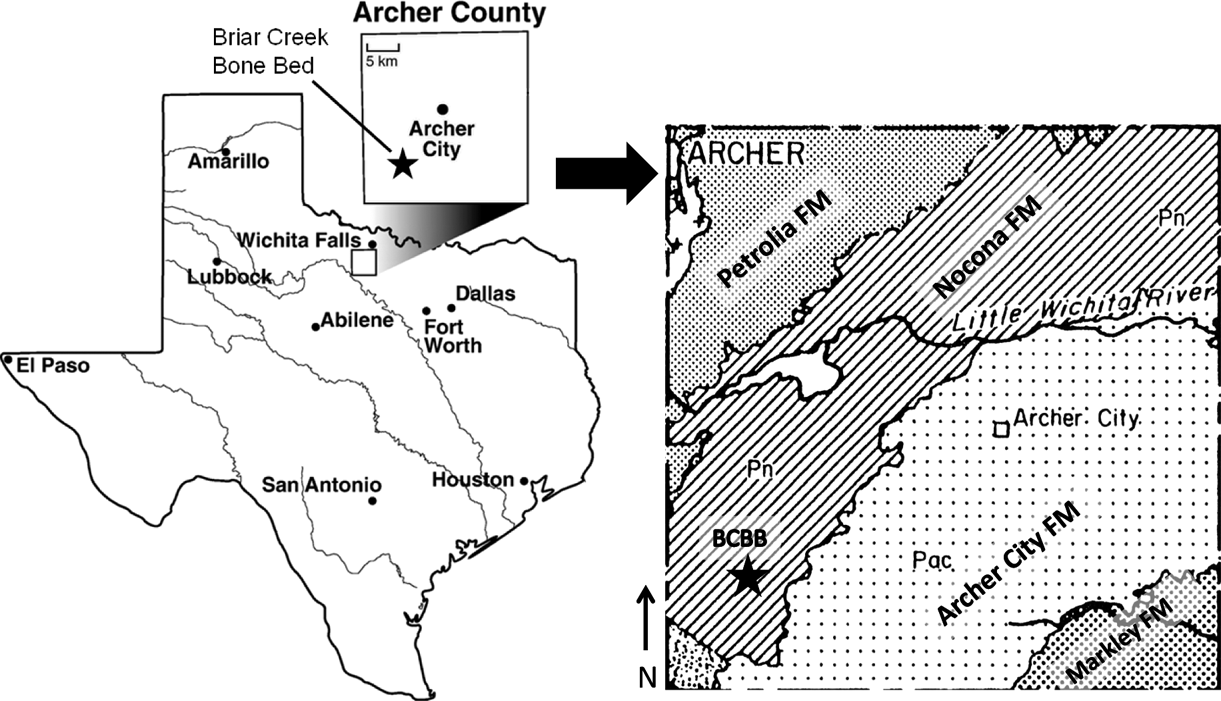
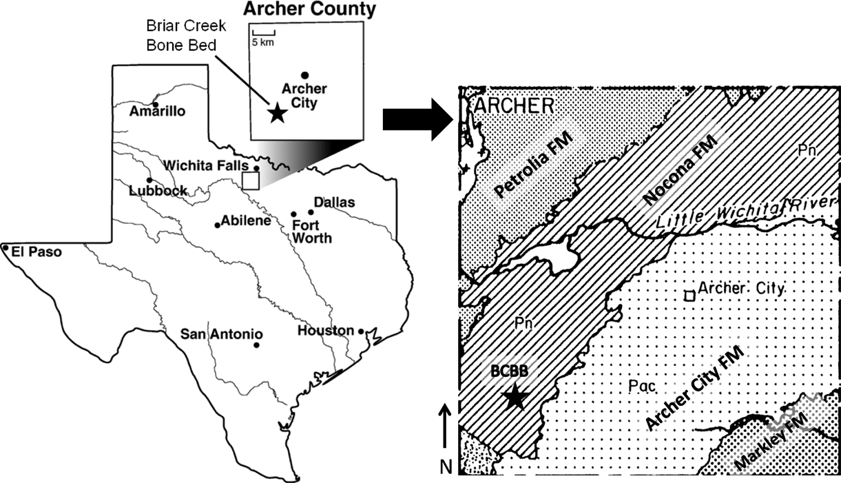
The Briar Creek Bonebed (BCBB) (Case Reference Case1915, who spelled it “Brier Creek”) in the Artinskian Nocona Formation of Archer County, Texas, USA (Fig. 1) is one of the major Lower Permian bonebeds, and contains a large volume of material and variety of taxa (Case Reference Case1915). When E. C. Case, of the University of Michigan, originally found the BCBB in 1912, he hypothesised that this area represented a pool or swamp that functioned as a “macerating tank” in that some of the bones appear to have rotted before preservation. He hypothesised that carcasses may have been washed in by a stream or river, resulting in an accumulation of unsorted and disarticulated bones, that pertain to reptiles and amphibians as well as some fish remains (coprolites, teeth and cartilage). A. S. Romer, of Harvard University, began work at the BCBB in 1927, and he was the last to do an organised excavation at the site in 1972 (unpublished field notes), one year before his death. The bone-bearing layer is approximately 30 cm thick, and for nearly a century has produced large quantities of material held in the collections of the AMNH, FMNH, KUVP, MCZ, USNM, OMNH, UMMP and TMM (Romer & Price Reference Romer and Price1940, Reisz Reference Reisz1986).
Location and geologic map of Archer County, Texas, USA. This map shows the location of the Briar Creek Bonebed in the Nocona Formation; coordinates are available on request at the JJ Pickle Laboratory at the University of Austin, Austin, TX (modified from Hentz Reference Hentz1988; Labandeira & Allen Reference Labandeira and Allen2007).
The published literature (Romer & Price Reference Romer and Price1940, Reisz Reference Reisz1986) and records from museum collections suggest the presence of four Dimetrodon species (D. milleri, D. natalis, D. booneorum and D. limbatus) in the BCBB. It should be noted that D. milleri cannot be substantiated as occurring in the upper Wichita Group until diagnostic skull material is found.
It also should be noted that remains of another sphenacodontid, Secodontosaurus obtusidens Romer, Reference Romer1936 (body size identical to D. booneorum and postcrania morphologically similar to Dimetrodon in general), have been rarely recovered from the BCBB as well (Romer & Price Reference Romer and Price1940). This means that intermediate-sized isolated sphenocodontid long bones from this locality may either pertain to D. booneorum or, less likely, to Secodontosaurus. However, species validation of the small D. natalis and large D. limbatus will not be affected given the known size range of Secodontosaurus (Romer & Price Reference Romer and Price1940). To test Bakker's (Reference Bakker1982) ontogenetic series hypothesis, a representative sample of disarticulated sphenacodontid limb bones (humeri and femora), of different sizes for histologic sectioning, was collected from the BCBB for this study.
2. Materials and methods
2.1. Material
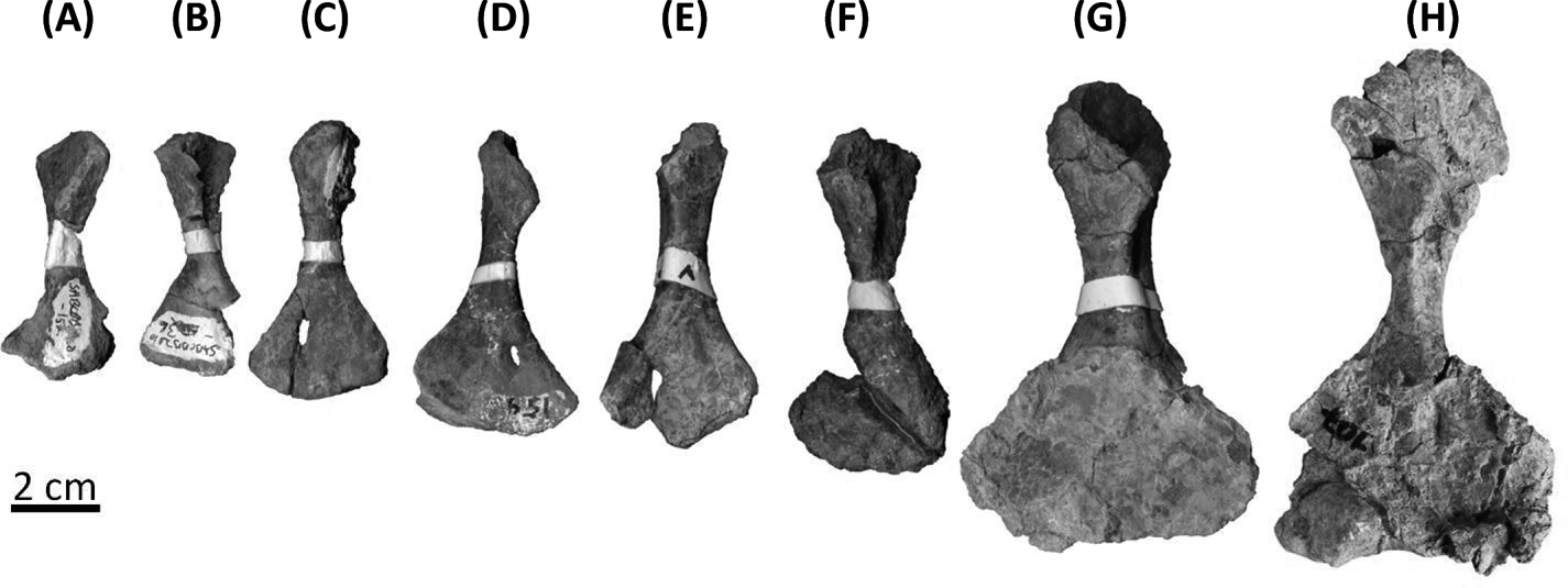
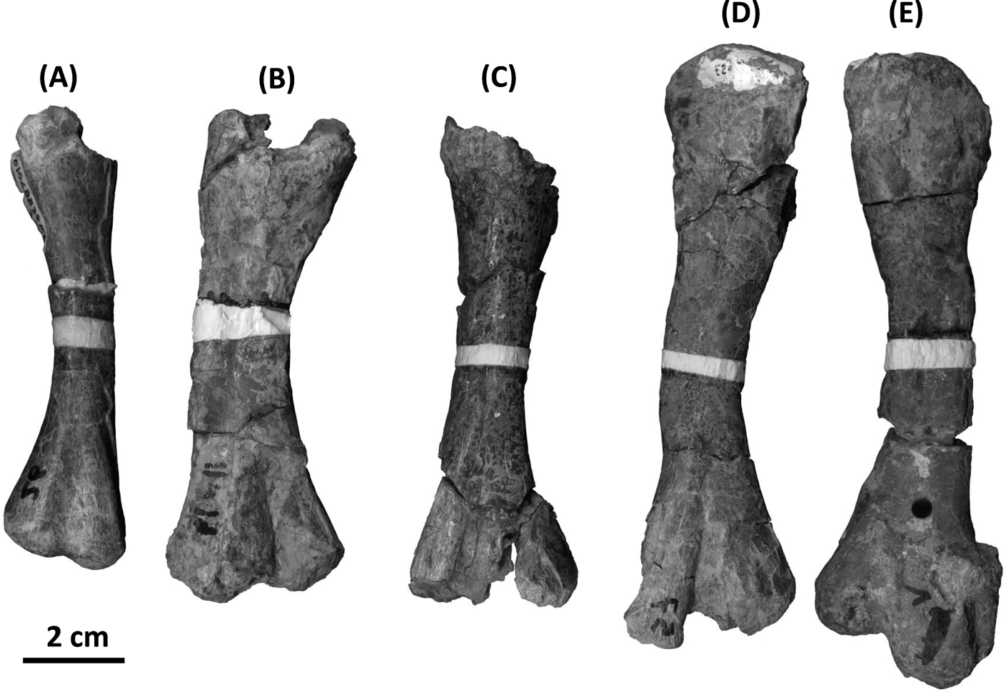
Vertebrate postcranial material was excavated from the BCBB in 2010 and 2011 for the sole purpose of histological analysis. Whilst numerous isolated bones of the zeugopodium were recovered as well, this study is based on two growth series made up of complete and partial sphencadontid humeri and femora (Figs 2, 3). This material was identified by us as Dimetrodon sp. by comparison to identified specimens in museum collections and the literature, primarily Romer & Price (Reference Romer and Price1940) and Reisz (Reference Reisz1986). However, the possibility that the material includes Secodontosaurus bones cannot be excluded. Thirteen long bones were sectioned histologically (Figs 2, 3; Table 1). The eight humeri studied ranged in size from 58 mm to 120 mm. The five femora studied ranged in size from 98 mm to 137 mm. Two femora were crushed dorsoventrally.
Ontogenetic series of Dimetrodon natalis humeri. All humeri were sectioned transversely at the mid-diaphysis. The sampled location was reconstructed with white plaster. Humeri are arranged by increasing length: (A) IPBSH-13 (48% maximum size); (B) IPBSH-25 (48% maximum size); (C) IPBSH-11 (53% maximum size); (D) IPBSH-14 (58% maximum size); (E) IPBSH-22 (67% maximum size); (F) IPBSH-5 (68% maximum size); (G) IPBSH-33 (94% maximum size); (H) IPBSH-4 (100% maximum size), photographed before sectioning. Ventral view, proximal end is at top.

Ontogenetic series of Dimetrodon natalis femora. All femora were sectioned transversely at the mid-diaphysis. The sampled location was reconstructed with white plaster. Femora are arranged by increasing size: (A) IPBSH-19 (72% maximum size); (B) IPBSH-6 (78% maximum size); (C) IPBSH-31 (79% maximum size); (D) SABCBB2010-57 (96% maximum size); (E) IPBSH-2 (100% maximum size). Dorsal side is facing up.
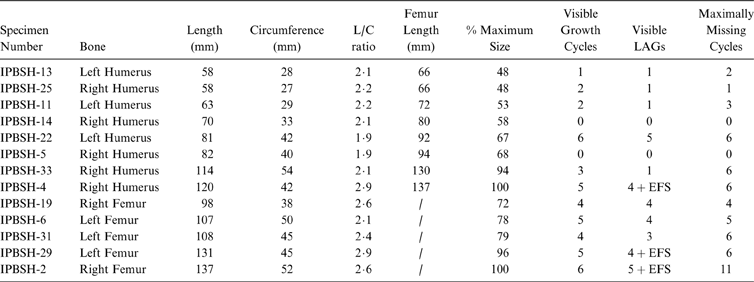
Physical dimensions and growth mark count of the sectioned small sphenacodontid long bones. Femur length for the humeri was calculated using the average length ratio (1·14) of articulated and associated D. limbatus humeri and femora material from various museum collections (SI 1). Abbreviations: C=circumference; EFS=external fundamental system; L=length; LAG=line of arrested growth.
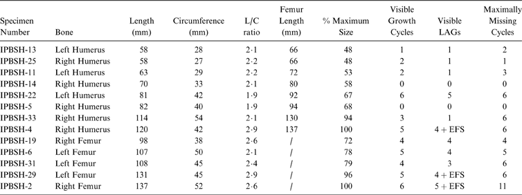
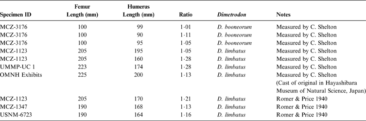
Femur length was used as a proxy for body size (cf. Carrano Reference Carrano, Carrano, Blob, Gaudin and Wible2006). This was constrained by assuming that the largest histologically sample sphenacodontid femur (IPBSH-2) represented the maximum size (100%). The humerus/femur ratio (of 1·14) was calculated from combined measurements of three D. booneorum and seven D. limbatus specimens, because there is insufficient articulated or associated D. natalis or Secodontosaurus material available from which to calculate a reliable ratio (SI 1). These data were obtained from the literature (Romer & Price Reference Romer and Price1940) and combined with measurements taken from specimens of different localities in the collections of the FMNH, MCZ, and OMNH (SI 1). Humerus length was standardised to femur length based on this ratio and given as a percentage of maximum size based on the longest femur (IPBSH-2) (Fig. 3; Table 1). Humerus size ranged from 48% to 100% maximum size (Fig. 2; Table 1), a greater relative size range compared to the femora (72% to 100% maximum size) (Fig. 3; Table 1).
Brinkman's (Reference Brinkman1988) ontogenetic stages were not applied to the long bones because necessary landmarks were damaged or missing. Therefore, we used arbitrary size classes to organise and describe the specimens. Small bones are considered to be those at 55% maximum size or less, intermediate bones are those between 56% and 80%, and large bones are 81% or more. The small humeri have a rough outer surface and unossified ends. The intermediate to larger humeri have a smooth surface with parallel micro-striations. All femora have a smooth outer surface. These size classes were combined with the morphological information available and the histological data found, in an attempt to ontogenetically classify the long bones as juvenile, subadult or adult. All fossil material is reposited at the Steinmann Institute of Geology, Mineralogy and Palaeontology, University of Bonn, Bonn, Germany (IPBSH).
Morphometric data (see below) were obtained from specimens excavated during the 2010 and 2011 campaigns and from BCBB material housed in the collections of the FMNH, MCZ and UMMP (SI 2: BCBB material attributed to D. milleri is listed here as Dimetrodon sp.).
2.2. Methods
2.2.1. Morphometrics
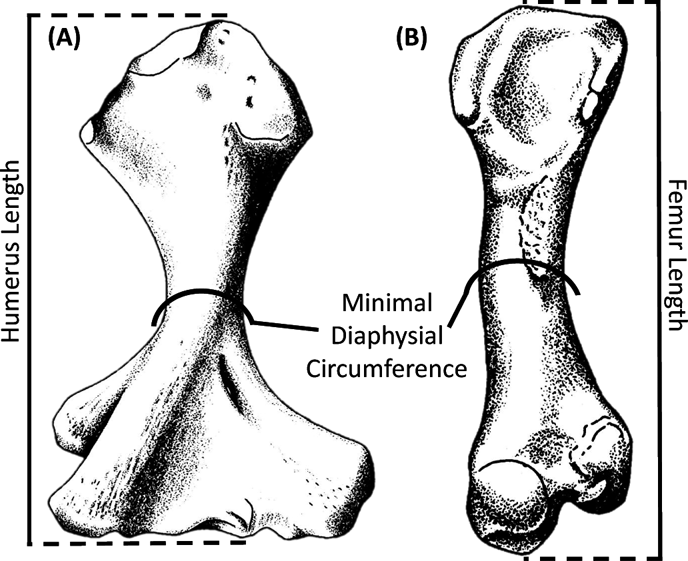
Morphometric analysis commonly needs to precede any histological work to procure the raw data before the bone is damaged in any way (for example, Sander & Klein Reference Sander and Klein2005; Sander et al. Reference Sander, Mateus, Laven and Knötschke2006; Klein & Sander Reference Klein and Sander2007). In the present case, simple morphometrics, i.e., plots of bone length vs. bone circumference, served two purposes. First, we wanted to detect size differences between the humeri and femora of BCBB sphenacodontid species (Dimetrodon spp. and Secodontosaurus) collected by us and earlier workers. Secondly, we wanted to detect possible species-specific clusters of length/circumference ratios. Thus, total length and minimal diaphysis circumference were recorded for each bone using standard analytical callipers and a metric measuring tape. Length was taken as the total distance between the termination of the proximal and distal ends. Circumference was taken at the mid-diaphysis (see Fig. 4).
Illustration of a sphenacodontid humerus (A) and a sphenacodontid femur (B), with indication of how and where total length and the minimal diaphysis circumference were taken. The mid-diaphysis was sectioned in order to see the best preserved growth record. Note that the bones shown here are D. limbatus (modified from Reisz Reference Reisz1986).
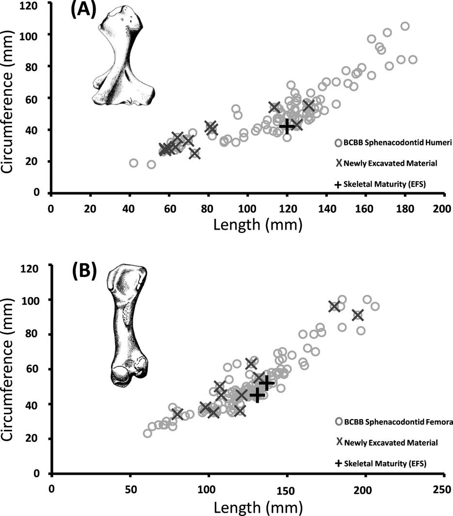
Microsoft Excel 2010 was used to construct a scatter plot to understand how the mid-diaphysis circumference relates to the length of the long bone (Fig. 5). This relationship is of interests because, especially in femora, it is assumed that long bone appositional growth is isometric (Bonnan Reference Bonnan2004; Lehman & Woodward Reference Lehman and Woodward2008; Kilbourne & Makovicky Reference Kilbourne and Makovicky2010; Sander et al. Reference Sander, Klein, Stein, Wings, Klein, Remes, Gee and Sander2011). As a long bone increases in length, it also increases in shaft circumference, and if this relationship is linear, cortical thickness increase can also be used as a proxy for (increasing) body size. This permits construction of growth curves based on percentage of maximum estimated cortical thickness (e.g., Lehman & Woodward Reference Lehman and Woodward2008). Only BCBB sphenacodontid (Dimetrodon spp. and Secodontosaurus) humeri and femora from the FMNH, MCZ and UMMP collections were measured and integrated with the data from the bones excavated (SI 2). One hundred and ten humeri and 131 femora were measured (Fig. 5). These data were not log transformed.
Minimal diaphysis circumference compared to length in the humerus and femur of Briar Creek Bonebed sphenacodontid species (Dimetrodon spp. and Secodontosaurus) humeri (A) and femora (B). Sample is from historic museum collections (FMNH, MCZ, UMMP), and from new material excavated during the 2010 and 2011 field seasons (SI 2). Sectioned Dimetrodon natalis long bones that possess an EFS have been marked with a red cross. No distinction was made between the different Dimetrodon species or Secodontosaurus.
2.2.2. Histological sampling, study and imaging
Silicon moulds (Provil Novo putty regular) of all long bone diaphyseal areas were created before sectioning. The damaged areas were cast in plaster for purposes of reconstructing and preserving the morphological and anatomical features of the original material. Next, each long bone mid-diaphysis was encased in a green epoxy resin (Technovit Universalliquid and Technovit 5071 Pulver) before being sectioned transversely with a rock saw equipped with a standard diamond tipped blade to prevent splintering of the cortex. The mid-diaphysis of the long bone is the region of the bone where the most complete record of growth is preserved. It also corresponds to the area of the smallest shaft circumference (Francillon-Vieillot et al. Reference Francillon-Vieillot, Buffrénil, Castenet, Géraudie, Meunier, Sire, Zylbergberg, Ricqlés and Carter1990; Currey Reference Currey2002). Humerus sections bisect the attachment of the coracobrachialis muscle and the femoral sections bisect the area of the adductor muscle attachment (Romer & Price Reference Romer and Price1940; Romer Reference Romer1969). After sawing, sections were ground to approximately 35 to 50 μm by hand on a glass plate with wet grit (600 and 800) and sealed with a cover slip using UV activated resin (Verifix LV 740). Sections were described, measured and photographed under nonpolarised and polarised light using a Leica DM2500LP compound microscope and Leica DFC420 digital camera, both manufactured in Germany. Thin sections were imaged with an EPSON V750 (manufactured in Japan) high-resolution transmitted light scanner in normal light (Figs 6, 7). Digital images were processed using the 2007 edition of Image Access Easy Lab7 software. Bone histological terminology follows Francillon-Vieillot et al. (Reference Francillon-Vieillot, Buffrénil, Castenet, Géraudie, Meunier, Sire, Zylbergberg, Ricqlés and Carter1990).
High-resolution images of the transverse sections were digitally reposited online for scholarly use at MorphoBank (http://MorphoBank.org). The project number is P845. All slides are reposited at the IPBSH.
Ontogenetic series of BCBB Dimetrodon natalis humeri sectioned transversely at the mid-diaphysis. Images were captured with a high-resolution transmitted light scanner in normal light. The organisation of the vascular canals (radial and longitudinal) gives the cortical bone a “spoked bicycle wheel” appearance. The smaller humeri have more longitudinal canals, and the larger humeri have more radial canals. However, the density of vascularisation appears to decrease through ontogeny. The medullary cavity of the humerus is occupied by a lattice work of secondary trabecular bone: (A) IPBSH-13 (48% maximum size); (B) IPBSH-25 (48% maximum size); (C) IPBSH-11 (53% maximum size); (D) IPBSH-14 (58% maximum size); (E) IPBSH-22 (67% maximum size), LAGs are visible in the cortex due to diagenetic staining; (F) IPBSH-5 (68% maximum size); (G) IPBSH-33 (94% maximum size), LAGs are visible in the cortex; (H) IPBSH-4 (100% maximum size), LAGs are visible in the cortex. Letters correspond to the specimens featured in Figure 2. Scale bars=5 mm.
Ontogenetic series of BCBB Dimetrodon natalis femora sectioned transversely at the mid-diaphysis circumference. Images were captured with a high-resolution transmitted light scanner in normal light. The cortical bone of the femora is thin. Vascularisation is similar to that seen in the humeri. Radial and longitudinal canals are organised such that they form a “bicycle wheel” pattern. Smaller femora have more longitudinal canals, and the larger femora have more radial canals but with a higher degree of anastomosis. The medullary cavity is open, and secondary trabecular bone is exclusively found on the inner lining: (A) IPBSH-19 (72% maximum size); (B) IPBSH-6 (78% maximum size), dorsoventrally crushed, LAGs are visible in the outer cortex; (C) IPBSH-31 (79% maximum size); (D) IPBSH-29 (96% maximum size), dorsoventrally crushed, LAGs are visible in the cortex; (E) IPBSH-2 (100% maximum size), slightly dorsoventrally crushed, LAGs and an EFS are visible in the cortex. Letters correspond to the specimens featured in Figure 3. Scale bars=5 mm.
2.2.3. Analysis of growth trajectory
Growth marks within long bones can be used to establish a growth curve (Bybee et al. Reference Bybee, Lee and Lamm2006; Klein & Sander Reference Klein and Sander2007; Cooper et al. Reference Cooper, Lee, Taper and Horner2008; Lee & Werning Reference Lee and Werning2008; Lehman & Woodward Reference Lehman and Woodward2008; Sander et al. Reference Sander, Klein, Stein, Wings, Klein, Remes, Gee and Sander2011) for studying aspects of life history. Growth marks develop annually (Castanet et al. Reference Castanet, Croci, Aujard, Perret, Cubo and de Margerie2004; Kohler et al. Reference Kohler, Marín-Moratalla, Jordana and Aanes2012) and include lines of arrested growth (LAGs) and entire growth cycles divided into a fast growing zone and a slow growing annulus (when the latter is present at all). As an animal nears skeletal maturity, bone growth rates decrease. As a result of this slowed growth, the periosteum produces an external fundamental system (EFS) on the periphery of the bone. The EFS is characterised by numerous rest lines laid down consecutively with very little space in between, and is composed of nearly avascular parallel-fibred or lamellar bone. Also, osteocyte lacunae are extremely flattened and oriented parallel to the bone surface. The amount of time represented by the EFS is often difficult to estimate (Erickson et al. Reference Erickson, Makovicky, Philip, Norell, Yerby and Brochu2004). As bones grow, expansion and remodelling of the medullary cavity destroys earlier growth marks, which must be determined to establish a reliable age. The missing growth cycles can be estimated by use of retrocalculation (e.g., Bybee et al. Reference Bybee, Lee and Lamm2006; Klein & Sander Reference Klein and Sander2007).
A growth trajectory for the sectioned sphenacodontid humeri and femora (Table 1) was constructed using Microsoft Excel 2010. Bone length (Fig. 4) was used as proxy for body size, and the number of growth cycles in each bone was used to determine the age of the individual at time of death (Fig. 10). First, each LAG (each of which represents one year of growth) visible in the cortex was counted under regular transmitted light. In some cases, a LAG was not visible, and the presence of a corresponding zone and annulus (visible under crossed plane-polarised light) was used to infer one year. Long bone lengths and age estimates are reported in Table 1. The EFS represents an unknown amount of time (Erickson et al. Reference Erickson, Makovicky, Philip, Norell, Yerby and Brochu2004). Its presence was noted (Table 1) if observed in the cortex, but it was not used to calculate the growth trajectory (Fig. 10).
The number of missing cycles (Table 1) for each long bone was estimated by utilising two methods of retrocalculation. The first method used was that of Klein & Sander (Reference Klein and Sander2007): the distance between the centre of the medullary cavity and the first visible LAG was measured and divided by the greatest distance between any two adjacent LAGs. This method will be referred to in the results section as RM1. It was applied to all humeri and to femora that show little to no crushing (IPBSH-19, IPBSH-31, IPBSH-2) (Figs 3, 7).
The same retrocalculation method was used on femora that are dorsoventrally crushed (IPBSH-6 and IPBSH-29) (Figs 3, 7), but with a modification to first estimate the distance from the centre of the medullary cavity to the first LAG. Given the circumference (c) of the outer cortex (Table 1), diameter (d) was first calculated (d=c/π). The average thickness of the cortical bone (ct) was calculated, doubled and subtracted from the diameter (d). The remaining number is the estimated diameter of the medullary cavity (dmc), calculated as dmc=d−(ct×2). The dmc was then divided in half. This gives an approximation of the distance from the centre of the medullary cavity to its margin (also known as the approximated radius of the medullary cavity) (rmc) had the bone not been crushed (rmc=dmc÷2). The distance from the margin of the medullary cavity to the first LAG was measured directly from the slide. This distance, when added to the approximated radius of the medullary cavity (rmc), gives the best estimated measurement for the distance from the centre of the medullary cavity to the first LAG. As in the first method, final calculation is processed by dividing this number by the greatest distance between any two adjacent LAGs. This method will be referred to in the results section as RM2.
In order to fully test the null hypothesis, an additional data point was added to the growth trajectory of the sectioned sphenacodontid femora. It was assumed that the femur reached a total length of 60 mm within the first year of growth after hatching, as this is the smallest femur measured, but not sectioned, from the collections (SI 2).
3. Results
3.1. Morphometrics
The scatter plots of the raw data of the minimal diaphysis circumference, as a function of length of the humeri and femora, show two major results. First, all BCBB sphenacodontid material follows a similar growth trajectory (Fig. 5). This means the long bones are lengthening at a rate similar to the apposition of cortical bone in the diaphysis. Moreover, the patterns produced by the scatter plots could be interpreted as resulting from the presence of a single species at the BCBB. Note, however, that this pattern does not exclude the presence of more than one species, as multiple species could also exhibit similar growth trajectories. Small-sized sphenacodontid long bones that possess an EFS have been specifically marked (Fig. 5).
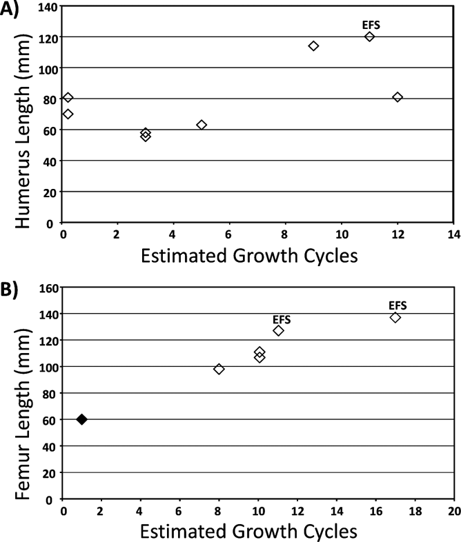
Retrocalculation of missing growth cycles was performed using either RM1 or RM2 (see section 2.2.3) for all sectioned long bones (Table 1). Some of the humeri (IPBSH-14: 70 mm and IPBSH-5: 82 mm) could not be retrocalculated, due to the fact that visible growth cycles or LAGs are not preserved in the cortical bone. Humeri that do have growth marks were analysed using RM1. Humeri IPBSH-13 (58 mm) and IPBSH-25 (58 mm) both came from an individual estimated to have been three years old. Humerus IPBSH-11 (63 mm) was estimated to be five years old. Humerus IPBSH-22 (81 mm) was estimated to have belonged to an individual twelve years old. Humerus IPBSH-33 (114 mm) was estimated to have been nine years old. Finally, Humerus IPBSH-4 (120 mm) was determined to be approximately eleven years old, with six maximally missing annual growth cycles.
Femora crushed dorsoventrally were analysed using RM2 (IPBSH-6 and IPBSH-29) All other femora were analysed using RM1. Femur IPBSH-19 (98 mm) came from an individual estimated to have been about eight years old. Femora IPBSH-6 (107 mm) and IPBSH-31 (108 mm) were both estimated to have been approximately ten years old at time of death. Femur IPBSH-29 was estimated to be eleven years old. The largest femur, IPBSH-2, was determined to be at least seventeen years old, with a maximum of eleven missing annual growth cycles.
The growth trajectory produced by plotting humerus length against total observed and estimated growth cycles (Fig. 10A; Table 1) revealed no obvious developmental pattern that would indicate the presence of a single species. However, there is an apparent variability in the sampled humeri with regards to size and age. This could be a reflection of developmental plasticity or sexual dimorpism, but most likely it is because some of these humeri are from juveniles or subadults of the larger BCBB sphenacodontids (Dimetrodon spp. and possibly Secodontosaurus).
The growth trajectory produced by plotting femur length against total observed and estimated growth cycles is shown in Figure 10B and Table 1.
3.2. Description of sphenacodontid humerus histology
The bone histology of the sampled sphenacodontid humeri (Figs 2, 6) is described below in order of increasing bone length. All histology is described from the mid-diaphysis (Fig. 4).
3.2.1. Small humeri
Left humerus IPBSH-13 (58 mm, 48% maximum size), right humerus IPBSH-25 (58 mm, 48% maximum size) and left humerus IPBSH-11 (63 mm, 53% maximum size) all have a rough outer bone surface, and unossified epiphyses. IPBSH-25 is slightly crushed dorsoventrally. The cortices are relatively thick and consist of a combination of parallel-fibred and woven-fibred bone (Fig. 8) and are well vascularised by radially arranged longitudinal and radial canals. The radial organisation of the canals gives the cortical bone a “bicycle wheel” appearance (Figs 6, 8). There was no further centripetal deposition of lamellar bone in the vascular canal. It only surrounds the outer circumference of the canal. Thus, these are incipient primary osteons (Fig. 8). The woven bone is located in between the canals. These regions remain dark at low magnification under polarised light and do not appear to exhibit any birefringence (Fig. 8E). When the stage is rotated 90 degrees under polarised light and a lambda filter, again there appears to be no change in birefringence of the matrix, but a colour change in the osteonal bone can be seen (Fig. 8C, D).
Histology of Dimetrodon natalis humeri from the Briar Creek Bonebed: (A) transverse section through the mid-diaphysis of juvenile humerus IPBSH-13 (48% maximum size) under polarised light. Radial and longitudinal canals extend from the medullary cavity to the outer cortex. Notice the ‘spoked bicycle wheel’ pattern formed by the arrangement of the vascular canals. Incipient primary osteons are visible throughout the cortex. Large erosion cavities can be seen in the medullary region. Secondary trabecular bone is present in the medullary cavity; (B) transverse section through the mid-diaphysis of the adult humerus IPBSH-4 (100% maximum size) under polarised light. Vascularisation of the cortex appears less dense when compared to the juvenile humerus in (A). However, there are more radial canals than longitudinal canals in the adult humeri. The dark areas between the vascular canals consist of woven bone matrix, brighter areas are parallel-fibred and lamellar bone lining the medullary cavity and vascular canals. Arrows indicate LAGs; (C) magnification of ventral cortical region in proximity to the coracobrachialis muscle attachment, indicated by the white box in (A). Cortex of IPBSH-13 viewed under polarised light and a lambda filter. Arrows indicate areas of colour change in the lamellar bone of the incipient primary osteons, from blue to yellow, when the stage is rotated 90 degrees. Areas between the vascular canals are woven bone and remain pink when the stage is rotated; (D) the same area pictured in (C), with the stage rotated 90 degrees. Note the colour change in the lamellar bone of the osteons and the areas of woven bone, indicated by the arrows, remain pink; (E) same view as in (C), but under polarised light without lambda filter. The crystallite orientation of the bone matrix is clearly visible. Woven bone between the vascular canals remains dark. Lamellar bone in the incipient primary osteons remains bright; (F) magnification of selected area in (E). Incipient primary osteon viewed under normal light. These osteons are present in both humeri and femora and retain this immature appearance throughout ontogeny. Note the large size, shape and orientation of the osteocyte lacunae within the lamellar bone. Abbreviation: WB=woven bone.
One LAG is visible in IPBSH-13 just below the bone surface, making the entire cortical area one growth cycle. IPBSH-25 has two growth cycles and one LAG mid-cortex. IPBSH-11 has two growth cycles and one LAG in the deep cortex near the resorption front. Vascularisation extends from the bone surface to the medullary region. The osteocyte lacuna shape is subangular to elliptical. Within the annuli, osteocyte lacunae are flat and oriented parallel to the bone surface. Mineralised Sharpey's fibres (highly birefringent under polarised light) are visible in IPBSH-13 (like those in Fig. 9B) on the anterior side and in IPBSH-25 on the dorsal and ventral sides. They are situated in between the vascular canals and are perpendicular to the bone surface. Sharpey's fibres were not observed in IPBSH-11.
Histology of Dimetrodon natalis femora from the Briar Creek Bonebed: (A) transverse section through the mid-diaphysis of the smallest femur IPBSH-19 (72% maximum size). Note the thin cortex. Vascular canals are concentrated in the zones of woven-fibred bone that are bordered by annuli consisting of parallel-fibred bone; (B) cortical bone with mineralised (white) and unmineralised (black) Sharpey's fibres. These are found all femora specimens; (C) magnification of dorsal cortex, indicated by white box in (A), viewed under polarised light. Bright areas are mostly crystallites extending parallel to the bone surface (parallel-fibred bone matrix), and the lamellar bone of the incipient primary osteons. Arrows indicate areas between the vascular canals that are woven bone; (D) the same area pictured in (C), rotated 45 degrees to demonstrate extinction patterns exhibited by the crystallites oriented obliquely to the surface. The parallel crystallites exhibit extinction and are mostly dark. Also, areas indicated by the arrows appear to remain dark. This is the woven bone matrix; (E) transverse section through the mid-diaphysis of the largest femur (IPBSH-2), viewed under normal light. Note the EFS. Arrows indicate LAGs; (F) Magnification of selected region in (E) (indicated by white box). Notice the closely spaced LAGs and reduction of vascularisation in the EFS. Diagenetic staining has obscured the view of the EFS in the outer most part of the cortex. Abbreviations: A=annulus; EFS=external fundamental system; MSF=mineralised Sharpey's fibres; PFB=parallel fibred bone; USF=unmineralised Sharpey's fibres; WB=woven bone; Z=zone.
The medullary region, in general, is similar in all three specimens. The cortex is separated from the medullary region by large erosion cavities due to resorption activity, more so in IPBSH-13. Endosteal lamellar bone, in the process of forming secondary trabecular bone (as seen from cross-cutting relationships of cementing lines), is present around the periphery of the medullary cavity, but in isolated areas. Secondary endosteal osteons are also visible in the medullary margin (Fig. 8). The medullary cavity is filled with broken trabeculae that have been displaced due to diagenetic crushing of the bone.
3.2.2. Intermediate humeri
The intermediate humeri include right humerus IPBSH-14 (70 mm, 58% maximum size), left humerus with broken epiphyses IPBSH-22 (81 mm, 67% maximum size), and right humerus IPBSH-5 (82 mm, 68% maximum size). The outer bone surface of these humeri is smooth with parallel micro-striations. An exception is IPBSH-5, which is slightly crushed dorsoventrally, and is covered by a thin layer of iron stone. Only IPBSH-14, slightly crushed on the dorsal side, has unossified epiphyses in combination with a smooth surface and parallel micro-striations visible with the naked eye. The cortex of these humeri is thick, and consists of a combination of parallel-fibred and woven bone vascularised by longitudinal and radial canals. This gives the cortex the “bicycle wheel” pattern. There is no further centripetal deposition of lamellar bone in the vascular canals compared to that seen in the smaller humeri. The vascular canals remain incipient primary osteons (Fig. 8). The thinnest cortical region is on the ventral side.
Growth cycles were not observed in IPBSH-14 or IPBSH-5. IPBSH-22 had six growth cycles, and five LAGs with subcycles (lighter, evenly spaced growth marks or rest lines in between adjacent LAGs) that are visible in the darker stained regions under nonpolarised light. All bones lack an external fundamental system (EFS). The vascular canals in all three specimens extend from the bone surface to the medullary region, with the exception of IPBSH-22, where vascularisation decreases after the fourth LAG. Osteocyte lacunae are subangular to elliptical and are randomly oriented, except for those in the annuli which are flatter and oriented parallel to the bone surface. A few mineralised Sharpey's fibres were only observed in IPBSH-14 under polarised light in the posterior dorsal region oriented perpendicular to the bone surface, and extending to the medullary cavity parallel to the vascular canals. The medullary region in all three specimens contains a network of secondary trabecular bone as seen from cross-cutting relationships of cementing lines. Endosteal bone surrounds most of the medullar cavity. Large erosion cavities and secondary endosteal osteons are present throughout the medullary margin.
3.2.3. Large humeri
There are two humeri in this category; one is a right humerus with crushed proximal end IPBSH-33 (114 mm, 94% maximum size). This bone has an outer smooth surface with parallel micro-striations. The other large humerus is right humerus IPBSH-4 (120 mm, 100% maximum size). This bone has a rough surface due to weathering. The cortex of both bones consists of parallel-fibred and woven bone, and is well vascularised by radially arranged longitudinal and radial canals exhibiting the “bicycle wheel” pattern (Fig. 8B). There is no further centripetal deposition of lamellar bone in the vascular canals, which are incipient primary osteons (Fig. 8). Vascularisation remains constant throughout the cortex, but appears to decrease just below the bone surface. A few mineralised Sharpey's fibres were observed in IPBSH-33 under polarised light in the posterior dorsal region oriented perpendicular to the bone surface, and extending to the medullary cavity parallel to the vascular canals. Sharpey's fibres were not observed in IPBSH-4.
IPBSH-33 has only three growth cycles (zones and annuli visible under polarised light) but only one LAG is visible under nonpolarised light and correlates with the first annulus. The cortical bone of IPBSH-4 has five growth cycles and four LAGs (Fig. 8B). The beginning of an external fundamental system (EFS) is visible in the outer cortex. The EFS confirms that this individual had reached skeletal maturity before death. It is unknown how much time is represented by the EFS (Erickson et al. Reference Erickson, Makovicky, Philip, Norell, Yerby and Brochu2004), but two rest lines with possible subcycles are visible. After the fourth growth cycle and first LAG, vascularisation decreases except between the dorsal and anterior side. In both humeri, osteocyte lacunae are subangular to elliptical in shape. Within the EFS, osteocyte lacunae are flattened.
For both humeri, the medullary region is separated from the cortex by lamellar endosteal bone, large erosion cavities and secondary endosteal osteons. The medullary cavity is occupied by secondary trabecular bone.
3.3. Description of sphenacodontid femur histology
The bone histology of the sampled sphenacodontid femora (Figs 3, 7) is described below in order of increasing overall length (Table 1). All histology is described from a transverse section made at the mid-diaphysis (Fig. 4).
3.3.1. Intermediate femora
Right femur IPBSH-19 (98 mm, 72% maximum size), left femur IPBSH-6 (107 mm, 78% maximum size stage), and left femur IPBSH-31 (108 mm, 79% maximum size) have ossified distal epiphyses, but the proximal ends are damaged. All bones have a smooth outer surface. IPBSH-6 is extremely crushed dorsoventrally. In all specimens, the cortex is relatively thin and consists of parallel-fibred and woven-fibred bone and is well vascularised by radially arranged longitudinal canals exhibiting a high degree of anastomosis (Figs 7, 9). Radial canals are present, but to a lesser degree than that seen in the humeri. Vascularisation is concentrated in the zones, where the woven bone is present (Fig. 9C, D). There is no further centripetal deposition of lamellar bone in the vascular canals, which remain as incipient primary osteons (Fig. 9C, D).
The cortex of IPBSH-19 contains four growth cycles and four LAGs. IPBSH-31 also contains four growth cycles, but only 3 LAGs. IPBSH-6 contains five growth cycles and four LAGs. Osteocyte lacunae are shaped subangular to elliptical. Those within the annuli are flattened and oriented parallel to the bone surface. Under polarised light, mineralised Sharpey's fibres appear white and extend from the bone surface to the medullary cavity (Fig. 9B). Under normal light, black unmineralised Sharpey's fibres extend from the bone surface to the middle of the cortex (Fig. 9B).
The medullary region is separated from the cortex by lamellar endosteal bone forming secondary trabeculae as seen from cross-cutting relationships of the cementing lines. Large erosion cavities are present due to resorption activity of the osteoclasts. Secondary endosteal osteons are present in the medullary margin. The medullary cavity is open; trabecular bone is only present around the outer margins.
3.3.2. Large femora
The material consists of a left femur IPBSH-29 (131 mm, 96% maximum size) that is extremely crushed dorsoventrally, and a right femur IPBSH-2 (137 mm, 100% maximum size) that is slightly crushed dorsoventrally. The cortex is relatively thin and consists of parallel-fibred and woven-fibred bone (Fig. 9) and is well vascularised by radially arranged longitudinal canals exhibiting a high degree of anastomosis. Vascularisation decreases just before reaching the bone surface where the EFS is located. There is no further centripetal deposition of lamellar bone in the vascular canals, which are incipient primary osteons (Fig. 9C, D).
The cortex of IPBSH-29 contains five growth cycles and four LAGs. IPBSH-2 contains six growth cycles and five LAGs, the deepest of which has almost been completely destroyed by expansion of the medullary cavity. The outermost cortex of both femora contains an EFS (Fig. 9E, F). The amount of time represented by the EFS is unknown (Erickson et al. Reference Erickson, Makovicky, Philip, Norell, Yerby and Brochu2004); however there are more visible rest lines in IPBSH-2 than in IPBSH-29. The distance between the LAGs decreases approaching the bone surface. The amount of vascularisation also decreases, and is almost non-existent in the EFS. In both specimens, osteocyte lacuna shape is subangular to elliptical and is extremely flat in the EFS. Mineralised and unmineralised Sharpey's fibres are visible throughout the cortex.
The medullary region is open and separated from the cortex by a thin layer of lamellar endosteal bone. Crushing has obscured much of this region in specimen IPBSH-29, but large erosion cavities and secondary endosteal osteons are still visible in the medullary margin. Secondary trabecular bone is present around the outer rim of the medullary cavity. IPBSH-2 has only a few small erosion cavities, and endosteal osteons are nonexistent in the medullary margin. Trabecular bone is not visible in the medullary cavity, which might be due to deformation of the diaphysis.
3.4. Summary and comparison of anatomy and histology of sphenacodontid humeri and femora
The mid-diaphysis of the humerus has a triangular to subtriangular cross section (Fig. 6), and femora are more round or oval (Fig. 7). Cortical bone in the humerus is relatively thick compared to that of the femora, both having the thinnest region of the cortex located on the ventral side. The cortex of the humeri and femora consists of a combination of two types of bone matrix: parallel-fibred bone and woven-fibred bone, either as annuli and zones or whole region of the bone (Figs 8, 9). In the humeri, vascularisation of the cortex consists of highly organised radially arranged longitudinal and radial canals extending from the medullary cavity to the bone surface. This gives the cortex a “bicycle wheel” appearance (Figs 6, 8). The femora have a vascularisation consisting of radially arranged longitudinal canals with a higher degree of anastomosis (Figs 7, 9), but less organised and less dense than what is seen in the humeri. Density of the vascularisation appears to decrease from younger to older individuals, especially in humeri. There is no centripetal deposition of lamellar bone in the vascular canals, which are identified as incipient primary osteons (Figs 8, 9). This immature appearance does not change with ontogeny. Osteocyte lacunae are subangular or star-shaped to elliptical, especially those in the annuli that are flat and oriented parallel to the bone surface. They sometimes follow the direction of the vascular canals (Figs 8, 9). Osteocyte lacunae can be seen encircling the incipient primary osteons (Fig. 9F). LAGs were observed in all but three of the humeri (Table 1). LAGs were observed in all femora. Superficially, this gives the cortical bone, laid out in the femora, a faux lamellar zonal pattern when viewed at low magnification (Fig. 9C, D). This is an artefact resulting from the fast and slow deposition of the bone tissue. The EFS is seen in the largest humerus (IPBSH-4) and the two largest femora (IPBSH-29 & IPBSH-2) (Fig. 9; Table 1). The amount of time represented by each EFS is unknown. Mineralised and unmineralised Sharpey's fibres are present in the cortex of both the humeri and femora, but more so in the femora (Fig. 9B).
In both humeri and femora, the medullary region is separated from the cortex by large erosion cavities due to resorption activity of the osteoclasts. Secondary endosteal osteons are also common in the medullary margin. Endosteal lamellar bone, in the form of secondary trabecular bone, as seen from cross-cutting relationships of cementing lines, is often present lining the medullary cavity. A network of secondary trabecular bone is found throughout the medullary cavity in the humeri. The medullary cavity in the femora appears to only have trabecular bone around the periphery. Most of the cavity is open.
4. Discussion
4.1. Ontogeny of the sphenocodontid humeri and femora
The humeri growth series (Fig. 2) represents a time span of nine to twelve years (Fig. 10B; Table 1). IPBSH-13, IPBSH-25 (both are at 48% maximum size), and IPBSH- 11 (53% maximum size) have been classified as juvenile because of their size and morphology, which is evident of an early ontogenetic stage (Fig. 2). In addition, growth marks were not visible in the cortex of IPBSH-14 (58% maximum size) nor in IPBSH-5 (68% maximum size), thus they are considered to be juveniles as well, possibly from a larger species. IPBSH-14 does have unossified epiphyses. However, the degree of ossification of the epiphyses of IPBSH-5 could not be observed due to the iron stone concretion encasing the entire bone (Fig. 2F).
Dimetrodon natalis growth trajectory was visualised by plotting length and estimated number of growth cycles (both observed and calculated) for each humerus (A) and femur (B) listed in Table 1. With regards to the humeri, a high variability between size and age is seen. These discrepancies within the growth series can be explained by developmental plasticity or sexual dimorphism (Romer & Price Reference Romer and Price1940). However, the most parsimonious explanation is that the sampled humeri are a mix of early ontogenetic material from multiple sphenacodontid species (Dimetrodon spp. and the more rare Secodontosaurus). Note that IPBSH-13 (58 mm) and IPBSH-25 (58 mm) have the same number of growth cycles. Also, IPBSH-14 (70 mm) and IPBSH-5 (82 mm) have no growth marks preserved in the cortex which is necessary for retrocalculations. IPBSH-4 (120 mm) possesses an external fundamental system (EFS) and has been indicated on the graph. With regards to femora, in order to test the null hypothesis, it was assumed that 60 mm was obtained within the first year of growth, after hatching, as this is the smallest Dimetrodon femur measured from the museum collections (indicated by the solid diamond) (see SI 2). All other data points correspond to the femora listed in Table 1. Note that IPBSH-6 (107 mm) and IPHBSH-31 (108 mm) have the same number of estimated growth cycles. Also, IPBSH-29 (131 mm) and IPBSH-2 (137 mm) both possess an EFS and have been labelled on the graph.

Humeri with parallel micro-striations on the bone surface may indicate that these individuals reached a level of sexual maturity (cf. Tumarkin-Deratzian et al. Reference Tumarkin-Deratzian, Vann and Dodson2006; Bickelmann & Sander Reference Bickelmann and Sander2008; Klein Reference Klein2010). IPBSH-22 is considered a subadult, in addition to having parallel micro-striations on the bone surface (IPBSH-14 also has the same parallel surface micro-striations but we still consider it a juvenile). The number of growth marks and the mid-diaphysis circumference are similar to that of the largest humerus measured (IPBSH-4), but an EFS is not present in the cortex. Also, humerus IPBSH-22 has damaged and missing epiphyses, therefore the length of the bone is shorter than what it was in life (Table 1).
Although IPBSH-33 was 94% maximum size, we observed only one LAG and three growth cycles in the cortex, and no sign of an EFS. Alternatively, this specimen could be an earlier ontogenetic stage (perhaps juvenile) of a larger sphenacodontid species. The shaft circumference is also larger than IPBSH-4 by a 12 mm difference, but IPBSH-33 is only 6 mm shorter than IPBSH-4. This could be due to the extreme crushing of the epiphyses in IPBSH-33, and this prevents us from properly ascertaining the extent of ossification of the epiphyses.
The presence of an external fundamental system (EFS) in the outermost layer of the cortex of the largest humerus (IPBSH-4: 100% maximum size) allows us to propose that this bone belonged to a fully grown adult of a small sphenacodontid species. Using retrocalculation to estimate the number of missing growth cycles, we have determined the age of this sphenacodontid, at time of death, to have been approximately 11 years old (Fig. 10a; Table 1). This individual could be even older than this, given the unknown amount of time represented by the EFS (Erikson et al. 2004). Although the EFS indicates a slowdown in skeletal growth, this is not simply a thick annulus, after the deposition of which fast growth would resume. No such structure or condition has been observed in any of the large taxa that we have examined. This is the first time an EFS has been observed in ‘pelycosaurs’.
Due to discrepancies between size and histology of specimens within the humeri growth series, we have to consider the possibility that the variability we are observing could be due to several factors, including developmental plasticity or sexual dimorphism (Romer & Price Reference Romer and Price1940). However, the most parsimonious explanation is that the sampled humeri are a mix of early ontogenetic material from multiple sphenacodontid species (Dimetrodon spp. and the more rare Secodontosaurus).
The femora growth series (Fig. 3) represents a time span of 11 years (Fig. 10A; Table 1). No identifiable juvenile femora were sampled. Although IPBSH-19 is the smallest femur sampled, we believe it is not a juvenile, as the epiphyses are ossified and not unfinished. The smallest femur, IPBSH-19 (72% maximum size), is from an individual at least eight years old based on visible growth cycles in the cortex and retrocalculation of maximally missing cycles (Fig. 10; Table 1). Subadult stage is also assigned to femora IPBSH-6 (78% maximum size) and IPBSH-31 (79% maximum size).
The EFSs observed in the two largest femora (IPBSH-29: 96% maximum size and IPBSH-2: 100% maximum size) allows us to propose that these bones belonged to fully grown adults of a small sphenacodontid species. Using retrocalculation to estimate the number of missing growth cycles, we have determined the age of these sphenacodontids, at time of death, to have been approximately 11 and 17 years old respectively (Fig. 10B; Table 1). They could be even older than this, given the unknown amount of time represented by the EFS (Erikson et al. 2004).
4.2. Multiple sphenacodontid species in the Briar Creek Bonebed
The information from the scatter plot (circumference vs. length) alone suggests that there are alternative interpretations of species composition. Either a single species of Dimetrodon occurs in this bonebed, possibly together with Secodontosaurus, or perhaps there was more than one species but with similar limb proportions (Bakker Reference Bakker1982) (Fig. 5). However, histological data (presence of the EFS) confirms at least two species of Dimetrodon, possibly with Secodontosaur, present in the BCBB. The smallest sphenacodontid species we attribute to D. natalis, whose body size compliments the small but fully grown long bones we studied histologically (IPBSH-4, IPBSH-29, IPBSH-2) (Figs 2, 3, 8, 9), and the fact that this is the only named small species of Dimetrodon occurring in the North Texas Permian redbeds (Romer & Price Reference Romer and Price1940). Secodontosaurus can be excluded from consideration because of its larger body size (cf. Romer & Price Reference Romer and Price1940). The present authors are aware that a proper assignment of their material to D. natalis would require its comparison with the holotype from the Geraldine Bonebed locality and identification of autapomorphies, but this work is clearly beyond the scope of this study, which was designed to test Bakker's (Reference Bakker1982) ontogenetic series hypothesis.
The results thus partially refute Bakker's (Reference Bakker1982) hypothesis, that the bones of D. natalis, D. booneorum and D. limbatus only represent an ontogenetic series of a single species, which may in turn disprove the juvenile/adult habitat shift hypothesis. Juveniles and adults of D. natalis are found in the same bonebed. However, the findings are insufficient for fully testing the habitat shift hypothesis. The results support Brinkman (Reference Brinkman1988), as well as the morphological classification used by Romer & Price (Reference Romer and Price1940) discussed earlier (see section 1.1).
4.3. Dimetrodon natalis long bone histology and its implications for growth strategy
Incipient fibro-lamellar bone (IFLB) is present in the postcranial skeleton of D. natalis and remains so throughout ontogeny. Despite the appearance of the tissue at low magnification as having a superficially lamellar-zonal pattern, we choose to use the more descriptive IFLB because of the combination of incipient primary osteons and an interstitial matrix of highly vascularised woven-fibred bone. Enlow & Brown (Reference Enlow and Brown1957) observed the same histology in an Ophiacodon long bone, referring to it as “protohaversian-like”. This is misleading, however, because it implies that the tissue in question is secondary whereas, in fact, there is no secondary tissue in the cortical bone of Dimetrodon at all.
The presence of IFLB in D. natalis suggests that it had a slightly faster metabolism than that seen in modern reptiles. Many reptiles and amphibians possessing dorsal sails coexisted at this time. The main function of the dorsal sail is hypothesised to have been a thermoregulatory organ, in addition to other functions such as sexual display (Hotton et al. Reference Hotton, MacLean, Roth and Roth1986; Tracey et al. Reference Tracey, Turner, Huey, Hotton, MacLean, Roth and Roth1986; Florides et al. Reference Florides, Wrobel and Kalogirou1999, Reference Florides, Kalogirou, Tassou and Wrobel2001; Tomkins et al. Reference Tomkins, LeBas, Witton, Martill and Humphries2010). If this is true, we would expect to find a similar tissue in edaphosaurids. In addition, as has been observed in modern mammals and sauropodomorphs (Bromage et al. Reference Bromage, R. S. Lacruz, Hogg, Goldman, McFarlin, Dirks, Perey-Ocha, Smolyar, Enlow and Boyde2009), osteocyte lacunae density (OLD) (Oc/mm3) is higher in the youngest femur IPBSH-19 (47413 Oc/mm3) than in the largest adult femur IPBSH-2 (34364 Oc/mm3), suggesting a faster growth strategy in the younger D. natalis (OLD taken from Stein (Reference Stein2011), IPBSH-2 and IPBSH-19 are identified by their field numbers SABCBB2010-26 and SABCBB2010-1 respectively. For method of OLD calculations please refer to this study). Vascularisation appears to decrease through ontogeny, but was not quantified (Figs 6–9).
The present of sphenacodontid long bones thus suggests that the early stages of evolution of the fibro-lamellar complex or fibrolamellar bone (FLB) can be placed as far back as the Lower Permian. IFLB tissue existed in these mammal-like reptiles during a time soon after the reptile and mammal lines split. Further evidence of this is the existence of true FLB in the non-mammalian therapsids that appear during the Upper Permian, after the extinction of the sphenacodontid line of the ‘pelycosuars’. IFLB is an intermediate between lamellar zonal bone (LZB) that is mostly found in modern reptiles and small (<10 kg) mammal species, and true FLB that exists in modern mammals (>10 kg), in addition to its occurrence in therapsids and many archosaurs (de Ricqlès Reference Ricqlès1974a; Francillon-Vieillot et al. Reference Francillon-Vieillot, Buffrénil, Castenet, Géraudie, Meunier, Sire, Zylbergberg, Ricqlés and Carter1990; de Ricqlès et al. Reference Ricqlès, Meunier, Castanet, Francillon-Viellot and Hall1991; Chinsamy-Turan Reference Chinsamy-Turan2005; Castanet Reference Castanet2006).
5. Conclusion
It has been shown through analysis of long bone histology that at least two species of Dimetrodon can be found in the Briar Creek Bonebed and, by extension, in the Nocona Formation. The smaller species have been attributed to D. natalis. The external fundamental systems observed in the largest humerus and the two largest femora confirm that D. natalis is not the juvenile of a larger species. The presence of the EFS in the cortex of their long bones unquestionably indicates that these animals had attained skeletal maturity. This is the first time an EFS has been reported for ‘pelycosauris’. Validation of the sympatric Dimetrodon species D. booneorum and D. limbatus will require similar histologic work, in particular the identification of an ESF in significantly smaller specimens than the known maximum size of D. limbatus. Additionally, similar histological studies to determine the species validity of D. booneorum will be inconclusive without proper sampling of Secodontosaurus postcrania. The presence of Secodontosaurus in the BCBB cannot be ignored, due to the similarities in size and morphology between these contemporaneous species (Romer & Price Reference Romer and Price1940).
Humeri were uninformative with regards to age approximation, because not all specimens have distinguishable growth marks, and the sample set included ontogenetic material of more than one sphenacodontid species in addition to D. natalis. However, it did help to illustrate the variability observed between bone size and histology. Histological analysis of additional humeri is required to better understand sphenacodontid species diversity in the BCBB.
According to the total number of missing and calculated growth cycles, the femora growth series represents a time frame of nine years. The smallest D. natalis femur (IPBSH-19) comes from an individual that was at least eight years old when it died. The oldest adult D. natalis, represented by femur IPBSH-2, is estimated to have lived approximately 17 years. This approximation may be more refined with additional analysis from juvenile femora that were not available for this study.
It was found that incipient fibro-lamellar bone is present in the postcranial skeleton of D. natalis throughout ontogeny. This tissue was called incipient fibro-lamellar bone, because of the combination of highly vascularised woven and parallel-fibred bone coupled with incipient primary osteons. According to Francillon-Vieillot et al. (Reference Francillon-Vieillot, Buffrénil, Castenet, Géraudie, Meunier, Sire, Zylbergberg, Ricqlés and Carter1990), bone histology is a continuum of intermediate situations regarding bone matrix organisation and vascularity. IFLB tissue is between the end members of LZB tissue and FLB tissue. IFLB existed in these mammal-like reptiles during a time soon after the split of the reptiles and synapsids.
6. Acknowledgments
We would wholeheartedly like to thank Jack and Marie Loftin of Archer City, Texas, for their help and hospitality; without it, this study would not have been possible. From the Texas Memorial Museum Vertebrate Paleontology Laboratory, Austin, TX, we especially would like to recognise and thank Wann Langston Jr. for his dedication to the science and his unyielding persistence to continue in the education of fledgling vertebrate palaeontologists. Also from the TMM, we thank Ernest Lundelius and Lyn Murray for their support and assistance with storage and shipping of fossil material. From the Steinmann Institute, University of Bonn, we would like to thank George Oleschinski for photography and figure preparation; Kayleigh Wiersma and Jessica Mitchell for helping with preparation; Katja Waskow and Olaf Dülfer for thin sectioning; and Kay Heitzplatz for administrative assistance. We thank Robert Bakker (Houston Museum of Natural History) for his personal communications and exchange of information. Also, we thank Don Brinkman (Tyrell Museum of Palaeontology) for his willingness to share data. We thank the following people for allowing access to their collections: Farish Jenkins Jr. and Jessica Cundiff (Museum of Comparative Zoology); William Simpson and Jörg and Nadia Frobisch (The Field Museum); Jeffrey Wilson and Gregg Gunnell (University of Michigan Museum of Paleontology); Richard Cifelli, Jennifer Larson, Kyle Davies (Sam Noble Oklahoma Museum of Natural History); and Mark Norell and Carl Mehling (American Museum of Natural History). We thank Donald and Brenda Shelton of Iowa Park, TX, for their assistance and hospitality and Steven Tudor of the Sam Noble Foundation in Ardmore, OK, for his assistance. Finally we thank Jeff Lindeman for granting us permission to excavate on his land. The authors would also like to thank Sarah Werning and Adam Huttenlocker for reviewing this paper. This project was funded by DFG grant SA 469/34-1 and the University of Bonn.
7. Appendices
7.1. Appendix 1. Specimens used to calculate femur/humerus ratio 1·14
7.2. Appendix 2. BCBB humeri and femora used to create scatter plots of the minimal diaphysis circumference plotted against length
Listing is in order of increasing length.