Introduction
Water is a major component of cells and tissues, and its movement across cell plasma membranes is a fundamental property of life. The movement and proper regulation of fluids across cell membranes are also important aspects of reproduction. There are two mechanisms by which water moves across cell membranes: simple diffusion across lipid bilayer and facilitated process by use of various channels (Verkman et al., Reference Verkman, Hoek, Ma, Frigeri, Skach, Mitra, Tamarappoo and Farinas1996). The transport of water primarily occurs by simple diffusion through lipid bilayer membrane. However, in the cells like RBC and renal tubular epithelial cells, high water permeability cannot be explained by simple diffusion because the lipid bilayer is hydrophobic (Preston et al., Reference Preston, Jung, Guggino and Agre1993).
Aquaporins (AQPs) are a family of small membrane-spanning monomer and hydrophobic integral membrane channel proteins that are expressed at plasma membranes of many cell types involving fluid transport (Agre et al., Reference Agre, King, Yasui, Guggino, Ottersen, Fujiyoshi, Engel and Nielsen2002). Being among the water channels, AQPs have recently been discovered and characterized in animals, humans, and plants (Preston et al., Reference Preston, Jung, Guggino and Agre1993; Li et al., Reference Li, Yu and Koide1994; Ishibashi et al., Reference Ishibashi, Kuwahara, Kageyama, Tohsaka, Marumo and Sasaki1997; Frigeri et al., Reference Frigeri, Nicchia, Verbavatz, Valenti and Svelto1998; Page et al., Reference Page, Winterfield, Goings, Bastawrous and Upshaw-Earley1998; Beitz et al., Reference Beitz, Kumagami, Krippeit-Drews, Ruppersberg and Schultz1999; Shanahan et al., Reference Shanahan, Connolly, Tyson, Cary, Osbourn, Agre and Weissberg1999).
To date, 13 isoforms (AQP0–AQP12) have been identified in mammals (Agre et al., Reference Agre, King, Yasui, Guggino, Ottersen, Fujiyoshi, Engel and Nielsen2002; Agre and Kozono, Reference Agre and Kozono2003; Huang et al., Reference Huang, He, Sun, Zhang, Meng and Ma2006) and 11 isoforms (AQP0–AQP10) have been identified in male and female reproductive systems (Huang et al., Reference Huang, He, Sun, Zhang, Meng and Ma2006).
Several mammalian AQPs (such as AQP0, 1, 2, 4, 5, 6, and 8) appear to be highly selective for passage of water, whereas AQP3, 7, 9, and 10 (recently termed as aquaglyceroporins) transport not only water but also neutral solutes, including glycerol, urea, and other small non-electrolytes (Ishibashi et al., Reference Ishibashi, Sasaki, Fushimi, Uchida, Kuwahara, Saito, Furukawa, Nakajima, Yamaguchi, Gojobori and Marumo1994). Several studies have shown that AQPs play pathophysiological roles in mammalian reproductive systems by mediating fluid transport (Huang et al., Reference Huang, He, Sun, Zhang, Meng and Ma2006). The Aqp3 genes are mainly distributed in the uterus, ovary, placenta, fetal membrane, and embryo (Damiano et al., Reference Damiano, Zotta, Goldstein, Reisin and Ibarra2001; Huang et al., Reference Huang, He, Sun, Zhang, Meng and Ma2006; Mann et al., Reference Mann, Ricke, Yang, Verkman and Taylor2002). Aquaporin 7 is also present in the ovary, testis, and embryo (Huang et al., Reference Huang, He, Sun, Zhang, Meng and Ma2006).
Edashige et al. (Reference Edashige, Sakamoto and Kasai2000) found that mRNAs of Aqp3 and Aqp7 are expressed in unfertilized mouse oocytes and embryos at all stages from 1-cell up to the blastocyst stage, but Aqp8 and Aqp9 are expressed only in blastocysts. Richard et al. (Reference Richard, Gao, Brown and Reese2003) observed expression of Aqp8 mRNAs in the inner cell mass and Aqp9 mRNAs in the mural trophectoderm of the implanting blastocysts. In addition, AQP7, 8, and 9 were found in rat granulosa cells which might help in moving water and/or glycerol into antral follicles (McConnell et al., Reference McConnell, Yunus, Gross, Bost, Clemens and Hughes2002). It was suggested that water permeability mediated by AQPs can control the rate of apoptosis in granulosa cells (Jablonski et al., Reference Jablonski, Webb, McConnell, Riley and Hughes2004).
In the cryopreservation of mouse oocyte, controlled expression of AQP3 might improve the permeability of water, glycerol, and various cryoprotectants (Edashige et al., Reference Edashige, Ohta, Tanaka, Kuwano, Valdez, Hara, Jin, Takahashi, Seki, Koshimoto and Kasai2007). It might be important that the change of AQP3 expression level could influence oocyte quality and subsequent embryo development in mice. In immature rat oocytes, Aqp9 transcripts were found to be present at the germinal vesicle stage but not in mature metaphase II oocytes (Ford et al., Reference Ford, Merot, Jawerbaum, Gimeno, Capurro and Parisi2000).
To date, there have been no reports with regard to AQP expressions in immature and in vitro matured oocytes. Therefore, our aim in this study was to investigate whether Aqp3 mRNAs are expressed in immature oocytes and altered during the oocyte maturation process. Furthermore, variable expressions of Aqp3 mRNAs were investigated according to developmental stage of immature oocytes.
Materials and Methods
Retrieval of immature oocyte and in vitro maturation
Five- to 6-week-old female ICR mice (Orient Co, Seoul, Korea) were used in this experiment. Animal care and use were in accordance with the institutional guidelines established by the Animal Care and Use Committee of Seoul National University Bundang Hospital. Sixty mice were randomly allocated into five experimental groups.
Immature oocytes were obtained without priming and after priming for 24 h or 48 h with 7.5 IU equine chorionic gonadotropin (eCG; Sigma). The mice were sacrificed by cervical dislocation, and both ovaries were excised and placed in 500 μl of washing medium (modified mouse tubal fluid, mMTF) supplemented with 0.4% human serum albumin (HSA; Sigma). The cumulus-enclosed oocytes (CEOs) covered with compact cumulus cells were collected by puncture of antral follicles. Immature oocytes were then retrieved as denuded state by treating with 85 IU/ml hyaluronidase (Cook).
A part of the immature oocytes was obtained 48 h after eCG priming and then matured in vitro using maturation medium for 17 to 18 h. Maturation medium consisted of a commercial TCM199 (Invitrogen) supplemented with 20% fetal bovine serum (FBS; Invitrogen) and recombinant FSH/hCG (75 mIU/ml and 0.5 IU/ml) (Serono, Geneva, Switzerland). In all cultures, groups of up to 10 oocytes were placed in 50 μl microdrops of medium under mineral oil (Sigma) in 35 × 10 mm Petri dishes (Falcon; Becton Dickinson) and were placed at 37°C in humidified 5% CO2 in air for 17 to 18 h. At the end of IVM, all CEOs were denuded completely by treating with 85 IU/ml hyaluronidase, and nuclear maturation of oocytes was assessed. The first polar body extrusion was used as the maturation criterion under an inverted microscope (×200).
In vitro fertilization of in vitro matured oocytes
Parts of immature oocytes obtained without priming and after priming for 24 h or 48 h with eCG were subjected to IVM and subsequently IVF to clarify their developmental competence. Only matured oocytes with the first polar body were transferred to fertilization medium (mMTF supplemented with 0.8% HSA). The epididymal spermatozoa were retrieved from the cauda epididymis of 8- to 10-week-old ICR mice, and the sperm suspensions were preincubated for 1.5 h in capacitation medium (mMTF supplemented with 0.8% HSA). The matured oocytes were then inseminated by sperms at a final dilution of 2 × 10⁶/ml and incubated at 37°C in humidified 5% CO2 in air. Inseminated oocytes were washed away from sperms by gently pipetting 6 h later and then placed in embryo maintenance medium (mMTF supplemented with 0.4% HSA). Fertilization was assessed by the formation of two cells on day 1 (the day after insemination). The cleaved embryos were transferred to new embryo maintenance medium and development to blastocyst was recorded on day 5 after insemination.
Collection of in vivo matured oocytes
The mice were treated with 7.5 IU eCG and 7.5 IU hCG (Sigma) given 48 h apart. The mice were sacrificed by cervical dislocation 17 to 18 h later and the oviducts were collected. The oviducts were dissected and placed into a Petri dish containing washing medium. The CEOs were released by tearing the ampulla of the oviducts. The cumulus cells were removed enzymatically using 85 IU/ml hyaluronidase and by mechanical dissociation using a glass pipette. The denuded oocytes were then washed three times in washing medium. Only morphologically normal mature MII oocytes, as judged by the presence of a first polar body, were used in our study.
RT-PCR analysis
Total RNAs were extracted from 80 to 150 oocytes in each experiment group using the RNeasy Mini Kit (Qiagen), and cDNAs were synthesized by the Reverse Transcription System (Promega) according to the manufacturer's instructions.
One microgram of total RNA was reverse transcribed using AMV-reverse transcriptase (Promega) and oligo dT primers. We used nested PCR amplifications; in the first PCR amplification, 1.5 μl of reverse-transcription product were amplified by PCR with primers specific for Aqp3 (outer set; sense primer 5′-GGCTTCCTCACCATCAACTT-3′ and anti-sense primer 5′-GATCTGCTCCTTGTGCTTCA-3′) (GenBank accession no. D17695). In the second round, one out of 20 of the first PCR products was used as template. One microlitre of the first PCR product was amplified by PCR with primers specific for Aqp3 (inner set; sense primer 5′-ATCAAGCTGCCCATCTACAC-3′ and anti-sense primer 5′- GGGCCAGCTTCACATTCTC-3′) (GenBank accession no. D17695). We used mRNA for actin as a housekeeping gene (sense primer 5′-GTACTTGCGCTCAGGAGGAGC-3′ and anti-sense primer 5′-CGGGGTCACCCACACTGTGCC-3′). One microgram of total RNA obtained from the mouse kidney was used as a positive control. Cumulus cells from immature CEOs were considered as negative control because our preliminary experiments showed that Aqp3 mRNA was not detected by RT-PCR.
The PCR was carried out in a 20 μl reaction mixture containing 2 μl 10× PCR buffer (containing 2.5 mM Mg2+), 2 μl dNTP (each 2.5 mM), 1 μl sense and anti-sense primer (10 μM), 0.2 μl Nova Taq (Nova clean Taq, Genenmed), and 10 μl cDNA sample. The PCR was conducted in the following profile: 94°C for 30 s, 60°C for 60 s, and 72°C for 30 s (40 cycles). For the positive control, the condition for PCR reaction was set as follows: 94°C for 15 s, 55°C for 30 s, and 72°C for 60 s (35 cycles). The PCR products were analysed by 2% agarose gel electrophoresis and stained with ethidium bromide.
Quantitative real-time PCR
Total RNA was extracted using RNeasy Mini Kit (Qiagen), and cDNAs were synthesized by the Reverse Transcription System (Promega) according to the manufacturer's instructions. Real-time PCR was performed with a 7500 Real Time PCR system with SYBR Green I (Applied Biosystems). Real-time PCR was carried out in a 30 μl reaction volume containing 15 μl 2× Universal master mix, 1.5 μl 20 × TaqMan probe (TaqMan gene expression probe: Mm00507977_g1 for Aqp3, Mm02619580_g1 for Actb), 10 μl cDNA, and 3.5 μl distilled water. Data were analysed by the comparative threshold cycle method in all experiments (Schefe et al., Reference Schefe, Lehmann, Buschmann, Unger and Funke-Kaiser2006). When the standard quantity of the Aqp3 mRNA in in vivo matured oocytes was considered as 1.0, the level of Aqp3 mRNA in kidney was 2758.9 ± 1047.4. Real-time PCR was repeated twice using entirely different sets of oocytes obtained from different mice, and the values were averaged.
Statistical methodology
Data were analysed with MedCalc Software (V6.1). The mean values and standard deviations were calculated in each group, but we did not perform a statistical comparison because the experiment was repeated only twice.
Results
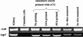
A site of 559 bp, which represents the Aqp3 band marker, was detected in mouse kidney (positive control), in in vivo as well as in vitro matured oocytes; Aqp3 bands were also detected in immature oocytes obtained with no eCG priming and 24 h or 48 h after priming (Fig. 1). Aqp3 mRNA was not detected by RT-PCR in cumulus cells obtained from immature CEOs.
The mRNAs encoding actin (upper part, band size 500 bp) and AQP3 (lower part, band size 559 bp) were analysed by RT-PCR followed by agarose gel electrophoresis in various developmental stages of denuded mouse oocytes. The Aqp3 mRNA was detected in kidney (positive control), immature oocytes obtained with no priming, 24-h- and 48-h-primed by equine chorionic gonadotropin (eCG). The Aqp3 mRNA was also detected in in vitro and in vivo matured oocytes, but not in cumulus cells obtained from immature cumulus–oocyte complexes (negative control).

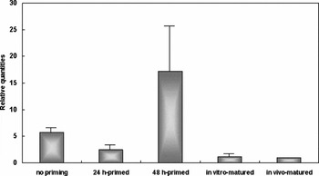
The relative expression levels of Aqp3 mRNA were different in three groups of immature oocytes according to eCG priming. Quantitative real-time PCR revealed that the level of Aqp3 mRNA expression was lowest in immature oocytes obtained 24 h after priming but highest in immature oocytes obtained 48 h after priming (Fig. 2). The expression level in the 48-h-primed group was approximately seven times higher than in the 24-h-primed group. However, the expression level was dramatically decreased after maturation in vitro, which is similar to in vivo matured oocytes. Both in vitro and in vivo matured oocytes showed reduced levels of Aqp3 mRNA compared with all of the immature oocyte groups.
Expression levels of Aqp3 mRNA in three types of immature oocytes, as well as in in vitro and in vivo matured mouse oocytes. Quantitative real-time PCR analysis revealed that a higher Aqp3 expression level was detected in immature oocytes obtained 48 h after eCG than those with no priming and those obtained 24 h after eCG priming. The standard quantity of the Aqp3 mRNA in in vivo matured oocytes was considered 1.0 and the experiment was repeated two times (no priming group; 5.7 ± 0.8, 24-h-primed group; 2.5 ± 0.8, 48-h-primed group; 17.2 ± 8.6, in vitro matured group; 1.2 ± 0.5, in vivo matured group; 1.0 ± 0.0, mean ± SD).

As expected, the in vitro developmental experiment demonstrated that 48-h-primed immature oocytes had a higher maturation (49.6%) and fertilization rate (61.0%) compared with no priming (33.6%, 47.2%) or the 24-h-primed group (44.4%, 50.0%).
Discussion
Expression of AQP3 was previously demonstrated in mature mouse oocyte (Edashige et al., Reference Edashige, Sakamoto and Kasai2000; McConnell et al., Reference McConnell, Yunus, Gross, Bost, Clemens and Hughes2002; Jablonski et al., Reference Jablonski, McConnell, Hughes and Huet-Hudson2003; Meng et al., Reference Meng, Gao, Xu, Dong, Sheng, Sheng and Huang2008). In the present study, we demonstrated for the first time that Aqp3 mRNA is also present in immature and in vitro matured mouse oocytes. In addition, we confirmed that Aqp3 mRNA is present in in vivo matured mouse oocytes but not in cumulus cells, which is in good agreement with previous studies (Edashige et al., Reference Edashige, Sakamoto and Kasai2000; Meng et al., Reference Meng, Gao, Xu, Dong, Sheng, Sheng and Huang2008).
We employed a mouse model designed specifically to approximate the cytoplasmic maturity. Follicular development in mice is more synchronized than in humans; therefore, to generate a model of cytoplasmic maturity, we retrieved immature oocytes from mice that were not primed and that were primed with eCG for 24 and 48 h. It can be assumed that immature oocytes retrieved 48 h after eCG priming are developmentally more competent compared with those obtained 24 h after priming or without priming. This was evident in that a higher maturation and fertilization rate was noted in immature oocytes retrieved 48 h after eCG.
Three types of immature oocytes according to priming showed quite different levels of Aqp3 mRNA expression; the level of Aqp3 mRNA was much higher in immature oocytes obtained 48 h primed compared with no priming or 24 h of priming. This result suggests that levels of Aqp3 mRNA are altered during development of immature oocytes and AQP3 is certainly needed for acquisition of immature oocytes’ full growing potential within antral follicles. Under gonadotropin stimulation, antral expansion occurs rapidly and follicles as well as oocytes may require a quick and massive transport of water. Thus, the increase of AQP3 expression during follicular development might represent cytoplasmic maturity in more competent immature oocytes.
Most interestingly, Aqp3 mRNA expression dramatically decreased during the maturation process in the present study. This finding indicates that AQP3 expression is closely related to the oocyte maturation process; however, the reason for the inverse relation between Aqp3 expression and nuclear maturity is unclear. Aquaporin 3 might no longer be required in matured oocytes; otherwise, it might be replaced by other types of water channel. Further study will be needed to clarify this topic.
In the previous rat study, Aqp9 mRNA disappeared in mature oocytes and at the same time the water permeability was reduced (Ford et al., Reference Ford, Merot, Jawerbaum, Gimeno, Capurro and Parisi2000). These findings demonstrated that immature oocytes had higher water and mannitol permeabilities than do mature ones. The presence of a broad selectivity to water and neutral solute during oocyte maturation may have important implications in their adaptation to osmotic stress; hence, future experiments will be warranted to clarify the link between expression and the function of oocyte water channels.
Another interesting finding was the similar expression of Aqp3 mRNA between in vitro and in vivo matured oocytes. In general, in vivo matured oocytes have better embryonic developmental potential and higher efficiency after cryopreservation than do in vitro matured oocytes. Therefore, our finding supports the insignificant role of AQP3 in the process of in vitro fertilization and cryopreservation of oocytes. A definite conclusion, however, cannot be drawn because the possibility exists that in vitro culture condition might change the expression of Aqp3 mRNA and we did not examine AQP3 expression in subsequent embryos or cryopreserved–thawed oocytes.
It has been reported that AQP3 expression is regulated by magnesium in Caco-2 cells, a colonic cell line (Okahira et al., Reference Okahira, Kubota, Iguchi, Usui and Hirano2008), retinoic acid in human skin (Bellemère et al., Reference Bellemère, Von Stetten and Oddos2008), nickel in lung epithelial cells (Zelenina et al., Reference Zelenina, Bondar, Zelenin and Aperia2003), copper in human bronchial epithelial cell line (Zelenina et al., Reference Zelenina, Tritto, Bondar, Zelenin and Aperia2004), and insulin in Caco-2 cells (Higuchi et al., Reference Higuchi, Kubota, Iguchi, Usui, Kiho and Hirano2007). Further studies on the regulation of AQP3 and other types of AQP in reproductive systems will be necessary for the development of efficient in vitro maturation and in vitro fertilization methods.
Further studies on AQP3-null mice would clarify the role of AQP3 within reproductive systems. Previous studies demonstrated that the growth and phenotype of AQP3-null mice are grossly normal but have impairment of urinary-concentrating ability (Ma et al., Reference Ma, Song, Yang, Gillespie, Carlson, Epstein and Verkman2000), as well as impairment of skin hydration, elasticity, and wound healing (Hara et al., Reference Hara, Ma and Verkman2002). The reproductive phenotypes in these AQP3-null mice are currently unknown. A recent study revealed that AQP4-null mice display subfertility as evidenced by a lower rate of pregnancy and decreased litter size (Sun et al., Reference Sun, Zhang, Fan, Ding, Sha and Hu2009).
In conclusion, we demonstrate for the first time that Aqp3 mRNA is expressed in immature oocytes as well as in vitro matured oocytes in mice. The expression of Aqp3 mRNA increased during development of immature oocytes, but decreased after in vitro maturation. The AQP3 is certainly needed for the acquisition of immature oocytes’ full growing potential within antral follicles.
Acknowledgement
This work was carried out at the oocyte research centre of Seoul National University Bundang Hospital and supported by grant No. 02-2008-008 from the Seoul National University Bundang Hospital Research Fund.