Introduction
Over the past decades, assisted reproductive technology (ART) has markedly evolved from an experimental approach to a routine procedure which has resulted in the delivery of more than 9 million babies (Steptoe and Edwards, Reference Steptoe and Edwards1978; Niederberger et al., Reference Niederberger, Pellicer, Cohen, Gardner, Palermo, O’Neill, Chow, Rosenwaks, Cobo, Swain, Schoolcraft, Frydman, Bishop, Aharon, Gordon, New, Decherney, Tan and Paulson2018). One of the major challenges for IVF scientists has always been the selection of the most competent embryo to transfer with the goal of obtaining a high pregnancy rate and reducing the incidence of multiple pregnancies (Sullivan et al., Reference Sullivan, Wang, Hayward, Chambers, Illingworth, McBain and Norman2012). Embryos are evaluated by non-invasive, morphological assessment, which essentially has not changed since the early days of IVF in the late 1970s (Steptoe and Edwards, Reference Steptoe and Edwards1978). This practice relies on a single microscope observation and on a few parameters, including the number and size of blastomeres, regularity of cell division, degree of fragmentation and multinucleation (Arce et al., Reference Arce, Ziebe, Lundin, Janssens, Helmgaard and Sørensen2006; Braude, Reference Braude2013; ESHRE Guideline Group on Good Practice in IVF Labs, Reference De los Santos, Apter, Coticchio, Debrock, Lundin, Plancha, Prados, Rienzi, Verheyen, Woodward and Vermeulen2015). With the establishment of a more physiological culture medium, it became more common to extend the culture to the blastocyst stage (day-5/day-6), which increases both uterine and embryonic synchronicity and aims to obtain a better pregnancy outcome (Gardner and Schoolcraft, Reference Gardner and Schoolcraft1999; De Vos et al., Reference De Vos, Van Landuyt, Santos-Ribeiro, Camus, Van de Velde, Tournaye and Verheyen2016). Blastocyst assessment is mainly based on the grading system initially proposed by Gardner and Schoolcraft, which has been efficient in defining the presence and volume of the inner cell mass (ICM), the cohesiveness of trophectoderm (TE) cells and the degree of expansion of the blastocoel cavity (Gardner and Schoolcraft, Reference Gardner and Schoolcraft1999). Recently, we have seen technical innovations with the introduction of a new incubator design with integrated time-lapse monitoring (TLM) system. This approach was first applied by Lewis and Gregory and lately by Payne and colleagues (Lewis and Gregory, Reference Lewis and Gregory1929; Payne et al., Reference Payne, Flaherty, Barry and Matthews1997). Currently, TLM has been introduced in several IVF laboratories. This technology allows the evaluation of the dynamic process of embryo development from the zygote to the blastocyst and combines three elements: a microscope, an incubator and imaging software. One of the benefits of this technology is that it enables continuous monitoring of embryo development (Meseguer et al., Reference Meseguer, Herrero, Tejera, Hilligsøe, Ramsing and Remohí2011, Reference Meseguer, Rubio, Cruz, Basile, Marcos and Requena2012; Basile et al., Reference Basile, Nogales, Bronet, Florensa, Riqueiros, Rodrigo, García-Velasco and Meseguer2014; Aparicio-Ruiz et al., Reference Aparicio-Ruiz, Basile, Pérez Albalá, Bronet, Remohí and Meseguer2016; Zaninovic et al., Reference Zaninovic, Nohales, Zhan, de Los Santos, Sierra, Rosenwaks and Meseguer2019; Sciorio et al., Reference Sciorio, Herrer Saura, Thong, Esbert Algam, Pickering and Meseguer2020a, Reference Sciorio, Thong, Thong and Pickering2021; Table 1) without the need to remove the embryos from the incubator, exposing them to non-physiological culture conditions (Zhang et al., Reference Zhang, Li, Peng, Guo, Heng and Tong2010; Wale and Gardner, Reference Wale and Gardner2016; Sciorio and Smith, Reference Sciorio and Smith2019). Much literature has been produced on the application of TLM in the embryology laboratory (Table 2); however, limited information is available on the global usage of this novel practice. Therefore, Dolinko and colleagues reported the results of a survey of 294 IVF centres in the USA. The authors noted that of 162 centres contacted, only 35 of them were using time-lapse technology (Dolinko et al., Reference Dolinko, Farland, Kaser, Missmer and Racowsky2017). Similar data were reported in a French study: 30 units out of 78 reported using TLM clinically (Boueilh et al., Reference Boueilh, Reignier, Barriere and Freour2018). Several studies have been published aiming to identify whether specific embryo characteristics could be associated with blastocyst formation and pregnancy outcomes (Meseguer et al., Reference Meseguer, Herrero, Tejera, Hilligsøe, Ramsing and Remohí2011, Reference Meseguer, Rubio, Cruz, Basile, Marcos and Requena2012; Aparicio-Ruiz et al., Reference Aparicio-Ruiz, Basile, Pérez Albalá, Bronet, Remohí and Meseguer2016; Sciorio et al., Reference Sciorio, Thong, Thong and Pickering2021). Wong and collaborators reported that development to the blastocyst stage was associated with specific timing, such as the duration of the first cleavage, and the time between the second and third divisions (Wong et al., Reference Wong, Loewke, Bossert, Behr, De Jonge, Baer and Reijo Pera2010). In 2011, Meseguer and collaborators noted that embryo implantation was linked to distinct cell division timing features, introducing the term ‘morphokinetics’ (Meseguer et al., Reference Meseguer, Herrero, Tejera, Hilligsøe, Ramsing and Remohí2011). More recently, Sciorio and collaborators in a retrospective study evaluated 664 single-blastocyst transfers resulting in an ongoing pregnancy, and reported a positive and significant correlation between blastocyst width and blastocyst area to pregnancy outcomes (Sciorio et al., Reference Sciorio, Thong, Thong and Pickering2021). Indeed, several studies have identified different aspects of embryo development as poor prognosis factors, such as direct, irregular or reverse cleavages (Aguilar et al., Reference Aguilar, Motato, Escribá, Ojeda, Muñoz and Meseguer2014; Desai et al., Reference Desai, Ploskonka, Goodman, Austin, Goldberg and Falcone2014; Liu et al., Reference Liu, Chapple, Feenan, Roberts and Matson2015) or blastocyst collapse(s) (Marcos et al., Reference Marcos, Pérez-Albalá, Mifsud, Molla, Landeras and Meseguer2015; Sciorio et al., Reference Sciorio, Herrer Saura, Thong, Esbert Algam, Pickering and Meseguer2020a, Reference Sciorio, Thong and Pickering2020b) and the occurrence of those events might be used as negative selection criteria. However, the application of TLM remains still questionable (Armstrong et al., Reference Armstrong, Bhide, Jordan, Pacey, Marjoribanks and Farquhar2019, Kovacs and Lieman, Reference Kovacs and Lieman2019). Therefore we highlight here some advantages of this novel practice, in particular, assessing the blastocyst collapse(s) event and blastocyst morphometric parameters. We also delineate other concerns provided by TLM as the morphokinetic and dynamic aspects and undisturbed culture conditions. An extensive search was performed by retrieving relevant literature from the PubMed search engine up to July 2022. The search strategies and keywords used were ‘time-lapse technology’, ‘embryo culture’ or ‘single-blastocyst transfer’ and returned 216 papers. In total, 49 original articles were used for data extraction, and their major findings are summarized in this manuscript.
Abnormal features observed with time-lapse imaging. Adapted with permission from Sciorio et al. (Reference Sciorio, Herrer Saura, Thong, Esbert Algam, Pickering and Meseguer2020a)

Some studies published in the last decade that have used the time-lapse technology. Adapted with permission from Sciorio et al. (Reference Sciorio, Herrer Saura, Thong, Esbert Algam, Pickering and Meseguer2020a)

Time-lapse technology
The goal of ART is to obtain in a short time a single healthy pregnancy, avoiding the incidence of multiple gestations, which are correlated with a higher risk of adverse perinatal and maternal outcomes [European IVF-Monitoring Consortium (EIM) et al., Reference Kupka, D’Hooghe, Ferraretti, de Mouzon, Erb, Castilla, Calhaz-Jorge, De Geyter and Goossens2016]. Indeed, even though we have seen several advancements in ART over the last decades, the selection criteria by which embryos are chosen remain elusive and still based on the morphology assessment. It is, therefore, still a main challenge to identify the single embryo with the highest implantation potential from a cohort of embryos. Since the birth of Louise Joy Brown, the first baby born following IVF on July 1978 (Steptoe and Edwards, Reference Steptoe and Edwards1978), standard morphological evaluation has been applied as the prevalent method for embryo selection. Albeit embryo morphology has been associated with implantation and pregnancy rates, its accuracy has proved restricted, mainly due to the subjectivity of the embryologist (Arce et al., Reference Arce, Ziebe, Lundin, Janssens, Helmgaard and Sørensen2006; Braude, Reference Braude2013). Furthermore, a single standard observation provides only an image of the embryo at that particular time, ignoring what happens during the intervals between two observations, and probably abnormal events remain unidentified (Cruz et al., Reference Cruz, Garrido, Herrero, Pérez-Cano, Muñoz and Meseguer2012). An alternative approach to this concern has led to the development of TLM, which allows the evaluation of the embryo morphology and every dynamic event during its development, and in addition, maintains stable culture conditions, which are critical to assure further embryo development (Meseguer et al., Reference Meseguer, Herrero, Tejera, Hilligsøe, Ramsing and Remohí2011, Reference Meseguer, Rubio, Cruz, Basile, Marcos and Requena2012; Basile et al., Reference Basile, Nogales, Bronet, Florensa, Riqueiros, Rodrigo, García-Velasco and Meseguer2014; Aparicio-Ruiz et al., Reference Aparicio-Ruiz, Basile, Pérez Albalá, Bronet, Remohí and Meseguer2016; Manna et al., Reference Manna, Nanni, Lumini and Pappalardo2013; Zaninovic et al., Reference Zaninovic, Nohales, Zhan, de Los Santos, Sierra, Rosenwaks and Meseguer2019; Sciorio et al., Reference Sciorio, Herrer Saura, Thong, Esbert Algam, Pickering and Meseguer2020a, Reference Sciorio, Thong, Thong and Pickering2021). Currently, there are different time-lapse systems: some models enclose a digital inverted microscope with a built-in camera to acquire embryo images, while others are equipped with a camera placed in a traditional large incubator. Regarding the software used for embryo annotations, several systems have been recently applied, such as semi-automatic or fully automated registration of the main events of embryo development (Cruz et al., Reference Cruz, Garrido, Herrero, Pérez-Cano, Muñoz and Meseguer2012; Kirkegaard et al., Reference Kirkegaard, Agerholm and Ingerslev2012; Chen et al., Reference Chen, Tan, Suraj, Reijo Pera and Shen2013, Manna et al., Reference Manna, Nanni, Lumini and Pappalardo2013). In addition, artificial intelligence (AI), which is the science and engineering of making intelligent machines, has been currently applied in ART, helping embryologists to manage thousands of time-lapse digitalized images and videos of embryo development. Indeed, TLM involves manual annotations, which implies a wide inter-operator and intra-operator variation (Sundvall et al., Reference Sundvall, Ingerslev, Breth Knudsen and Kirkegaard2013; Storr et al., Reference Storr, Venetis, Cooke, Kilani and Ledger2017; Apter et al., Reference Apter, Ebner, Freour, Guns, Kovacic, Le Clef, Marques, Meseguer, Montjean, Sfontouris, Sturmey and Coticchio2020). The incorporation of AI can provide automated annotations, which is critical to overcoming the problem of subjectivity (Danuser, Reference Danuser2011). Despite the fact that AI is not the main topic of this review paper, it is worth noting that several studies have been recently published investigating novel approaches able to predict the developmental potential of human embryos with an accuracy between 70% and 90% (Manna et al., Reference Manna, Nanni, Lumini and Pappalardo2013; Khosravi et al., Reference Khosravi, Kazemi, Zhan, Malmsten, Toschi, Zisimopoulos, Sigaras, Lavery, Cooper, Hickman, Meseguer, Rosenwaks, Elemento, Zaninovic and Hajirasouliha2019; Tran et al., Reference Tran, Cooke, Illingworth and Gardner2019; Bormann et al., Reference Bormann, Kanakasabapathy, Thirumalaraju, Gupta, Pooniwala, Kandula, Hariton, Souter, Dimitriadis, Ramirez, Curchoe, Swain, Boehnlein and Shafiee2020; Bori et al., Reference Bori, Paya, Alegre, Viloria, Remohi, Naranjo and Meseguer2020; Chavez-Badiola et al., Reference Chavez-Badiola, Flores-Saiffe-Farías, Mendizabal-Ruiz, Drakeley and Cohen2020; Alegre et al., Reference Alegre, Del Gallego, Bori, Loewke, Maddah, Aparicio-Ruiz, Palma-Govea, Marcos and Meseguer2021). Bori and co-workers, in a retrospective study counting 637 patients who underwent elective single-blastocyst transfer (eSBT), described how the use of AI might be applied in predicting implantation potential with an accuracy of 77% (Bori et al., Reference Bori, Paya, Alegre, Viloria, Remohi, Naranjo and Meseguer2020). Also, Liao and collaborators using time-lapse videos, report an AI model that can predict blastocyst formation (accuracy of 71.9%) and usable blastocysts (accuracy of 79%) (Liao et al., Reference Liao, Zhang, Feng, Huang, Xu, Tian, Liu, Yu, Guo, Liu, Huang, Ma, Ai, Xu and Li2021). Those early studies are truly promising. In the future, including an automated system and AI in the embryology laboratory could improve IVF efficiency by decreasing both the time spent making annotations and variations among embryologists as might be applied to anticipate successful pregnancy.
Time-lapse incubator: benefits of uninterrupted culture and morphokinetics
Human embryogenesis implicates multiple chemical intracellular signals among gametes that result in the evolution of viable embryos able to generate a successful pregnancy. The process depends on specific and physiological culture conditions, as embryos are fragile and unprotected as they lack epithelial surfaces. Embryos exposure to un-physiological culture conditions causes the generation of physical and chemical stressors (Wale and Gardner, Reference Wale and Gardner2012). Also, in vitro exposure to increased oxygen concentration is correlated with the production of reactive oxygen species (ROS), which might modify embryo metabolism, and gene expression and reduce implantation potential (Fischer and Bavister, Reference Fischer and Bavister1993; Meintjes et al., Reference Meintjes, Chantilis, Douglas, Rodriguez, Guerami, Bookout, Barnett and Madden2009, Bontekoe et al., Reference Bontekoe, Mantikou, van Wely, Seshadri, Repping and Mastenbroek2012; Sciorio and Smith, Reference Sciorio and Smith2019; Gardner et al., Reference Gardner, Kuramoto, Tanaka, Mitzumoto, Montag and Yoshida2020; Sciorio et al., Reference Sciorio, Tramontano, Rapalini, Bellaminutti, Bulletti, D’Amato, Manna, Palagiano, Bulletti and Esteves2023). In that context, a considerable benefit of TLM is that it provides undisturbed culture conditions, avoiding embryo exposure to an non-physiological environment, such as pH or temperature changes, as well as gas concentrations, which are extremely critical to ensure future embryo development and viability (Fischer and Bavister, Reference Fischer and Bavister1993; Sciorio and Smith, Reference Sciorio and Smith2019; Gardner et al., Reference Gardner, Kuramoto, Tanaka, Mitzumoto, Montag and Yoshida2020). Using TLM as a non-invasive procedure ensures not only stable culture conditions but allows the continual recording of morphological changes during the in vitro embryo culture, therefore enabling embryologists to analyse every single morphokinetic change that cannot be seen (Basile et al., Reference Basile, Vime, Florensa, Aparicio-Ruiz, Garcia-Velasco, Remohi and Meseguer2015; Zaninovic et al., Reference Zaninovic, Irani and Meseguer2017). Furthermore, novel parameters of embryo development have been described by several authors: at the fertilization process (Coticchio et al., Reference Coticchio, Mignini Renzini, Novara, Lain, De Ponti, Turchi, Fadini and Dal Canto2018), during the first three cell cleavages (Meseguer et al., Reference Meseguer, Herrero, Tejera, Hilligsøe, Ramsing and Remohí2011; Liu et al., Reference Liu, Chapple, Feenan, Roberts and Matson2015; Aparicio-Ruiz et al., Reference Aparicio-Ruiz, Basile, Pérez Albalá, Bronet, Remohí and Meseguer2016) at the compaction and morula stage (Coticchio et al., Reference Coticchio, Lagalla, Sturmey, Pennetta and Borini2019, Reference Coticchio, Ezoe, Lagalla, Shimazaki, Ohata, Ninomiya, Wakabayashi, Okimura, Uchiyama, Kato and Borini2021) and finally at the blastocyst formation (Dal Canto et al., Reference Dal Canto, Coticchio, Mignini Renzini, De Ponti, Novara, Brambillasca, Comi and Fadini2012; Marcos et al., Reference Marcos, Pérez-Albalá, Mifsud, Molla, Landeras and Meseguer2015; Sciorio et al., Reference Sciorio, Herrer Saura, Thong, Esbert Algam, Pickering and Meseguer2020a, Reference Sciorio, Thong and Pickering2020b). Motato and colleagues made a retrospective analysis of cleavage times for 7483 embryos (Motato et al., Reference Motato, de Los Santos, Escriba, Ruiz, Remohí and Meseguer2016). Several parameters were investigated, such as time of cleavage from two to nine cells (t2, t3, t4, t5, t6, t7, t8, and t9), time to the morula stage (tM), blastocyst formation (tB) when the blastocoel cavity filled the embryo and the ICM and trophectoderm cells were clearly detectable, as well as the time of expanded blastocyst (tEB) and hatching of the blastocyst (tHB), when the trophectoderm starts herniating through the zona pellucida. The authors noticed that some of these features were significantly associated with blastocyst formation and implantation. In particular, the most predicting parameters for blastocyst formation were the time of transition from five to eight cells (t8-t5: ≤8.78 h) and the time of morula formation (tM: 81.28–96.0 h after ICSI). Also, Dal Canto and co-workers, in a retrospective study, including 4915 embryos, evaluated several morphokinetic parameters such as time to pronuclear fading (tPNf), t2, t3, t4, t5 and t8. The authors reported that all parameters were reached earlier in embryos producing live birth (LB) than in non-live birth embryos (NLB). Although retrospective, those data illustrate that embryos resulting in a successful pregnancy with live birth reach tPNf (LB: 22.5 vs. NLB 23.9; P < 0.0001), t2 (LB 25.0 vs. NLB 26.8; P < 0.0001), t3 (LB 36.1 vs. NLB 37.6; P < 0.0001), t4 (LB 36.8 vs. NLB 39.0; P < 0.0001), t5 (LB 48.8 vs. NLB 50.0; P = 0.009) and t8 (LB 52.7 vs. NLB 55.0; P = 0.0002) earlier than embryos that do not lead to an LB (Dal Canto et al., Reference Dal Canto, Bartolacci, Turchi, Pignataro, Lain, De Ponti, Brigante, Mignini Renzini and Buratini2021). Those findings are in agreement with previous studies published by others (Meseguer et al., Reference Meseguer, Rubio, Cruz, Basile, Marcos and Requena2012; Mizobe et al., Reference Mizobe, Oya, Iwakiri, Yoshida, Sato, Miyoshi, Tokunaga and Ezono2016; Motato et al., Reference Motato, de Los Santos, Escriba, Ruiz, Remohí and Meseguer2016). Finally, as mentioned earlier, several authors using TLM have identified atypical phenotypes and been considered as poor prognosis factors and, therefore, might be used as deselection criteria (Aguilar et al., Reference Aguilar, Motato, Escribá, Ojeda, Muñoz and Meseguer2014; Desai et al., Reference Desai, Ploskonka, Goodman, Austin, Goldberg and Falcone2014; Liu et al., Reference Liu, Chapple, Feenan, Roberts and Matson2015; Marcos et al., Reference Marcos, Pérez-Albalá, Mifsud, Molla, Landeras and Meseguer2015; Mizobe et al., Reference Mizobe, Oya, Iwakiri, Yoshida, Sato, Miyoshi, Tokunaga and Ezono2016; Sciorio et al., Reference Sciorio, Herrer Saura, Thong, Esbert Algam, Pickering and Meseguer2020a, Reference Sciorio, Thong and Pickering2020b). In this vein, a retrospective multicentre cohort study involving 651 embryos, described four atypical phenotypes, including abnormal syngamy (AS), abnormal first cytokinesis (A1-cyt), abnormal cleavage (AC) and chaotic cleavages (CC). The authors noticed that those atypical features are quite frequent in human embryos: AS 25.1%, A1-cyt 31.0%, AC 18% and CC 15%. In addition, these embryos displayed a significantly lower blastocyst formation compared with their control groups: AS 21.5% vs. 44.9%, A1-cyt 21.7% vs. 44.6%, AC 11.7% vs. 43.1%, and CC 14.0% vs. 42.3% (Athayde Wirka et al., Reference Athayde Wirka, Chen, Conaghan, Ivani, Gvakharia, Behr, Suraj, Tan and Shen2014). Comparable results have been reported by another multicentre study published by Rubio and colleagues. The goal of the study, including 5,225 embryos, was to assess embryos with direct cleavage (≤5 h) from two to three cells and correlate this feature to the implantation rate. They found that 13.7% (715/5,225) of the embryos showed a direct cleavage from two to three cells, and those embryos if transferred, resulted in a statistically significantly lower implantation rate compared with embryos with a normal cleavage pattern (1.2% vs. 20.2%, P < .0001 respectively) (Rubio et al., Reference Rubio, Kuhlmann, Agerholm, Kirk, Herrero, Escribá, Bellver and Meseguer2012).
Blastocyst selection and the collapse event
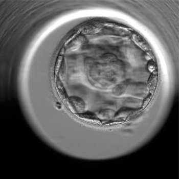
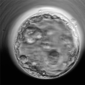
Extended in vitro culture to the blastocyst stage has been considered a challenge in ART cycles, and has resulted in higher pregnancy outcomes and LB rates compared with the transfer of cleavage-stage embryos (Glujovsky et al., Reference Glujovsky, Farquhar, Quinteiro Retamar, Alvarez Sedo and Blake2016; Revelli et al., Reference Revelli, Canosa, Carosso, Filippini, Paschero, Gennarelli, Delle Piane and Benedetto2019). Blastocyst-stage transfer seems to be a good approach to increase the policy of single-embryo transfer (Vega et al., Reference Vega, Zaghi, Buyuk and Jindal2018; Freeman et al., Reference Freeman, Hinds, Howard, Howard and Hill2019). Different studies have reported a correlation between various time-lapse features and blastocyst development. As mentioned earlier, some morphokinetic features, such as the synchrony between the 3-cell and 4-cell stages, or time to morula formation and the time from 5-cell to 8-cell embryos, might be used to anticipate the blastocyst formation and implantation potential (Meseguer et al., Reference Meseguer, Herrero, Tejera, Hilligsøe, Ramsing and Remohí2011, Reference Meseguer, Rubio, Cruz, Basile, Marcos and Requena2012; Valeri et al., Reference Valeri, Pappalardo, De Felici and Manna2011; Hashimoto et al., Reference Hashimoto, Kato, Saeki and Morimoto2012; Hlinka et al., Reference Hlinka, Kaľatová, Uhrinová, Dolinská, Rutarová, Rezáčová, Lazarovská and Dudáš2012; Conaghan et al., Reference Conaghan, Chen, Willman, Ivani, Chenette, Boostanfar, Baker, Adamson, Abusief, Gvakharia, Loewke and Shen2013; Kirkegaard et al., Reference Kirkegaard, Kesmodel, Hindkjær and Ingerslev2013; Basile et al., Reference Basile, Nogales, Bronet, Florensa, Riqueiros, Rodrigo, García-Velasco and Meseguer2014; Aparicio-Ruiz et al., Reference Aparicio-Ruiz, Basile, Pérez Albalá, Bronet, Remohí and Meseguer2016; Zaninovic et al., Reference Zaninovic, Nohales, Zhan, de Los Santos, Sierra, Rosenwaks and Meseguer2019; Sciorio et al., Reference Sciorio, Thong, Thong and Pickering2021). According to the current literature, it seems reasonable to state that a correlation exists between the timing of the early development and blastocyst formation, but if this correlation is preferable to morphological assessment remains to be proved. Therefore, it might be critical to have a more objective parameter which correlates with blastocyst quality and implantation potential. In that vein, spontaneous blastocyst collapse has been suggested as a novel element of embryo viability and pregnancy outcome (Figures 1–4). This feature was described for the first time in the rabbit model in 1929 (Lewis and Gregory, Reference Lewis and Gregory1929) and recently has also been seen in other mammals, including mice (Niimura, Reference Niimura2003), domestic cats (Kij et al., Reference Kij, Kochan, Fryc, Niżański, Prochowska, Gabryś, Nowak and Bugno-Poniewierska2020) and human (Marcos et al., Reference Marcos, Pérez-Albalá, Mifsud, Molla, Landeras and Meseguer2015; Sciorio et al., Reference Sciorio, Herrer Saura, Thong, Esbert Algam, Pickering and Meseguer2020a, Reference Sciorio, Thong and Pickering2020b). During the 5 or 6 days of development, the fertilized oocyte generates the blastocyst that will implant into the uterine cavity. The blastocyst consists of the trophectoderm cells surrounding the blastocoel, a fluid-filled lumen, and a group of cells which will form the fetus: the ICM. Blastocyst formation begins when the compacted morula initiates to secrete a fluid; the small cavity steadily acquires fluids in its blastocoel and grows by actions of the sodium pump, which raises the salt concentration within the embryo, attracting water through osmosis (Biggers, Reference Biggers1998). This action induces a rise in pressure and blastocyst expansion, subsequently, TE cells secrete lysine that implicated the zona pellucida (ZP) thinning followed by the hatching process, which is mandatory for the process of implantation (Biggers, Reference Biggers1998). Recently, using TLM, blastocyst collapse has been observed in detail, and studies have reported that weak contractions appear to be beneficial to the hatching process, although strong collapse may negatively affect hatching (Gonzales et al., Reference Gonzales, Jones, Pinyopummintr, Carnevale, Ginther, Shapiro and Bavister1996). Marcos and collaborators have first illustrated the event in human embryos. They have defined the collapse event when there is a separation of at least 50% of the trophoblast from the ZP, while a weak contraction is when the detachment involves less than 50% of the trophoblast (Figures 1–4). The authors found that other features, including time to the morula stage and blastulation, were significantly shorter in blastocysts showing collapse(s) episodes. In addition, following the embryo transfer, results showed a decline in implantation rate from 48.5% to 35.1% if blastocysts that displayed a collapse episode were transferred (Marcos et al., Reference Marcos, Pérez-Albalá, Mifsud, Molla, Landeras and Meseguer2015). Another retrospective trial illustrated that blastocysts presenting collapse during development have a significantly reduced probability of generating a pregnancy compared with embryos that do not report any collapse (Sciorio et al., Reference Sciorio, Thong and Pickering2020b). Sciorio and collaborators, in a multicentre trial involving four different units, found comparable results. In this study, almost 1300 embryos cultured in a time-lapse incubator were analyzed. Blastocyst assessment was performed between 112 and 118 h post insemination, at that time if a collapse(s) event was seen during the development it was annotated. It needs to be mentioned that the slide containing the embryos was inserted in the time-lapse device at the time of fertilization check and never removed from the incubator until the morning on day 5 before the ET. The authors reported that ∼20% of embryos showed one or two collapses during the development and following eSBT, a significant reduction in pregnancy rate (61.0% vs 45.2%; P < 0.0001) and ongoing pregnancy rate (51.9% vs 37.5%; P < 0.0001) was noticed when collapsed blastocysts were replaced compared with cycles in which blastocysts displayed no collapse event (Sciorio et al., Reference Sciorio, Herrer Saura, Thong, Esbert Algam, Pickering and Meseguer2020a). However, the molecular mechanism of blastocyst collapse is still not completely elucidated. It has been hypothesized that the variation of the mechanical force and the change of pressure during the collapse event might damage the gap junctions between the TE cells. Therefore, once the blastocyst recovers, it might have a defective TE (Togashi et al., Reference Togashi, Kumagai, Sato, Shirasawa, Shimoda, Makino, Sato, Kumazawa, Omori and Terada2015). Finally, the re-expansion process requires energy. Therefore, if the blastocyst collapses twice or more, it might need a considerable amount of energy to recover and complete the hatching process, a deficiency of which may have a detrimental effect (Baltz et al., Reference Baltz, Smith, Biggers and Lechene1997). Further studies are required to clarify the blastocyst collapse event in humans. In addition, although these are retrospective studies with associated limitations, the utility of blastocyst collapse episodes as a predictor of implantation and pregnancy outcome is suggested for the first time. Therefore, it may be an additional and useful parameter to consider when selecting a single blastocyst to transfer, especially in cases in which there are several blastocysts available to choose from for transfer.
Time-lapse monitoring of a cultured human blastocyst. Arrow shows a weak contraction (volume reduction less than 50%). Adapted with permission from Sciorio et al. (Reference Sciorio, Herrer Saura, Thong, Esbert Algam, Pickering and Meseguer2020a).


Time-lapse monitoring of a cultured human blastocyst showing a collapse event with volume reduction more than 50%. Adapted with permission from Sciorio et al. (Reference Sciorio, Herrer Saura, Thong, Esbert Algam, Pickering and Meseguer2020a).


Video processing, morphometric blastocyst parameters and pregnancy outcome
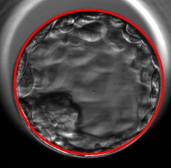
In humans, the embryo genome activation takes place at approximately the 8-cell stage; therefore, extending the culture to the blastocyst stage, it might be indicated to select the embryo with high implantation potential compared with the cleavage stages (day 2/day 3) (Braude et al., Reference Braude, Bolton and Moore1988). The novel use of TLM provides relevant details, including morphometric parameters, which allow embryologists to be more confident when selecting the single blastocyst to transfer. The debate in the literature on which elements between ICM, TE and expansion have the most predictive value is mainly unsolved. Studies have reported a solid correlation between blastocyst quality and high implantation rates, as well as poor ICM, which has been associated with reduced pregnancy outcomes (Gardner et al., Reference Gardner, Lane, Stevens, Schlenker and Schoolcraft2000). Chimote and co-authors noticed that the hatching process might be considered an important parameter for a successful pregnancy. In a prospective study of 146 patients undergoing their first IVF treatment, the authors compared the developmental potential and pregnancy outcome of spontaneously hatching blastocysts against expanded blastocysts. They found a significantly higher pregnancy rate (59.4% vs. 45.1%, P = 0.0173) and LB rate (36.8% vs. 22.8%, P = 0.003) when hatching blastocysts were transferred compared with the expanded blastocyst group (Chimote et al., Reference Chimote, Chimote, Nath and Mehta2013). Similar results have been observed by Yoon and co-workers, who noted a significantly higher pregnancy rate in women with one or more hatching blastocysts transferred on day-6 compared with patients who received non-hatching blastocysts on day-5 (Yoon et al., Reference Yoon, Yoon, Son, Im and Lim2001). Conversely, studies have suggested blastocyst expansion as the major feature in embryo selection (Subira et al., Reference Subira, Craig, Turner, Bevan, Ohuma, McVeigh, Child and Fatum2016), such as the study by Van den Abbeel and collaborators, who analyzed the pregnancy outcome of 618 ICSI embryos with mandatory eSBT on day-5, and concluded stating that the selection of the best quality blastocyst to transfer in a fresh cycle should be mainly based on the expansion and hatching stage (Van den Abbeel et al., Reference Van den Abbeel, Balaban, Ziebe, Lundin, Cuesta, Klein, Helmgaard and Arce2013). In contrast, there are studies reporting that TE grade was significantly associated with implantation potential and LB rates (Ahlström et al., Reference Ahlström, Westin, Reismer, Wikland and Hardarson2011; Hill et al., Reference Hill, Richter, Heitmann, Graham, Tucker, DeCherney, Browne and Levens2013; Van der Weiden, Reference Van der Weiden2013). Hill and co-workers evaluated the effect of the trophectoderm and ICM morphology grade on implantation and LB in 694 single-blastocyst transfers. Live birth rates were 57%, 40%, and 25% for TE grades A, B, and C, respectively. Whereas for the ICM grades A, B, and C LB rates were 53%, 52%, and 0%, respectively. The authors performed a multiple logistic regression analysis which demonstrated that only TE quality and patient age were significantly correlated with LB, whereas ICM grade was not significantly associated with pregnancy outcome (Hill et al., Reference Hill, Richter, Heitmann, Graham, Tucker, DeCherney, Browne and Levens2013). Comparable results were detected by Ahlström and co-authors in a retrospective study of 1117 fresh day-5 eSBT. They noticed that TE was the only independent predictor of LB outcome. They concluded that, despite ICM being essential, a solid and robust TE is important to enable the hatching process and further implantation (Ahlström et al., Reference Ahlström, Westin, Reismer, Wikland and Hardarson2011). Considering those contrasting results among the most important features associated with a successful pregnancy, it is mandatory to include more objective parameters in blastocyst gradings, such as blastocyst area or blastocyst maximum width and try to establish a correlation with successful pregnancy (Figures 5 and 6). This was the main goal of a recent study published by Sciorio and collaborators (Sciorio et al., Reference Sciorio, Thong, Thong and Pickering2021), who investigated morphometric blastocyst parameters, involving a specific measurement in microns (μm) of the maximum blastocyst diameter and the entire area occupied by the blastocyst expressed in square microns (μm2). They correlated those novel features with ongoing pregnancy rates. The study, including 664 single eSBT on day-5, was performed between ∼112–118 h following insemination. Before the transfer, blastocyst morphometric assessment was performed using the EmbryoViewer™ software (Vitrolife) and the embryo maximum width expressed in microns (μm) was determined (Figure 5), as already described by others (Almagor et al., Reference Almagor, Harir, Fieldust, Or and Shoham2016). Similarly, as previously illustrated by Huang and colleagues, a measurement in square micrometres (μm2) of the blastocyst area, excluding the area occupied by the ZP, was taken (Huang et al., Reference Huang, Chinn, Kosasa, Ahn and Kessel2016). The results of this study illustrated a statistically significant association between clinical pregnancy and maximum blastocyst width, following the transfer of one blastocyst. In women who resulted to a successful pregnancy, the transferred blastocyst width was larger (184 μm) compared with non-pregnant patients (160 μm). A comparable correlation was observed evaluating the blastocyst area. Results showed a significantly larger blastocyst area (26,099 μm2) transferred in women who obtained a positive ongoing pregnancy, compared with non-pregnant women (22,251 μm2; Sciorio et al., Reference Sciorio, Thong, Thong and Pickering2021). Although this was a retrospective study with associated limitations, the results of this analysis are in line with other papers previously published that investigated the association between blastocyst diameter and clinical pregnancy, as well as the importance of the blastocyst expansion to obtain a successful pregnancy (Della Ragione et al., Reference Della Ragione, Verheyen, Papanikolaou, Van Landuyt, Devroey and Van Steirteghem2007; Mio and Maeda, Reference Mio and Maeda2008; Kresowik et al., Reference Kresowik, Sparks and Van Voorhis2012; Thompson et al., Reference Thompson, Onwubalili, Brown, Jindal and McGovern2013; Iwata et al., Reference Iwata, Yumoto, Sugishima, Mizoguchi, Kai, Iba and Mio2014; Barrie et al., Reference Barrie, Homburg, McDowell, Brown, Kingsland and Troup2017).
Blastocyst maximum width 205 µm.

Blastocyst area 33,549 µm2.

Concluding remark
In conclusion, the application of TLM in the modern ART laboratory has provided novel morphokinetic and morphometric parameters, supporting the embryologist team when selecting a single embryo to transfer, to reduce complications associated with multiple gestations. Nevertheless, another aspect that needs to be mentioned is the high cost of acquiring a time-lapse incubator, which remains a concern. For the moment, TLM technology is still very expensive, and laboratory managers should prioritize the allocation of resources. Additionally, regarding the dishes to use, each system used only one type of culture dish compatible with the incubator chamber. The price of slides might be higher than the variety of IVF culture dishes available on the market. Indeed, it is worth noting, as suggested by the Human Fertilization and Embryology Authority (HFEA: UK Fertility Regulator), that time-lapse incubation includes still conflicting evidence from randomized controlled trials to show that it is effective in increasing the chances of having a baby for most infertile couples. Time-lapse being undisturbed while they grow may improve the quality of the embryos, but there is certainly not enough evidence to show that this novel approach is effective at improving the chance of having a successful pregnancy following ART. Finally, TLM combines more stable culture conditions and allows embryologists to identify unknown or undetectable aspects of embryo development, including atypical phenotypes such as direct or reverse cleavages, which might result in a negative pregnancy. Indeed, good IVF laboratory practice should be in place and already optimal before time-lapse technology can be considered. Presumably, TLM will be routinely applied in the embryology laboratory to select the single embryo to transfer for couples undergoing ART treatments.
Compliance with ethical standards/Human and animal rights
All procedures performed in the studies involving human participants were in accordance with the ethical standards of the institution and with the 1964 Helsinki Declaration and its later amendments. For this type of study, formal consent is not required.
Conflict of interest
The authors have no conflict of interest to declare.