Aortic coarctation is defined as the restriction of blood flow in the distal aortic arch as a consequence of mechanical obstruction occurring distal to the left subclavian artery, appearing in 0.2–0.6/1000 newborns. In 6–8% of patients with congenital heart defects, coarctation of the aorta is present.Reference Hoffman and Kaplan 1 , Reference Wilson, Corres-Villaenor, Loffredo and Ferenc 2 Clinical signs and symptoms depend on the degree of stenosis. Patients become symptomatic in the newborn period, with signs of severe congestive heart failure due to the pressure strain of the left ventricle. The typical clinical finding is a difference of blood pressure and pulse status between the upper and lower limb.Reference Schumacher 3 The mortality rate of a typical coarctation in the developed countries is currently low, and long-term outcome has improved.Reference Kaushal, Patel and Patel 4 , Reference Pedersen, Røpcke and Hjortdal 5 In contrast to typical coarctation, a spontaneous aortic arch thrombosis is very rare, but potentially a life-threatening event with a high rate of mortality.Reference Ahmadi, Fürste and Pringsheim 6 – Reference Uva, Serraf and Lacour-Gayet 12
Thrombotic complications in childhood are very rare. Neonates and infants <1 year of age are mainly affected. In most cases, the thrombosis is triggered by an external risk factor, in particular asphyxia, sepsis, dehydration, or maternal diabetes.Reference Chalmers 13 , Reference Münchow, Kosch, Schobess, Junker, Auberger and Nowak-Göttl 14 Another risk factor is the presence of a central venous line or umbilical catheter.Reference Revel-Vilk and Ergaz 15 In the Canadian registry, the incidence of clinically apparent neonatal thrombosis was 2.4 per 1000 admissions to neonatal intensive care units, and in the German registry it was 5.1 per 100,000 births.Reference Chalmers 13 The annual incidence of thrombosis in childhood was reported to be 0.7 to 1.4 per 100,000 children.Reference Nowak-Göttl, Kurnik, Manner and Kenet 16 , Reference Young, Albisetti and Nowak-Göttl 17 In all available registries – Canada, Germany, and Denmark – the majority of thrombosis concerned the venous system with 66–88%, whereas arterial thrombosis was only diagnosed in 12–34% of the cases.Reference Chalmers 13 , Reference Tuckuviene, Christensen, Helgestad, Johnsen and Kristensen 18 To date, no reliable data exist on the prevalence, incidence, morbidity, mortality, or underlying thrombophilic disorder in case of aortic arch thrombosis. To generate more reliable information about this rare disease, we reviewed the literature and analysed the data of all identified patients with aortic arch thrombosis.
Methods
Literature search was made of the electronic databases MEDLINE – results range from March, 1953 to February, 2010. The following search terms were used: coarctation, aorta, aortic arch thrombosis, and neonatal aortic thrombosis. The full text of all case reports, studies, and reviews of possible relevance were obtained. There was no language restriction. We analysed the clinical course, associated comorbidities, associated thrombotic events, underlying thrombotic risk factors, and outcome of all reported cases of neonatal aortic arch thrombosis. Statistical analysis included the two reported patients. For statistical analysis of potential differences in survival, Fisher's exact test (two sided) was used.
Case reports and Results
Case 1
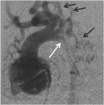
Prenatal routine ultrasound in uneventful pregnancy suggested the presence of aortic coarctation without abnormalities of other organs. The girl was born in the 38 + 5 week of gestation. Prostaglandin E1 infusion was started immediately after birth. Echocardiography demonstrated an interruption of the aortic arch, atypical for classical coarctation, therefore suggesting a thrombotic occlusion of the aortic arch. No other cardiac abnormalities could be found. Owing to the atypical presentation, angiography was performed before operation, showing a long-distance interruption of the aortic arch. Multiple large collateral vessels suggested a rather long-standing neonatal thrombosis (see Fig 1). The patient was successfully operated upon on day 9. Intra-operative findings confirmed the suspected thrombosis mimicking congenital aortic coarctation. Externally, the aorta looked normal. The thrombus was removed after incision, and the aortic isthmus was resected, although no coarctation was present. The aorta was reconstructed by end-to-end anastomosis without patch augmentation. Patient evaluation for thrombophilic risk factors resulted in heterozygous state of a factor V Leiden mutation – non-prothrombin mutation – and protein C, protein S, antithrombin were normal for the age. After successful operation, oral anticoagulation with phenprocoumon was started and no further thrombotic events were reported after resection of the thrombus.
Picture shows the angiography of the ventricle and aortic arch of patient 1. The angiography revealed a long-distance interruption of the aortic arch (white arrow) close to the outlet of the left carotid artery, already partially compensated by large collateral vessels (black arrows).

Case 2
The boy was born at 33 + 5 weeks of gestation by Caesarean section because of the HELLP syndrome in the mother. After normal initial adaptation, a considerable difference of pre- and post-ductal blood pressure and transcutaneous oxygen saturation was noticed. The initial echocardiographic examination suggested critical coarctation of the aortic arch. Treatment with prostaglandin E1 was started immediately. The routinely performed cerebral ultrasound showed a large intracranial bleeding in both hemispheres. The magnetic resonance imaging scan showed a large infarction of the right hemisphere with diffuse haemorrhagic transformation of the brain and hypoperfusion of the right internal carotid artery, as well as occlusion of the peripheral branches of the medial cerebral artery. The left hemisphere also showed several small areas of infarction.
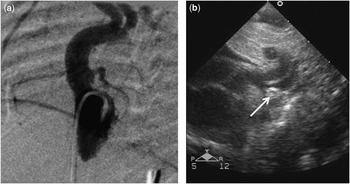
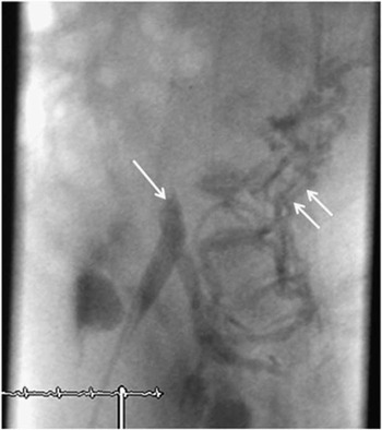
Similar to Case 1, the long-distance interruption of the aortic arch, visible in the echocardiogram, was indicative of a thrombotic occlusion. Thus, angiography was performed in order to clarify the exact anatomy. Angiography revealed a long-distance interruption of the aortic arch (see Fig 2). In addition, occlusion of the inferior cava vein was found (see Fig 3). Owing to the intracranial infarction and haemorrhage, interventional treatment of aortic coarctation with recanalisation and stent implantation was judged to be too risky, because of the necessity of post-interventional high-dose anticoagulation and consecutive high risk of further intracranial bleeding. For the same reason, operation of the aortic arch with the necessity of cardiopulmonary bypass was omitted. Further coagulation studies showed a heterozygous state of factor V Leiden mutation and positive antiphospholipid antibodies (with a pathological plasma mixed test). The test for prothrombin mutation was negative, and proteins C and S, as well antithrombin III, were normal for age. Owing to the poor neurologic prognosis and the absence of therapeutic options, the patient received palliative care. The boy died at the age of 6 days after the prostaglandin E1 infusion was stopped. The post-mortem examination confirmed the thrombotic occlusion of the aortic arch.
Angiography and corresponding echocardiography of patient 2. The angiography ( a ) and the echocardiography ( b ) demonstrate the interrupted aortic arch. Angiography reveals many collateral vessels, indicating a long-standing occlusion. On echocardiography, the thrombus can be seen below the outlet of the left carotid artery (marked with an arrow).

Picture shows the spontaneous connatal thrombotic closure of the inferior caval vein (one arrow) close to the confluence of the iliacal veins, already compensated by collateral vessels (two arrows).

Results
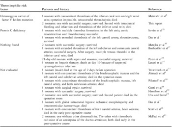
Literature search retrieved 51 articles from March, 1953 to February, 2010. In these reports, we were able to identify 19 patients with neonatal aortic arch thrombosis (see Table 1). Of these 19 patients, 11 (58%) died. The mortality rate of patients with multiple thromboses was 78% (7/9) compared with 20% (2/10) for patients with isolated aortic arch thrombosis. In 9 of the 19 (47%) patients, the authors reported the results of the potential underlying thrombophilic risk factors. In three of these nine patients (33%), a thrombophilic risk factor could be demonstrated. There were two patients with protein C deficiency and one patient with heterozygous state of factor V Leiden mutation and antiphospholipid antibodies. Statistical analysis of the patients, including the two patients from our centre, was performed. The mortality rate of patients with multiple thromboses was 80% (8/10) versus 18% (2/11) in the group with isolated aortic arch thrombosis. This difference was statistically significant on using the two-sided Fisher exact test (p = 0.009). The combined post-operative and intra-operative mortality rate was 33% (5/15). All patients (100%, 6/6) who did not receive an operation, including the two patients treated with high-dose heparin – one with low-molecular-weight heparin – died.
Reported patients with aortic arch thrombosis in the literature.

Discussion
A spontaneous aortic arch thrombosis mimicking aortic coarctation is a very rare but potentially life-threatening event with a high rate of mortality, especially if other thrombotic complications occur.Reference Ahmadi, Fürste and Pringsheim 6 – Reference Uva, Serraf and Lacour-Gayet 12 Only in 8 of the 16 (50%) patients, the authors reported results of underlying thrombophilic risk factors, but only in three of these eight patients (38%) a thrombophilic risk factor could be demonstrated. The case series reported by Nagel et al listed a total number of 148 patients with neonatal aortic thromboses. In all, 78% of the thrombotic events were related to a history of arterial umbilical catheterisation and only 33 cases occurred spontaneously.Reference Nagel, Tuckuviene, Paes and Chan 9 A typical localisation for a thrombotic occlusion caused by an arterial umbilical catheter was the descending aorta. According to this case series and other reports, the thrombotic occlusion of the descending aorta seems to be much more common than the thrombotic occlusion of the aortic arch.Reference Nagel, Tuckuviene, Paes and Chan 9 , Reference Kawahira, Kishimoto and Lio 19 – Reference Sherman, Münster, Govendrageloo, Harrisberg and Levin 24
The two reported patients from our centre were carriers of a heterozygous factor V Leiden mutation. One had an additional thrombophilic risk factor: he was positively tested for antiphospholipid antibodies. In only a small proportion of patients with aortic arch thrombosis reported in the literature, thrombophilic risk factors were detected (Table 1): two were reported to be suffering from protein C deficiency. Only one more patient is reported to be suffering from heterozygous factor V Leiden mutation.Reference Das, Sahoo and Ivy 7 , Reference Metsvaht, Hermlin, Kern, Kahre and Starkopf 8 , Reference István, Krisztina, Eva, Valéria, Judit and Károly 25 In many patients, no risk factors could be identified, and the cause for thrombosis remained unclear.Reference Matejka, Hucin, Marek, Chaloupecky and Skovranek 26 – Reference Pozzi, Taylor and de Leval 28 The role of thrombophilic risk factors in the development of aortic arch thrombosis is therefore still not clear.
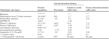
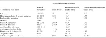
Aside from the Methylenetetrahydrofolate Reductase 677 TT genetic polymorphism, which is present in 10% of the Caucasian population, factor V Leiden mutation with 8% is the most common thrombophilic risk factor in the normal population (Table 2).Reference Münchow, Kosch, Schobess, Junker, Auberger and Nowak-Göttl 14 , Reference Kenet, Lütkhoff and Nowak-Göttl 29 – Reference Ehrenforth, Junker and Koch 31 Both mutations are associated with a relatively low risk for arterial or venous thromboembolic events. The risk for venous thromboses increases if two or more genetic thrombophilic risk factors coexist (odds ratio 9.5) – antithrombin deficiency (odds ratio 9.4), protein C deficiency (odds ratio 7.7), or protein S deficiency (odds ratio 5.8; see Table 2).Reference Nowak-Göttl, Kurnik, Manner and Kenet 16 , Reference Young, Albisetti and Nowak-Göttl 17
Incidence of thrombotic risk factors in the normal population and odds ratio for arterial or venous thrombosis in children.
MTHFR = methylenetetrahydrofolate reductase
*acquired
Arterial thromboses with 12–34% are far less common than venous thromboses.Reference Chalmers 13 , Reference Tuckuviene, Christensen, Helgestad, Johnsen and Kristensen 18 Most of the data regarding arterial thromboses are related to arterial ischaemic strokes. Comparing these events with venous thromboembolism, risk factors differ significantly. The highest risk for arterial ischaemic stroke is described for patients suffering from protein C deficiency (odds ratio 11), followed by antiphospholipid antibodies (odds ratio 6.53) and high lipoprotein A levels (>30 mg/dl; odds ratio 6.53). Similar to patients with venous thromboembolism, patients with two or more genetic traits (odds ratio 18.75) have the highest risk for arterial thromboembolism. In contrast, a single heterozygous factor V Leiden mutation showed a relatively low odds ratio of 3.7.Reference Kenet, Lütkhoff and Nowak-Göttl 29 Balci et al reported 50 children with non-stroke arterial thrombosis – 10% of all thrombosis in children in the study. In all, 50% of these patients had a history of arterial catheterisation. Other predisposing factors were cardiac disease and infections. They found all known thrombotic risk factors to be elevated in comparison with the normal population (see Table 2). In all, 50% of patients presented two or more of these thrombophilic risk factors.Reference Balci, Unal, Gumruk, Cetin, Ozkutlu and Gurgey 32
These findings fit the reported thrombophilic risk factors described in the few cases with aortic arch thrombosis. Of the five reported patients, two suffered from protein C deficiency.Reference Das, Sahoo and Ivy 7 , Reference István, Krisztina, Eva, Valéria, Judit and Károly 25 Only one patient is reported to have factor V Leiden mutation.Reference Metsvaht, Hermlin, Kern, Kahre and Starkopf 8 A comparison of our two patients showed that in the patient with additional positive findings for antiphospholipid antibodies thrombosis of aortic arch was complicated by stroke and thrombotic occlusion of the inferior caval vein. In the literature, patients with initial multiple thromboses had a poor prognosis.Reference Ahmadi, Fürste and Pringsheim 6 , Reference Metsvaht, Hermlin, Kern, Kahre and Starkopf 8 , Reference Pilossoff, Schöber, Müller, Schumacher, Sebening and Babic 10 , Reference Bonhoeffer, Bonnet, Sidi and Kachaner 27 Sheridan-Pereira et alReference Sheridan-Pereira, Porreco, Hays and Burke 33 and Tuohy and HarrisonReference Tuohy and Harrison 34 reported cases of neonatal aortic thromboses associated with lupus anticoagulant. Bhat et alReference Bhat, Kawoosa, Bhat and Ali 35 showed a neonate femoral artery thrombosis mediated by antiphospholipid antibodies and sepsis. Boffa and Lachassinne described 16 cases of perinatal thromboses, of which 13 were arterial, but not of the aortic arch, with positive test for antiphospholipid antibodies. Most patients exhibited a second risk factor, either prenatal (pre-eclampsia and/or intrauterine growth retardation) or perinatal (asphyxia, sepsis, arterial or venous catheter, and congenital thrombophilia).Reference Boffa and Lachassinne 36 Some of these criteria were also present in our second patient: he was born to a mother with pre-eclampsia and was found to be positive for antiphospholipid antibodies in addition to the heterozygous factor V Leiden mutation. In summary, antiphospholipid antibodies seem to be associated with an increased risk for both arterial and venous thrombosis.
Summarising all reported patients in the literature and the two cases we reported on, the overall mortality rate of aortic arch thrombosis seems to be about 25-fold higher (11/21, 52%) compared with the reported 2% mortality rate of the typical coarctation.Reference Kaushal, Patel and Patel 4 In particular, the patients with aortic arch thrombosis complicated by other thrombotic events had a poor outcome. The mortality rate of patients with multiple thromboses, including the two reported cases, was 80% (8/10) compared with 18% (2/11) for patients with isolated aortic arch thrombosis. The combined intra- and post-operative mortality rate was 33% (5/15), reflecting the critical status of the patients before and after the operation. All patients who did not undergo operation died (6/6), including two patients treated with high-dose anticoagulation.
With regard to the potential underlying thrombophilic risk factors, the analysis of the literature revealed protein C deficiency and factor V Leiden mutation to be the most common risk factors for aortic arch thrombosis.
There are some limitations in this study. All data are based on case reports and each case differs in clinical history, diagnosis, and treatment. The high mortality of neonatal aortic arch thrombosis could be caused by a possible trend to publish preferable patients with a complex clinical course and fatal outcome. Therefore, the incidence of associated comorbidities and mortality may be overestimated. Moreover, not all authors have reported on negative results of the potential thrombophilic risk factors. For that reason, the conclusions regarding the underlying thrombophilic risk factors have to be interpreted with care.
Conclusion
A spontaneous aortic arch thrombosis mimicking aortic coarctation is a very rare but life-threatening event. The analysis of all identified patients found in the literature demonstrated that compared with typical coarctation thrombotic occlusion had a higher mortality rate, especially if patients suffered from associated thrombotic complications. Different congenital thrombophilic risk factors such as factor V Leiden mutation or protein C deficiency seem to play an important role in the development of this fatal disease.