
Introduction
Diatoms[Reference Round, Crawford and Mann1–Reference Bowler, De Marino and Falciatore3] are unicellular, eukaryotic microalgae. With about 250 genera and an estimated number of about 100,000 species or more, diatoms occur in nearly all aquatic habitats including sea and fresh water. Most diatoms are photoautotrophic and contribute significantly to the global carbon fixation. Their organic constituents like carbohydrates, fatty acids, lipids, and vitamins make them an important primary food source for higher organisms. After cell death and decomposition, diatom biosilica sediments at the sea floor thus forming huge amounts of so-called diatomaceous earth over geologic periods. Diatomaceous earth (diatomite) has long been used by the industry as an inexpensive raw material. It serves as an abrasive and filter material, sorbent, anti-caking agent, and insulation material. Furthermore, diatoms biosynthesize and accumulate lipids in the form of triacylglycerols. The resulting buoyancy prevents the cell from sinking to the ground. Diatoms are interesting as a potential renewable source for biofuel production because triacylglycerols are important lipid storage compounds.[Reference Stonik and Stonik4, Reference Raven, Evert and Eichhorn5] In addition, two other constituents of algal biomass are of special interest for industrial applications: carbohydrates could be used for ethanol production by fermentation and proteins for methane production via anaerobic gasification.[Reference Bozarth, Maier and Zauner6]
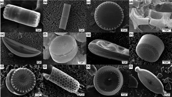
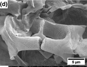
Diatoms are famous for their beautiful, micro- and nanopatterned silica-based cell walls (see Fig. 1). Cell wall morphogenesis takes place under genetic control. Diatom species are thus distinguishable by their specific cell wall structures. The biochemical and biophysical processes underlying cell wall formation are not fully understood and remain a subject of ongoing research.
SEM images of different diatom species. (a) Cyclotella sp., (b) Aulacoseira granulate, (c) Stephanodiscus niagarae, (d) Eucampia zodiacus, (e) Amphora perpusilla, (f) Thalassiosira pseudonana, (g) Cymatopleura solea, (h) Melosira varians, (i) Stephanodiscus minutulus, (j) Stephanopyxis turris, (k) Lindavia bodanica, (l) Caloneis sp. Reproduced from Ref. Reference Fischer7 (Fischer, 2017).

Diatom biosilica is recently considered for various technologic/nanotechnologic applications.[Reference Parkinson and Gordon8–Reference Ragni, Cicco, Vona and Farinola15] The “green” synthesis of highly structured micro- and nanopatterned silica materials by biologic self-assembly together with low expected costs for culturing make diatom biosilica an attractive raw material for industrial implementation. This material can be functionalized and tailored for special applications following two different approaches, namely in vitro and in vivo. Numerous in vitro modifications became meanwhile feasible, e.g., the conformal conversion of diatom biosilica preserving the characteristic shape and patterning into other materials, e.g., metals, metal oxides, carbons, or polymers.[Reference Fang, Berrigan, Cai, Marder and Sandhage16–Reference Fischer, Oschatz, Nickel, Leistenschneider, Kaskel and Brunner19] Other examples are nanoparticle (NP) decoration, e.g., for uses in catalysis[Reference Jantschke, Herrmann, Lesnyak, Eychmueller and Brunner20–Reference Fischer, Adam, Mueller, Sperling, Wustmann, van Pée, Kaskel and Brunner22] and staining to enhance/create special optical properties[Reference Fuhrmann, Landwehr, El Rharbi-Kucki and Sumper23, Reference Kucki and Fuhrmann-Lieker24]; just to name a few of them.
In addition to these various in vitro modifications, increasing efforts are made to functionalize diatom biosilica in vivo. In general, this is possible following two different ways: (i) by genetic modification and (ii) variation of the culture conditions like compositional changes of the growth medium. Genetic modifications are currently developed. An example is the in vivo incorporation of special proteins like enzymes into biosilica by the so-called Live Diatom Silica Immobilization (LiDSI) method.[Reference Poulsen, Berne, Spain and Kröger25, Reference Sheppard, Scheffel, Poulsen and Kröger26] Compositional changes of the growth medium can enhance the uptake and cell wall attachment of “foreign” elements or NPs, often metals, thus transforming biosilica into mixed materials or nanocomposites. The latter will be in the focus of the present review paper answering the following questions: (i) Which metals can be taken up by diatoms and are eventually processed into NPs? (ii) Are significant amounts of these metals incorporated into the biosilica thus resulting in mixed materials/nanocomposites? (iii) How can the obtained materials be useful for materials science?
In vivo modification of diatom biosilica by metal incorporation
Diatoms take up monosilicic acid from their environment via special transmembrane proteins, so-called silicon transport proteins.[Reference Hildebrand, Volcani, Gassmann and Schroeder27, Reference Hildebrand, Alverson and Thamatrakoln28] Chemically similar elements are taken up from the environment and inserted into biosilica as well. This is, e.g., true for germanium[Reference Azam, Hemmingsen and Volcani29, Reference Davis and Hildebrand30] in the form of Ge(OH)4 and was already exploited to synthesize GeO2-containing biosilica-based materials.[Reference Davis and Hildebrand30–Reference Ali, Divya, Gunasekaran and Thajuddin33] After silicon starvation and subsequent feeding with Si(OH)4 and Ge(OH)4, germanium is incorporated and can influence the cell wall structure depending on the species and Ge concentration. In analogy to the experiments with germanium, titanate was also admixed to the growth medium.[Reference Jeffryes, Gutu, Jiao and Rorrer34] After H2O2 treatment––which removes organic cell constituents––the samples still contained significant amounts of Ge or Ti as shown by inductively coupled plasma (ICP) analyses and scanning electron microscopy–energy dispersive spectroscopy (SEM–EDS) line measurements. It was concluded that GeO2 and TiO2 are incorporated into the biosilica. The authors suggest applications as dye-sensitized solar cells for enhanced light trapping and structured photocatalysts for such materials.[Reference Jeffryes, Gutu, Jiao and Rorrer31, Reference Jeffryes, Gutu, Jiao and Rorrer34] Further experiments on diatom biosilica with metabolically inserted Ge revealed electroluminescence and photoluminescence in the visible spectral range.[Reference Jeffryes, Solanki, Rangineni, Wang, Chang and Rorrer32]
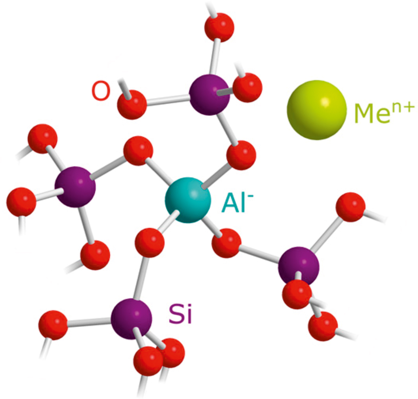
Other foreign elements such as Al,[Reference van Bennekom, Buma and Nolting35–Reference Köhler, Machill, Werner, Selzer, Kaskel and Brunner38] Ca,[Reference Godinho, Cabrita, Alves and Pinheiro39] Zn,[Reference Ellwood and Hunter40, Reference Ellwood and Hunter41] and Fe[Reference Ellwood and Hunter40, Reference Kaden, Brückner, Machill, Krafft, Pöppl and Brunner42] can also be incorporated into diatom biosilica. The amount of Al accumulated in biosilica is strongly enhanced by increasing the Al concentration in the growth medium.[Reference Machill, Köhler, Ueberlein, Hedrich, Kunaschk, Paasch, Schulze and Brunner37, Reference Köhler, Machill, Werner, Selzer, Kaskel and Brunner38] So far, the highest amount of silica-incorporated Al is observed in Stephanopyxis turris. It corresponds to a molar Si:Al ratio of 15 : 1 in the cell wall and was obtained by offering an initial Si:Al ratio of 1 : 1 in the growth medium.[Reference Machill, Köhler, Ueberlein, Hedrich, Kunaschk, Paasch, Schulze and Brunner37] Gehlen et al.[Reference Gehlen, Beck, Calas, Flank, van Bennekom and van Beusekom36] proposed Al incorporation into the silica network thus replacing Si atoms. The resulting negatively charged SiO−Al sites must then be compensated by positively charged ions such as Ca2+ or Na+ (see Fig. 2). Infrared (IR) spectroscopy indeed confirmed Al incorporation into the silica network.[Reference Machill, Köhler, Ueberlein, Hedrich, Kunaschk, Paasch, Schulze and Brunner37] Moreover, charge compensating cations are exchangeable by ammonium ions as demonstrated recently.[Reference Köhler, Machill, Werner, Selzer, Kaskel and Brunner38] Subsequent ammonia removal even results in the formation of catalytically active Brønsted acid sites. Aluminum-enriched diatom biosilica may therefore be a “green” raw material for future catalyst production.[Reference Köhler, Machill, Werner, Selzer, Kaskel and Brunner38]
Structure of aluminum in the silica framework with Men+ as counter ion. Reproduced from Ref. Reference Köhler, Machill, Werner, Selzer, Kaskel and Brunner38 (MDPI, 2017) (https://creativecommons.org/licenses/by/4.0/).

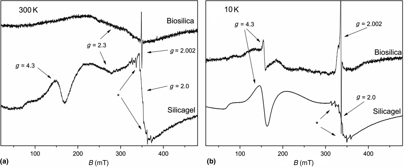
Zinc and iron are also incorporated into diatom biosilica[Reference Ellwood and Hunter40–Reference Kaden, Brückner, Machill, Krafft, Pöppl and Brunner42], but only at relatively low concentrations. The amount of biosilica-incorporated Zn nevertheless correlates with the amount of Zn2+ offered in the growth medium.[Reference Ellwood and Hunter40] In contrast, the amount of strongly biosilica-associated iron does not correlate with the Fe3+ concentration in the growth medium.[Reference Ellwood and Hunter40, Reference Kaden, Brückner, Machill, Krafft, Pöppl and Brunner42] For the diatom species S. turris, almost constant molar Si:Fe ratios of about 500 : 1 were usually observed independent of the iron concentration in the growth medium.[Reference Kaden, Brückner, Machill, Krafft, Pöppl and Brunner42] Even lower Fe concentrations were reported for Thalassiosira pseudonana biosilica.[Reference Ellwood and Hunter40] It is noteworthy that biosilica-attached iron almost completely occurs as Fe2O3 clusters or NPs as could be shown by electron paramagnetic resonance (EPR) spectroscopy in combination with 29Si magic angle spinning nuclear magnetic resonance (MAS NMR) spectroscopy (see Fig. 3). A minor fraction, i.e., <5% of the biosilica-attached Fe is dispersed[Reference Kaden, Brückner, Machill, Krafft, Pöppl and Brunner42] in contrast to the observations made for aluminum (see above).
Comparison of the EPR spectra of biosilica and iron-containing silicagel (pH 7) measured at (a) 300 K and (b) 10 K. Note the presence of a broad signal at g = 2.3 in biosilica which disappears after cooling to 10 K. This signal is due to Fe2O3 clusters. In contrast, Fe is mainly dispersed in silicagel and gives rise to signals at 4.3 and 2.0 which are also present at 10 K. Reproduced from Ref. Reference Kaden, Brückner, Machill, Krafft, Pöppl and Brunner42 (Springer Nature, 2017). * denotes a weak Mn2+ signal due to a minor impurity in the silicagel.

In vivo NP biosynthesis by diatoms
NPs exhibit physical, chemical, and biologic properties that are different from the bulk material.[Reference Peng, Manna, Yang, Wickham, Scher, Kadavanich and Alivisatos43–Reference Kagan, Lifshitz, Sargent and Talapin46] They are considered to play an essential role in the development of future electronics, optoelectronics, and sensors and have already found application in biomedicine, e.g., for drug delivery, diagnosis, and targeted chemotherapy. However, industrial syntheses of NPs often require organic solvents as well as harsh reducing and stabilizing agents. Often, the synthesis reactions are carried out at high temperatures and pressures. Increasing efforts are, therefore, made in order to develop “green,” environment-friendly synthesis routes.[Reference Narayanan and Sakthivel47–Reference Korbekandi, Iravani and Abbasi57] One possibility is the use of living organisms or molecules extracted from organisms for NP synthesis. Organisms such as plants, fungi, bacteria, and algae offer ecologically friendly alternatives to current NP syntheses.[Reference Beattie and Haverkamp49–Reference Patel, Berthold, Puranik and Gantar56] So far, most in vivo experiments describe gold and silver NP syntheses. NPs consisting of Cu, Fe3O4, ZnO, CdS, SiO2, and ZnS could also be produced.[Reference Korbekandi, Iravani and Abbasi57] Compared with other organisms, the exploration of diatoms for NP synthesis is still at its beginning––but gains increasing interest.[Reference Korbekandi, Iravani and Abbasi57]
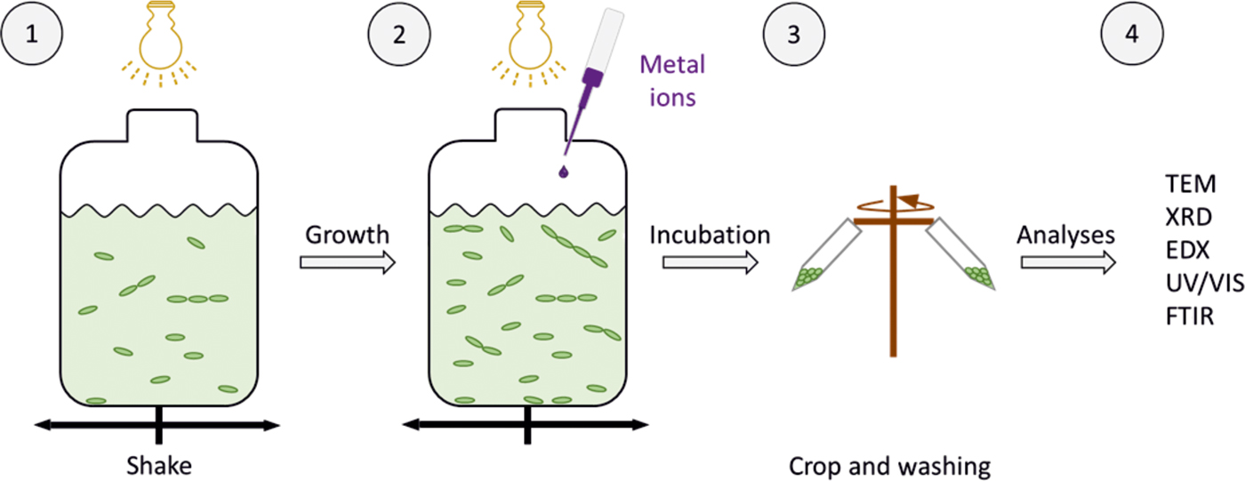
The commonly used scheme for in vivo NP biosynthesis by diatoms is demonstrated in Fig. 4. After cell growth in “normal” ASW, a metal salt solution is added to the culture. Cultures are harvested, washed, and prepared for further analyses after certain incubation times.
Schematic procedure for NP biosynthesis by diatoms.

Chakraborty et al.[Reference Chakraborty, Pal, Ramaswami, Nayak and Lahiri58] reported gold accumulation by diatoms from the growth medium without analyzing size, form, and composition of the reduced gold particles. Schröfel et al.[Reference Schröfel, Kratošová, Krautová, Dobročka and Vávra59] added tetrachloroaurate solution to diatom cultures and investigated the produced Au NPs by UV-Vis spectroscopy, x-ray diffraction, and different types of microscopy. Comparison of two different species revealed a species-dependent form and size distribution of the biosynthesized Au NPs. Microscopic and spectroscopic results indicated that these Au NPs occur either in the extracellular polysaccharides or close to the siliceous cell walls. Extracellular polysaccharides exhibit metal-binding sites where NPs in the medium can easily bind.[Reference Schröfel, Kratošová, Krautová, Dobročka and Vávra59]
Similar Au–SiO2 nanocomposites could be produced and separated from the cells by sonication in sodium citrate solution and subsequent centrifugation. The NPs showed a broad range of sizes between 5 and 45 nm with different shapes in transmission electron microscopy (TEM) images and were able to bind algal DNA. These diatom-based Au–SiO2 nanocomposites are proposed for biomedical applications such as photothermal therapy and multimodal imaging.[Reference Roychoudhury, Nandi and Pal60]
Another possible biomedical application is suggested for silver NPs synthesized from the aqueous extract of the diatom Amphora-46. These NPs possess antimicrobial activity against Gram-negative and Gram-positive bacteria. Diatom extracts only mediate NP synthesis in the presence of light.[Reference Jena, Pradhan, Dash, Panda and Mishra61] This observation indicates the involvement of at least one light-sensitive biomolecule in this process. After various extractions and separations, UV–Vis analyses finally revealed fucoxanthin as the responsible molecule for ion reduction to Ag NPs.[Reference Jena, Pradhan, Dash, Panda and Mishra61]
Gold NP synthesis by Nitzschia sp. diatoms was reported by Borase et al.[Reference Borase, Patil, Suryawanshi, Koli, Mohite, Benelli and Patil62] These NPs preferentially occur in close proximity to diatom cells thus indicating an interaction between NPs and the organism. Separation of biosynthesized Au NPs and subsequent Fourier transform IR spectroscopy indicate an important role of proteins and polysaccharides for the metal reduction and NP stabilization. Furthermore, Au NPs were successfully coupled with the antibiotics penicillin and streptomycin and showed an increased antibacterial activity.[Reference Poulsen, Berne, Spain and Kröger25]
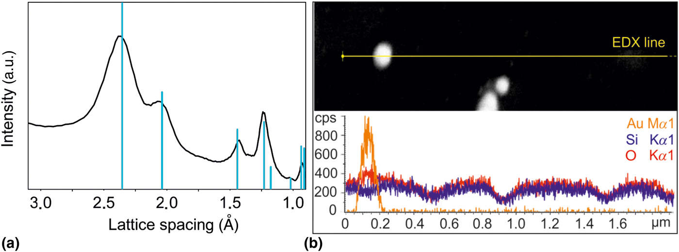
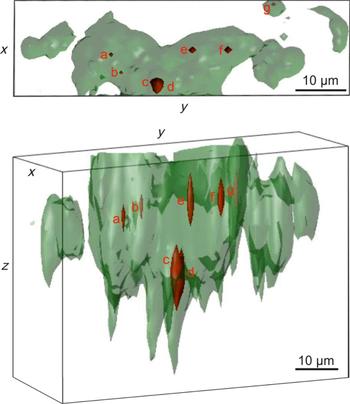
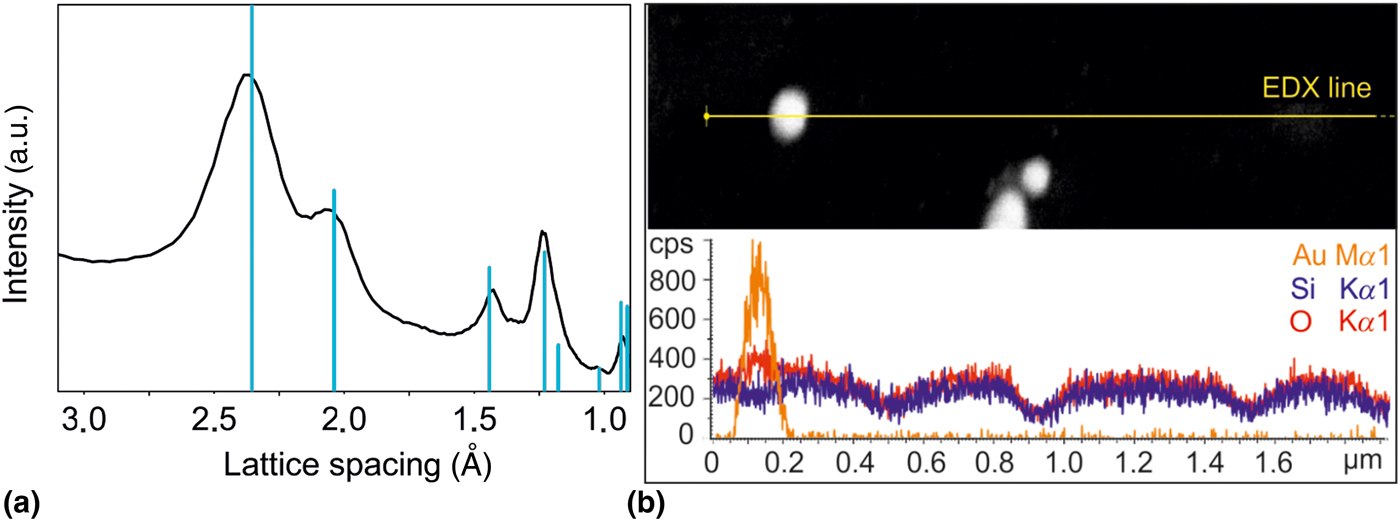
It is, however, difficult to answer the question where such biosynthesized NPs are located. Only few methods are capable of verifying the presence of NPs inside organisms. Methods such as thin cut preparation influence the state of the cell and are thus not decisive because the samples do not necessarily reflect the native state and the location of the NPs may change during the preparation procedure. In order to circumvent this problem, three-dimensionally resolved surface-enhanced Raman spectroscopy (SERS) can be used to localize biosynthesized Au NPs in diatom cells in vivo. This technique was established previously by Lahr and Vikesland to study the green alga Pseudokirchneriella subcapitata [Reference Lahr and Vikesland63] and could meanwhile also be applied to diatoms. Figure 5 shows the results of energy-dispersive x-ray spectroscopy (EDX) and selected area electron diffraction (SAED) measurements confirming the presence of Au NPs in an S. turris culture incubated with tetrachloroaurate as described above.[Reference Pytlik, Kaden, Finger, Naumann, Wanke, Machill and Brunner64] The SERS effect is a local phenomenon and occurs exclusively in the neighborhood of metal NPs. Based on this fact, spatially resolved measurements of SERS enhancements provide information about NP localization. False color images encoding the measured SERS effect proved that some of the biosynthesized NPs are indeed located inside the intact diatom cells (see Fig. 6).[Reference Pytlik, Kaden, Finger, Naumann, Wanke, Machill and Brunner64]
(a) SAED result of biosynthesized Au NPs (black) in comparison with the reflexes from Au crystals with the face-centered cubic space group Fm-3m (blue) and (b) EDX line measurement of biosynthesized Au NPs. Adapted from Ref. Reference Pytlik, Kaden, Finger, Naumann, Wanke, Machill and Brunner64 (Elsevier, 2017).
Three-dimensional image of an Stephanopyxis turris cell (22 µm × 67 µm × 45 µm) with schematic chloroplasts (green) and SERS spectra (red) from different perspectives. Adapted from Ref. Reference Pytlik, Kaden, Finger, Naumann, Wanke, Machill and Brunner64 (Elsevier, 2017).

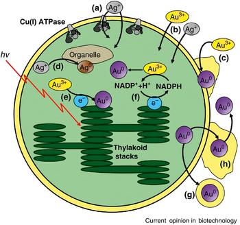
Jeffryes et al.[Reference Jeffryes, Agathos and Rorrer14] describe different possibilities for Ag and Au NP biosynthesis by photosynthetic microorganisms (see Fig. 7). As diatoms are photosynthetic microorganisms, they could follow one of the proposed pathways. So far, however, none of the suggested mechanisms was experimentally validated. For intracellular NP biosynthesis, metal ions must enter the cell before NP formation. They could either be taken up via the Cu(I)-ATPase pathway [Fig. 7(a)] or simply enter by diffusion [Fig. 7(b)]. Afterwards, metal ions can be reduced by enzymes in the cytoplasm [Fig. 7(d)], electrons of the photosynthetic electron transport chain [Fig. 7(e)], or by photosynthetically produced redox mediators [Fig. 7(f)]. Such intracellularly biosynthesized NPs may then bind to organelle membrane surfaces [Fig. 7(d)] or leave the cell [Fig. 7(g)] and subsequently the surrounding extracellular matrix [Fig. 7(h)].[Reference Jeffryes, Agathos and Rorrer14] On the other hand, metal ions could also be reduced extracellularly before entering the cell [Fig. 7(c)], e.g., by extracellular polymeric substances (EPS) containing suitable reducing agents. This poses the fundamental question whether or not such presynthesized extracellular NPs can subsequently be taken up by diatom cells, e.g., via endocytosis.
Possible ways for transport and synthesis of Ag and Au NPs by photosynthetic microorganisms. Uptake of Ag+ into the cell by (a) Cu(I)-ATPase or (b) diffusion. (c) Extracellular gold reduction by extracellular polymeric substances. (d) Binding of intracellular Au NPs to organelle membrane surfaces. Intracellular gold reduction by photosynthetically produced (e) electrons or (f) redox mediators. (g) Export of Au NPs and (h) release of the Au NPs from the extracellular matrix. Reproduced from Ref. Reference Jeffryes, Agathos and Rorrer14 (Elsevier, 2015).

Mechanisms for NP uptake
To date, the knowledge about possible NP transport mechanisms from the environment into the diatom cells is very limited, although various references indicate the intracellular presence of NPs. The main problem is the experimental difficulty to unequivocally determine the intracellular localization of NPs in intact cells (see above). Suggestions for possible NP uptake mechanisms in diatoms usually rely on the investigations of other organisms. In general, NP uptake by cells mainly involves two steps: (i) penetration through the cell wall; (ii) intracellular transport and processing. This chapter summarizes basic ideas for the uptake mechanisms derived from the investigations of various cell types. These ideas could possibly be relevant for diatoms as well.
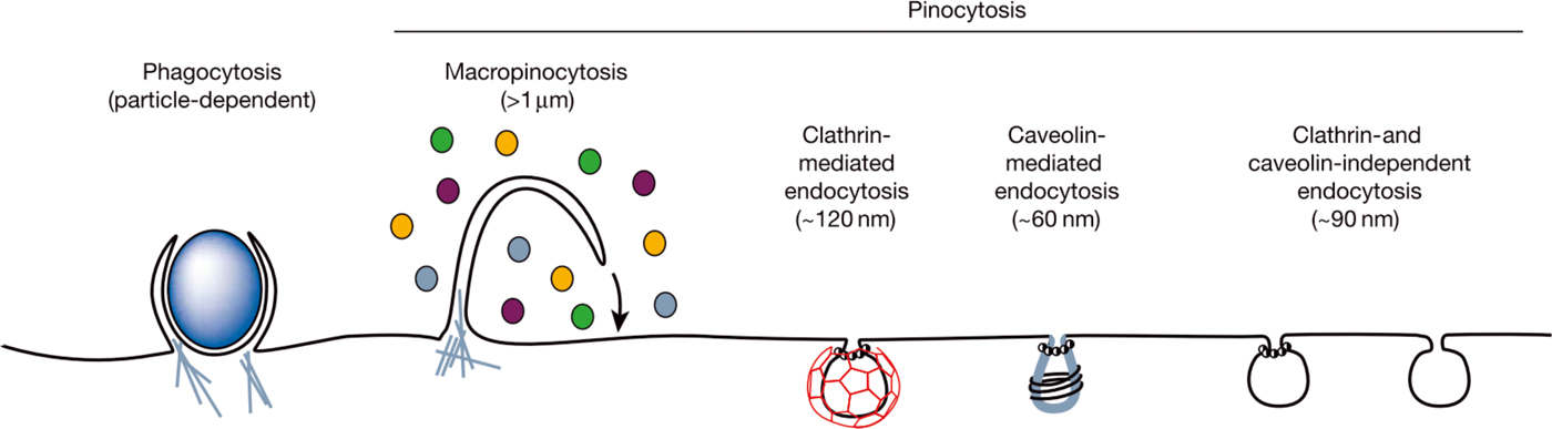
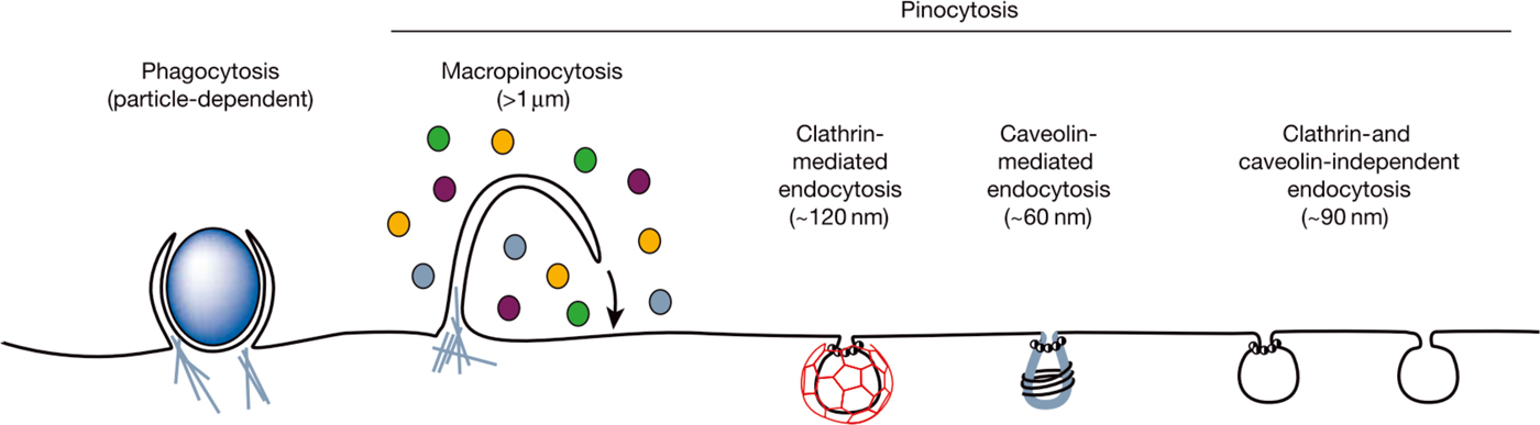
Conner and Schmid describe the plasma membrane as a dynamic structure separating the intracellular cytoplasm from the extracellular environment. It controls the uptake and disposal of various molecules of different sizes. Ions as well as small essential molecules like amino acids can cross this plasma membrane via membrane proteins acting as pumps or channels. Larger molecules, however, need vesicles to be inserted into the cell interior by endocytosis. A schematic overview over the fundamental endocytosis processes is presented in Fig. 8.[Reference Conner and Schmid65]
Different endocytic pathways for cellular uptake depending on the size, the nature of the cargo (ligands, receptors, and lipids), and the mechanism of vesicle formation. Reproduced from Ref. Reference Conner and Schmid65 (Springer Nature, 2003).
After endocytosis, the internalized compounds/NPs are delivered to early endosomes providing a network of tubules and vacuoles near the plasma membrane. Endosomes mature and fuse finally with hydrolase vesicles thus forming the endolysosome.[Reference Pollard, Earnshaw, Lippincott-Schwartz and Johnson66, Reference Lowe67]
The interactions between NPs and cells depend on the charges of the NPs and/or their stabilizing groups/ligands.[Reference Verma and Stellacci68–Reference Harush-Frenkel, Debotton, Benita and Altschuler70] Neutral NPs hardly interact with cells. Negatively charged NPs can be taken up by cells at least to a limited extent. Cationic NPs bind to the cell surface and are internalized much more efficiently than negatively charged or neutral NPs. The uptake of cationic NPs mainly follows the so-called clathrin-mediated pathway.[Reference Verma and Stellacci68–Reference Harush-Frenkel, Debotton, Benita and Altschuler70] However, the so-called caveolae-mediated pathway can also be chosen sometimes.[Reference Harush-Frenkel, Debotton, Benita and Altschuler70] NPs taken up via endocytosis remain mostly in endolysosomes. Localization in these membrane-bound compartments prevents the nanomaterial from entering the cytosol. Cationic NPs can also bind to negative groups on the cell surface and enter the cell by direct cell membrane penetration leading to the formation of holes in the lipid membrane bilayer. Consequently, the balance between intracellular and extracellular compounds can be disturbed leading to a visible toxic effect of the NPs on the cell.[Reference Verma and Stellacci68, Reference Huefner, Kuan, Barker and Mahajan71] However, it was also reported that NPs are able to enter the cell by penetration without visible hole formation. The proposed explanation for this observation includes some amphiphilic domains of NPs, which permit NP fusion with the cell membrane without disruption. These NPs are able to penetrate through the bilayer similar to some peptide internalization processes.[Reference Verma, Uzun, Hu, Hu, Han, Watson, Chen, Irvine and Stellacci72] While most NPs stay close to the cell wall in endolysosomes, several functional groups of NPs were shown to leave the endolysosome and enter the cell cytosol. Brust and co-workers demonstrated that NPs stabilized with cell-penetrating peptides entered the cytosol of human fibroblast cells. It was hypothesized that these NPs reach the cytosol by endosomal escape or by passing through the cell membrane.[Reference Nativo, Prior and Brust73]
Apart from the NP charge, many other factors are known to influence the rate and mechanism of NP uptake, depending on the specific experimental conditions. Not only the cell type, but also the molecular arrangement of surface chemical groups as well as shape and size of the NPs lead to different interactions.[Reference Verma, Uzun, Hu, Hu, Han, Watson, Chen, Irvine and Stellacci72, Reference Sahay, Alakhova and Kabanov74] As already described above, cells can also synthesize NPs from precursors in their environment. The uptake mechanism for such biosynthesized NPs is even more difficult to reveal as the stabilizing agents are not known in advance.
NP uptake by diatom cells
So far, different explanations for the toxicity of NPs were suggested (see above). Toxicity probably depends on the intracellular localization of NPs. The latter is, unfortunately, difficult to determine in native cells (see above). Branco et al. have washed a cell pellet with HNO3 to remove loosely bound Cd from the frustule. After sonication and digestion of the cell pellet, remaining Cd was considered as intracellular. It is supposed that the metal––once inside the cell––is transported into the vacuole via chelation with phytochelatins.[Reference Branco, Lima, Almeida and Figueira75] Other approaches to detect intracellular NPs follow similar pathways. Cells are washed with acids or metal chelators to remove the loosely bound metal from the cell wall. Supernatant fractions are either separated in subfractions via centrifugation or analyzed as one batch for their metal content. The detected metal is assumed to arise from the cell interior.[Reference Bielmyer-Fraser, Jarvis, Lenihan and Miller76, Reference Miao and Wang77]
Another interesting approach to demonstrate cellular uptake of presynthesized NPs is described by Pletikapić et al. They detected pore-like lesions in the valve region of diatom cells and assigned them to NP penetration sites. The occurrence of such lesions was interpreted as an evidence for the ability of NPs to directly pass through the cell wall.[Reference Pletikapić, Žutić, Vrček and Svetličić78]
Feurtet-Mazel et al.[Reference Feurtet-Mazel, Mornet, Charron, Mesmer-Dudons, Maury-Brachet and Baudrimont79] suggest that the driving force inducing NP biosynthesis from precursors in the growth medium (see above) is a detoxification mechanism initiated by the cell for self-protection against metal salt solutions. TEM images obtained from ultrathin cuts provide first hints for the presence of biosynthesized Au NPs also inside diatom cells, although the presence of gold in the described intracellular NPs has not yet been confirmed by elemental analysis.[Reference Feurtet-Mazel, Mornet, Charron, Mesmer-Dudons, Maury-Brachet and Baudrimont79] The same method has been applied previously for detecting presynthesized ZnO NPs inside Phaeodactylum tricornutum cells. Only few ZnO NPs were located inside the cell, while most of them were directly attached to the cell walls. TEM cuts were prepared by fixation of the diatom cells in glutaraldehyde/phosphate buffer, following treatment with OsO4, dehydration, and final cutting into 80 nm slices using an ultramicrotome.[Reference Peng, Palma, Fisher and Wong80] Furthermore, TEM allowed detection of metallic Cd in ultrathin cuts of P. tricornutum after incubation in Cd2+-containing growth medium. Intracellular Cd gave rise to electrodense granulations, which were interpreted as metal depositions sequestered by specific molecules used for detoxification.[Reference Torres, Cid, Herrero and Abalde81]
Toxicity of presynthesized NPs
To obtain information about interactions of diatom cells with NPs, presynthesized NPs are usually brought into contact with living diatoms. Effects on cell viability and transformations of the NPs under the influence of diatom cultures are monitored. In general, key factors determining the toxicity of NPs are their chemical composition, size, surface, degree of dissolution, self-assembly, concentration, and aggregation behavior. This multiplicity of factors explains why it is so far hardly possible to make clear and reliable predictions for the effects of different NPs upon organisms. Smaller NPs exhibit a higher specific surface area increasing their reactivity. Consequently, interactions with biomolecules and accumulation are enhanced, both reducing the surface to volume ratio.[Reference Chang, Zhang, Xia, Zhang and Xing82] As agglomeration affects NP toxicity, the cultivation medium––especially its salt content––also represents an important factor.[Reference García-Negrete, Blasco, Volland, Rojas, Hampel, Lapresta-Fernández, De Haro, Soto and Fernández83] The growth medium can also contain various chelating substances, which can bind metal ions. In this way, the NPs could be dissolved in the growth medium.[Reference Burchardt, Carvalho, Valente, Nativo, Gilliland, Garc, Passarella, Pedroni and Lettieri84] The following chapters focus on the influence of size and chemical composition of the NPs upon their toxicity.
Toxicity can arise from the generation of reactive oxygen species and resulting oxidative stress. Furthermore, dissolved NP ions can occupy binding sites in proteins and affect the protein folding process resulting in changed protein functions.[Reference Chang, Zhang, Xia, Zhang and Xing82] Manier et al. suggest that NPs in close contact with the cells influence the transport of nutrients and metabolites across the cell membrane. Spatial vicinity could also result in physical damage like abrasive effects or indirect membrane damage caused by oxidative stress.[Reference Manier, Bado-Nilles, Delalain, Aguerre-Chariol and Pandard85]
The influence of the chemical composition of NPs upon toxicity
An important factor influencing toxicity is the chemical composition of the NPs. Usually, gold exhibits a relatively low toxicity for organisms. However, nanostructured Au is more toxic because the Au atoms are highly exposed and accessible. Nevertheless, Au NPs have a lower toxicity compared with Ag NPs in general.[Reference Moreno-Garrido, Pérez and Blasco86] Bielmyer-Fraser et al.[Reference Bielmyer-Fraser, Jarvis, Lenihan and Miller76] compare the influence of various metal oxide (ZnO, AgO, and CuO) NPs as well as of their metal ions upon the diatom Thalassiosira weissflogii. Toxicity levels are similar for dissolved metals and their nanoparticulate forms, especially for Ag and Cu. After exposure to metal oxide NPs, the highest metal concentration was found in the diatom cell wall. After exposure to dissolved metals, the highest concentration was found in the organelles and the endoplasmic reticulum. The authors explain this observation by different mechanisms or rates of metal uptake. Dissolved metals could be able to pass the cell wall quickly through ion channels into the cell. In contrast, only few NPs are able to enter the cell interior due to size exclusion.[Reference Bielmyer-Fraser, Jarvis, Lenihan and Miller76]
Branco et al. have analyzed the cell response of various biochemical markers to the addition of Cd in different concentrations to the diatom Nitzschia palea. The increasing amounts of phytochelatins indicate that the cell initiates metal chelation as a protection mechanism against intracellular Cd. After chelation, Cd may be deposited in the vacuole thus preventing it from interference with the cell metabolism.[Reference Branco, Lima, Almeida and Figueira75] CeO2 NPs are not toxic for this diatom species in contrast to Cd. It is thus concluded that the studied diatom species possesses effective protection mechanisms deactivating CeO2 NPs. In addition to the afore-mentioned chelation by biosynthesized phytochelatins, two other possible mechanisms are suggested: (i) the diatom cell wall itself can act as a shield against toxic NPs. (ii) The enhanced production of EPS may enable effective adsorption of NPs thus preventing direct contact with the diatom cell constituents.[Reference Bour, Mouchet, Verneuil, Evariste, Silvestre, Pinelli and Gauthier87]
Verneuil et al.[Reference Verneuil, Silvestre, Mouchet, Flahaut, Boutonnet, Bourdiol, Bortolamiol, Baqué, Gauthier and Pinelli88] demonstrated that N. palea is not only immune against CeO2 NPs, but also against multi-walled carbon nanotubes (MWCNTs). Cell growth was inhibited in early developmental stages, but recovered at the end of the experiment. The authors explain this observation as follows. In the early incubation phase, the cell relies on protection processes and avoids cell division until the NPs are “disarmed.” Additionally, MWCNTs agglomerate on the EPS. This supports the idea that EPS are part of the protection mechanisms against NPs. It is suggested that EPS secretion also enables the diatom to escape from NP-rich environments.[Reference Verneuil, Silvestre, Mouchet, Flahaut, Boutonnet, Bourdiol, Bortolamiol, Baqué, Gauthier and Pinelli88] Note that EPS seem to be involved in Ag+ detoxification either by ion accumulation or by changing the subcellular metal distribution.[Reference Miao, Schwehr, Xu, Zhang, Luo, Quigg and Santschi89]
The influence of NP size upon toxicity
Various studies report size-dependent toxic effects of presynthesized NPs upon diatoms. Burchardt et al. observed that larger Ag NPs caused lower growth inhibition, and thus, less toxicity than smaller NPs. This was explained by the small pore size of the diatom cell wall, whereby only smaller NPs are expected to enter the cell interior. These intracellular NPs are believed to affect the cells more than extracellular ones. Another explanation is the reduced bioavailability of larger NPs caused by sedimentation.[Reference Burchardt, Carvalho, Valente, Nativo, Gilliland, Garc, Passarella, Pedroni and Lettieri84] In general, NP size is proven to affect especially the rate and mechanism of cellular uptake. Lower toxicity of larger NPs for diatoms is confirmed by other authors as well.[Reference Peng, Palma, Fisher and Wong80, Reference Clément, Hurel and Marmier90] However, it was also proven that NPs partly dissolved in the growth medium. It is, therefore, arguable, whether or not Ag NPs themselves are toxic for diatoms, because the toxicity may also be due to released silver ions.[Reference Burchardt, Carvalho, Valente, Nativo, Gilliland, Garc, Passarella, Pedroni and Lettieri84, Reference Miao, Schwehr, Xu, Zhang, Luo, Quigg and Santschi89]
Peng et al. studied the influence of ZnO NPs upon different diatom species and demonstrated that 4–5% of the NPs dissolve after 72 h independent of their concentration and morphology. According to the authors, this phenomenon is due to Ostwald ripening. It is, therefore, suggested that mainly the smaller NPs dissolve and the remaining NPs are thus on average larger and more homogeneous.[Reference Peng, Palma, Fisher and Wong80] Size-dependent solubility of NPs is explained by the Gibbs–Thomson effect.[Reference Taylor91–Reference de M. Donegá93] This effect provides another explanation for the observation that bigger NPs show a lower toxicity. Fast dissolving smaller NPs cause a higher toxicity for diatom cells.
Taken together, it is not yet clear whether or not the toxicity of NPs arises exclusively from released ions.[Reference Moreno-Garrido, Pérez and Blasco86] Furthermore, free ions have a high affinity for algal surfaces and extracellular extrudates. It is, therefore, likely that these ions are not detected by the measurements of the amount of free ions in the medium.[Reference Moreno-Garrido, Pérez and Blasco86] It should also be taken into account that toxicity effects could be different for free and adsorbed ions.
Conclusion
Diatoms are interesting as a potential renewable source for various purposes, e.g., food and biofuel production. In addition, algal carbohydrates could be useful for ethanol production by fermentation and proteins for methane production via anaerobic gasification. Given this fact, it is also interesting to evaluate the potential technologic uses of the siliceous cell walls of diatoms (biosilica). Considering the various possibilities to create biosilica with special, tailored functions described in this review, in vivo modified diatom biosilica forms a promising source for “green” material synthesis. Germanium- and titanium-enriched biosilica might, e.g., be useful as dye-sensitized solar cells for enhanced light trapping and structured photocatalysts. Aluminum-enriched biosilica might become a “green” raw material for acid catalyst production. Moreover, diatoms could be useful for noble metal NP or nanocomposite synthesis. The “green” synthesis of such highly structured micro- and nanopatterned materials by biologic self-assembly together with potentially low costs for culturing make diatom biosilica an attractive raw material for industrial implementation.
Acknowledgment
The authors thank the Deutsche Forschungsgemeinschaft for financial support (grants no. BR1278/22-1 and BR1278/25-3).
Statement of Responsibility
Nathalie Pytlik and Eike Brunner both did research and wrote this review paper.
Conflict of Interest Disclosure
The authors declare that they have no conflict of interest.