Introduction
Successful cryopreservation of gametes and embryos is crucial for conservation in a number of production and endangered species (Mara et al., Reference Mara, Casu, Carta and Dattena2013). Vitrification has become the method of choice for cryopreservation of human oocytes and embryos yielding high success rates (Balaban et al., Reference Balaban, Urman, Ata, Isiklar, Larman, Hamilton and Gardner2008; Vajta, Reference Vajta2013; Gomaa et al., Reference Gomaa, Baydoun, Sachak, Lapana and Soliman2016) under commercially standardized protocols (Balaban et al., Reference Balaban, Urman, Ata, Isiklar, Larman, Hamilton and Gardner2008; Desai et al., Reference Desai, Goldberg, Austin and Falcone2013). Cryopreservation, however, remains a challenge for oocytes and embryos with high lipid content such as those from pigs (Seidel, Reference Seidel2006; Maehara et al., Reference Maehara, Matsunari, Honda, Nakano, Takeuchi, Kanai, Matsuda, Matsumura, Hagiwara, Sasayama, Shirasu, Takahashi, Watanage, Umeyama, Hanazono and Nagashima2012). In vitro production also appears to exacerbate the sensitivity of embryos to cryopreservation, particularly in those embryos from ruminants and pigs (Rizos et al., Reference Rizos, Ward, Duffy, Boland and Lonergan2002, Reference Rizos, Gutiérrez-Adán, Pérez-Garnelo, de la Fuente, Boland and Lonergan2003; Vajta Reference Vajta2013) and special measures must be taken to ensure post-thaw survival of these embryos (Li et al., Reference Li, Murphy, Spate, Wax, Isom, Rieke, Walters, Samuel and Prather2009; Nakano et al., Reference Nakano, Matsunari, Nakayama, Ogawa, Kurome, Takahashi, Matsumoto, Murakami, Kaji and Nagashima2011; Sakagami et al., Reference Sakagami, Nishida, Misumi, Hirayama, Yamashita, Hoshi, Misawa, Akiyama, Suzuki and Yoshioka2016). Moreover, cryopreservation of complex tissues, and of large yolk-containing cells, such as fish oocytes or embryos, remains largely unsuccessful (Janik et al., Reference Janik, Kleinhans and Hagedorn2000; Brockbank & Taylor, Reference Brockbank, Taylor, Baust and Baust2007; Zhang et al., Reference Zhang, Rawson, Pekarsky, Lubzens, Babin, Cerdà and Lubzens2007). Clearly, new avenues must be explored to optimize cryopreservation results in some of these instances.
In general, faster cooling rates minimize chilling injury of biological tissues. Ultrafast cooling rates, however, such as those applied in the vitrification process also require adequate permeation to high concentrations of cryoprotectants in order to prevent intracellular ice crystal formation (Vajta, Reference Vajta2013). Given its relative lower toxicity, ethylene glycol is often used alone or in combination with other cryoprotectants for vitrification of mammalian embryos (Kasai & Mukaida, Reference Kasai and Mukaida2004). Rapidly permeating agents are most desirable not only to reduce the exposure time before freezing but, more importantly, they can diffuse out of the cell rapidly at thawing, a process that also helps prevent osmotic shock (Kasai, Reference Kasai1996; Kasai & Mukaida, Reference Kasai and Mukaida2004; Vajta, Reference Vajta2013). In mammals, permeability of oocytes and embryos to commonly used cryoprotectants is generally low and varies among different stages of embryo development, being lowest in early stages (Kasai, Reference Kasai1996; Pedro et al., Reference Pedro, Yokoyama, Zhu, Yoshida, Valdez, Tanaka, Edashige and Kasai2005; Jin et al., Reference Jin, Kawai, Hara, Takeda, Seki, Nakata, Matsukawa, Koshimoto, Kasai and Edashige2011, Reference Jin, Higashiyama, Nakata, Yonezawa, Xu, Miyake, Takahasi, Kiluchi, Yazawa, Mizobuchi, Niimi, Kitayama, Koshimoto, Matsukawa, Kasai and Edashige2013). Notably, membrane transport mechanisms for solutes such as those used in cryopreservation may vary depending upon the embryo developmental stage.
Transport of water and solutes across the plasma membrane can occur via pores formed by transmembrane protein channels termed aquaporins (AQPs; Denker et al., Reference Denker, Smith, Kuhajda and Agre1988; Sales et al., Reference Sales, Lobo, Carvalho, Moure and Rodrigues2013), and therefore these molecules may play an important role during cell cryopreservation (Edashige et al., Reference Edashige, Yamaji, Kleinhans and Kasai2003; Jin et al., Reference Jin, Kawai, Hara, Takeda, Seki, Nakata, Matsukawa, Koshimoto, Kasai and Edashige2011, 2103). At least 17 members of the AQP protein family have been identified in mammals diverging in substrate selectivity (King et al., Reference King, Kozono and Agre2004; Finn et al., Reference Finn, Chauvigné, Hlidberg, Cutler and Cerdà2014). Aquaglyceroporins, a subfamily within AQPs, are permeated by small non-charged solutes such as glycerol or urea, in addition to water (King et al., Reference King, Kozono and Agre2004). In fact, artificial expression of the aquaglyceroporin AQP3 in vertebrate oocytes and embryos enhanced their permeability to water and glycerol (Hagedorn et al., Reference Hagedorn, Lance, Fonseca, Kleinhans, Artimov, Fleischer, Hoque, Hamilton and Pukazhenthi2002; Edashige et al., Reference Edashige, Yamaji, Kleinhans and Kasai2003, Reference Edashige, Ohta, Tanaka, Kuwano, Valdez, Hara, Jin, Takahashi, Seki, Koshimoto and Kasai2007; Yamaji et al., Reference Yamaji, Valdez, Seki, Yazawa, Urakawa, Jin, Kasai, Kleinhans and Edashige2006; Seki et al., Reference Seki, Kouya, Hara, Valdez, Jin, Kasai and Edashige2007; Chauvigné et al., Reference Chauvigné, Lubzens and Cerdà2011). Moreover, overexpression of AQP3 improved the post-thaw survival of mouse oocytes cryopreserved in a glycerol–sucrose-based medium (Edashige et al., Reference Edashige, Yamaji, Kleinhans and Kasai2003), demonstrating a novel approach through which cell survival after freezing could be improved.
Overexpression of AQPs as an approach to facilitate cryoprotectant permeability is dependent upon not only the efficient expression of a given paralogue in the system of interest, but also upon any conditions altering AQP function during the freeze–thaw process, such as changes in pH (Zeuthen & Klaerke, Reference Zeuthen and Klaerke1999). Indeed, when the zebrafish (Danio rerio) duplicated paralogues DrAqp3a and DrAqp3b were overexpressed in Xenopus laevis oocytes through cRNA microinjection, the permeability to ethylene glycol was highest at pH 8.5 and abolished at pH 6.0 (Chauvigné et al., Reference Chauvigné, Lubzens and Cerdà2011). Point mutations in the amino acid sequence forming the water pore resulted in DrAqp3b-T85A, a mutant that displayed higher water and ethylene glycol permeability at both neutral and alkaline pH than the wild-type DrAqp3b when expressed in Xenopus oocytes (Chauvigné et al., Reference Chauvigné, Lubzens and Cerdà2011), or than human AQP3 when expressed in porcine oocytes (Morató et al., Reference Morató, Chauvigné, Novo, Bonet and Cerdà2014). These studies underlined the highly conserved nature of AQPs, permitting cross-species expression and functionality, but did not address whether these results would translate into increased resistance to cryopreservation. Therefore, the present research was designed as a proof-of-concept study to ascertain whether the expression of DrAqp3b-T85A in early mouse embryos would improve their post-thaw survival in an ethylene glycol–sucrose-based medium using a low cryoprotectant concentration and a relatively fast freezing rate in order to further stress any potential beneficial effects stemming from the expression of this fish AQP mutant.
Materials and methods
Mouse embryo collection
All procedures involving mice were performed following guidelines for animal care approved by the Animal Science Regulation Unit at the ‘Generalitat de Catalunya’ and monitored by the Institutional Animal Care Committee at the Parc de Recerca Biomèdica de Barcelona (PRBB). To obtain 1-cell zygotes, 8-week-old CD-1 female mice were superovulated by intraperitoneal injection of 10 IU equine chorionic gonadotropin (eCG; Sigma-Aldrich, Tres Cantos, Spain), followed by 10 IU of human chorionic gonadotropin (hCG; Sigma-Aldrich) 47 h later. Immediately following hCG injection each female was housed individually with one 2–6-month-old CD-1 male mouse, overnight for mating. The next morning, around 17–18 h later, females with vaginal plugs were euthanized by cervical dislocation and presumptive zygotes dissected from the oviduct. Cumulus cells were removed by pipetting the embryos in a bovine testis hyaluronidase solution (15 μg/ml; Sigma-Aldrich) in M2 medium (EmbryoMax® M2 Medium, Merck Millipore, Madrid, Spain) and washed three times with M2 medium. Then, zygotes identified by the presence of two pronuclei and/or two polar bodies were placed in droplets of pre-equilibrated KSOM (EmbryoMax® KSOM Medium with ½ amino acids; Merck Millipore) in an incubator at 37°C in a 5% CO2 in an humidified air atmosphere. Microinjection experiments began within 3 h of zygote collection.
cRNA preparation
The cDNAs encoding DrAqp3b-T85A (Chauvigné et al., Reference Chauvigné, Lubzens and Cerdà2011) and enhanced green fluorescent protein (eGFP) were amplified by PCR and each subcloned into a pT7TS plasmid containing the T7 promotor, 5′UTR and 3′UTR sequences from the Xenopus beta-globin and a minimal poly(A) tail, similarly to the plasmid described by Hansson et al. (Reference Hansson, Albert, González Somermeyer, Peco, Mejía-Ramírez, Montserrat and Izpisúa Belmonte2015). Amplified DrAqp3b-T85A and eGFP were used as templates as in Mandal & Rossi (Reference Mandal and Rossi2013) for in vitro transcription reactions using the MEGAscript® T7 Transcription Kit (AM1334 by Invitrogen, Barcelona, Spain) adding 4:1 anti-reverse cap analogue (AM8045 by Life Technologies, Carlsbad, CA, USA) for an optimal ratio of capped mRNA. Following transcription reactions, samples were treated with 1 μl of DNase I (Ambion by Invitrogen), heat inactivated and purified using the MegaClear kit (Ambion by Invitrogen) according to the manufacturer's directions. Final washes were performed with microinjection buffer (75 mM KCl, 20 mM HEPES, pH 7.0) instead of water. Then, cRNA was stored in single-use aliquots at a concentration of 1 μg/μl at −80°C. Prior to microinjection experiments, cRNA was thawed on ice and diluted with cold microinjection buffer to a final concentration of 0.4 μg/μl. Diluted aliquots were centrifuged at 1000 g for 10 min and the supernatant was carefully drawn and placed into a clean microcentrifuge tube to avoid any debris that may clog the microinjection capillary.
Zygote microinjection
Zygotes were placed in a 50-μl drop of FHM Mouse Embryo Culture Medium (MTI-GlobalStem by Tebu-bio, Le Perray-en-Yvelines, France). Manipulations were performed on an inverted Olympus IX71 microscope (Olympus Europa Holding GMBH, Hamburg, Germany) using Narishige micromanipulators (Narishige International Limited, London, UK) connected to an Eppendorf FemtoJet® Microinjector (Eppendorf AG, Hamburg, Germany) and a CellTram® Oil vacuum system (Eppendorf AG). The injection pipette, containing 0.4 μg/μl of either eGFP (control) or a mixture of eGFP + DrAqp3b-T85A was advanced into the cytoplasm of each zygote for injection (5–10 pl). Groups of zygotes were kept outside the incubator for no more than 30 min. About 10 min following the injection of the last zygote in a batch (to allow for membrane sealing), zygotes were placed in droplets of pre-equilibrated KSOM in an incubator at 37°C in a 5% CO2 in a humidified air atmosphere. Zygotes remained in culture overnight to allow sufficient time for cRNA translation prior to immunofluorescence staining, cryopreservation or RNA collection.
Embryo immunostaining
For these experiments, 1-cell zygotes were microinjected as above with a combination of eGFP + DrAqp3b-T85A so that eGFP fluorescence could be used as a reporter for successful intracytoplasmic microinjection. Immunodetection of successful DrAqp3b-T85A expression was performed using a rabbit-raised primary affinity-purified antibody against a synthetic peptide corresponding to the last 15 amino acids of the DrAqp3b C-terminus (Chauvigné et al., Reference Chauvigné, Lubzens and Cerdà2011). Following overnight culture (see above) 2-cell embryos were fixed in 2% paraformaldehyde for 1 h at room temperature (RT). Following fixation, embryos were washed for 5 min in each of three drops of M2 medium at RT. Then, embryos were incubated in blocking buffer (TBS + 6% donkey serum) for 30 min at RT. Incubation in the primary anti-DrAqp3b-T85A antibody (1:500) was performed in microdroplets of blocking buffer covered with mineral oil overnight at 4°C. Following three washes in blocking buffer, embryos were incubated in the same conditions as per the primary antibody using an anti-rabbit–Cy3 secondary antibody (1:100; Jackson ImmunoResearch Europe Ltd., Newmarket, Suffolk, UK). After two more washes, embryos were incubated in DAPI stain (1:10,000) for 10 min at RT. Embryos were mounted in a 35-mm dish with a glass bottomed centre well (P35G-1.5–10-C; MatTek Corporation, Ashland, MA, USA) to avoid flattening them with the cover-slip. Uninjected control embryos were also processed to ensure the specificity of DrAqp3b-T85A detection by the immunostaining procedure. Slides were examined using a Leica TCS SP5 AOBS spectral confocal microscope (Leica, Mannheim, Germany).
Embryo cryopreservation
The cryopreservation protocol used herein is a modification of those reported by Abas Mazni et al. (Reference Abas Mazni, Valdez, Takahashi, Hishinuma and Kanagawa1990) and Gutiérrez et al. (Reference Gutiérrez, Garde, Artiga, Muñoz and Pintado1993). In this study, the freezing medium was FHM with the addition of 5% FBS (Gibco by ThermoFisher Scientific, Madrid, Spain), 0.25 M sucrose (Sigma-Aldrich), and either 1.5 or 3 M ethylene glycol (Sigma-Aldrich). Thawing medium consisted of FHM with the addition of 5% fetal bovine serum (FBS) and 0.5 M sucrose. For embryo washing, FHM was supplemented with 5% FBS. For freezing, embryos were rinsed through 3 × 90-μl droplets of washing medium and then moved in groups of 5–10 embryos into 90-μl droplets of freezing medium. After 2–3 min, once embryos had sunk to the bottom of the droplet, these were loaded into 0.25-ml French straws (IMV by ThermoFisher Scientific). For loading, three columns consisting of washing medium, embryo-containing freezing medium, and washing medium, respectively, separated by air bubbles were consecutively aspirated into the straws. Straws were heat sealed. At exactly 5 min from the time embryos entered freezing medium, straws were placed on a rack positioned 1 cm above liquid nitrogen (LN2) for 2 min, at which time embryos were plunged into the LN2. Embryos were kept for no less than 1 h in LN2 prior to thawing. For thawing, straws were aired for 20 s at RT, followed by dipping for 20 s in a water bath at 37°C. After wiping off excess water, straws were emptied into a 400-μl drop of thawing medium. After 5 min, embryos were rinsed through three drops of washing medium and then placed in culture as above.
Embryo development evaluation
Only 2-cell embryos in which both blastomeres survived the freeze–thaw process were placed in culture for evaluation. Embryos were then evaluated at 24-h intervals for embryo development until all embryos in a group had degenerated (typically 6–7 days following embryo collection). Only embryos displaying morphologically normal cleavage, morulae compaction or blastocyst development were counted in the assessment of embryo development rates.
Assessment of injected cRNA degradation
To assess the rate of cRNA degradation, groups of five injected and uninjected contemporary embryos were placed in a 0.5 ml microcentrifuge tube in 100 μl of QIAzol Lysis Reagent (Qiagen by IZASA, L'Hospitalet de Llobregat, Spain) at 3, 24, 48, 72 and 96 h following injection. Samples were flash-frozen by plunging into LN2 and stored at −80°C until analysis. Total RNA was extracted with the RNeasy Micro Kit (Qiagen, Ref. 74004) following the manufacturer's instructions. For normalization, 50 pg (10 pg/embryo) of luciferase RNA (Ref. L4761; Promega, Alcobendas, Spain) were added as spike to the first homogenate prior to RNA extraction. Samples were treated with DNase I and final elution was performed in 14 µl. A 25-ng aliquot was reverse-transcribed using the QuantiTect Reverse Transcription Kit (Qiagen), following the manufacturer's instructions, for 1.5 h at 42°C. Real-time quantitative RT-PCR (qRT-PCR) was carried out using 5 μl of SYBR Green qPCR master mix (Life Technologies Corp.), 1 μl of cDNA undiluted (eGFP, Aqp3b) or of 1:10 diluted cDNA (Luciferase), and 0.5 μM of each primer. Primers were: for Aqp3b, 5′-CAGTCTTTGCCCCATTCATT-3′ (forward) and 5′-TTCTCTTTCCACCGCTTCTC-3′ (reverse); for eGFP, 5′-AAGGGCATCGACTTCAAGG-3′ (forward) and 5′-ATGCCGTTCTTCTGCTTGTC-3′ (reverse); and, for luciferase, 5′-GGAAAGACGATGACGGAAAA-3′ (forward) and 5′-TTCGGTACTTCGTCCACAAAC-3′ (reverse). Each sample was assayed in triplicate on 384-well plates using the ABI PRISM 7900HT sequence detection system (Applied Biosystems, Life Technologies Corp.). The amplification protocol consisted of an initial denaturation and activation step at 50°C for 2 min and 95°C for 10 min, followed by 40 cycles at 95°C for 15s and 63°C for 1 min and finally a temperature-determining dissociation step was carried out at 95°C for 15s, 60°C for 15s, and 95°C for 15s. The relative total aqp3b and egfp transcript level during embryo development was calculated by using a standard curve generated for each primer pair from five-fold serial dilutions of a pool of first-stranded cDNA template from embryos injected with both Aqp3b and green fluoresent protein (GFP). All curves exhibited correlation coefficients >0.95 and primer efficiency was between 1.8 and 2.1.
Statistical analysis
Statistical analyses were performed using SigmaPlot 13 (Systat software, Inc., San José, CA, USA) Means for post-thaw embryo survival and embryo development rates among groups were compared by analysis of variance (ANOVA). If significant differences were detected (P<0.05) means were compared by the Tukey test.
Results and Discussion
Herein we sought to analyze if expression of a genetically modified zebrafish aquaglyceroporin, DrAqp3b-T85A, would improve cryopreservation of early mammalian embryos. For this purpose, we subjected 2-cell mouse embryos to a suboptimal cryopreservation protocol, using a fast freeze–thaw rate and a low concentration of cryoprotectant. Aquaporins consist of six transmembrane helices connected by five loops. Two of such loops contain the highly conserved asparagine–proline–alanine (NPA) motifs that, when folded within the membrane and in opposition to each other, form the characteristic pore (King et al., Reference King, Kozono and Agre2004). Some teleosts, such as zebrafish, have a particular feature within the first NPA motif whereby the Ala is substituted by a Thr residue (i.e. NPT; Tingaud-Sequeira et al., Reference Tingaud-Sequeira, Calusinska, Chauvigné, Lozano, Finn and Cerdà2010). Genetic modification of DrAqp3b by replacing the first NPT by NPA yielded a mutant (i.e. DrAqp3b-T85A) that when expressed in Xenopus oocytes was more permeable to water and ethylene glycol at pH 7.5 and 8.5 than the wild-type DrAqp3b (Chauvigné et al., Reference Chauvigné, Lubzens and Cerdà2011). Moreover, DrAqp3b-T85A could also be expressed in porcine oocytes where it also conferred a higher permeability to water and ethylene glycol than that observed after ectopic expression of wild-type human AQP3 (Morató et al., Reference Morató, Chauvigné, Novo, Bonet and Cerdà2014).
Given the above premises, we first wanted to ascertain whether DrAqp3b-T85A would be successfully expressed in our system. In the aforementioned study, porcine oocytes were microinjected with 3–11 pg of DrAqp3b-T85A cRNA, yielding positive results in regards to both expression and changes in membrane permeability (Morató et al., Reference Morató, Chauvigné, Novo, Bonet and Cerdà2014). Given that the average calculated volume of a mouse oocyte would be ~35% smaller than that of a pig oocyte (Griffin et al., Reference Griffin, Emery, Huang, Peterson and Carrell2006) we hypothesized that injecting 2–4 pg would likely be sufficient to obtain adequate protein expression in early mouse zygotes. Indeed, all mouse embryos displaying reporter eGFP fluorescence at the time of examination also presented adequate plasma membrane localization of expressed DrAqp3b-T85A (Fig. 1). Hence, lack of DrAqp3b-T85A expression was due to microinjection failure rather than an inability of the mammalian system to successfully translate and translocate to the plasma membrane the zebrafish construct, as it was shown in porcine oocytes (Morató et al., Reference Morató, Chauvigné, Novo, Bonet and Cerdà2014). Binding of the antibody against DrAqp3b-T85A was specific as immunostained control uninjected oocytes displayed only DAPI fluorescence (not shown).
Expression of a zebrafish aquaporin mutant (DrAqp3b-T85A) in mouse embryos. In vivo-produced mouse zygotes were injected at the 1-cell stage with a combination of DrAqp3b-T85A and enhanced green fluorescent protein (eGFP) cRNA, the latter serving as a reporter for successful microinjection. Embryos were incubated for 18–20 h prior to immunostaining using a specific primary antibody raised against a synthetic peptide corresponding to the last 15 amino acids of DrAqp3b. (A) DAPI; (B) eGFP; (C) DrAqp3b-T85A + Cy3; (D) merged. Bar scale: 100 μm (large panels) or 50 μm (insets).

We next wanted to ascertain whether expression of the mutant zebrafish AQP in embryos would improve post-thaw survival rates. In order to appreciate any benefits brought about by expression of the AQP we purposely chose a cryopreservation method that may yield suboptimal results. Thus, the cryopreservation protocol used in this study consisted of a relatively fast-freezing rate that had yielded satisfactory results when used to cryopreserve morula-stage mouse embryos (Gutiérrez et al., Reference Gutiérrez, Garde, Artiga, Muñoz and Pintado1993). However, oocytes and early-stage embryos displayed a lower permeability to cryoprotectants and a higher volume to surface area ratio, both factors posing a higher susceptibility towards the cryopreservation process (Gao & Critser, Reference Gao and Critser2000; Pedro et al., Reference Pedro, Yokoyama, Zhu, Yoshida, Valdez, Tanaka, Edashige and Kasai2005; Jin et al., Reference Jin, Kawai, Hara, Takeda, Seki, Nakata, Matsukawa, Koshimoto, Kasai and Edashige2011, Reference Jin, Higashiyama, Nakata, Yonezawa, Xu, Miyake, Takahasi, Kiluchi, Yazawa, Mizobuchi, Niimi, Kitayama, Koshimoto, Matsukawa, Kasai and Edashige2013). The lower permeability to cryoprotectants of oocytes and early embryos was also potentially related to the localization of endogenous aquaporins in the cytoplasm rather than the plasma membrane (Edashige et al., Reference Edashige, Tanaka, Ichimaru, Ota, Yazawa, Higashino, Sakamoto, Yamaji, Kuwano, Valdez, Kleinhans and Kasai2006). For these reasons, we expected that the chosen cryopreservation protocol (Gutiérrez et al., Reference Gutiérrez, Garde, Artiga, Muñoz and Pintado1993) would not yield satisfactory results in early-stage mouse embryos. However, when we tested this fast-freeze method with medium containing 3M ethylene glycol and 2.5M sucrose, survival of 2-cell mouse embryos was around 87% (data not shown), a value that would allow for little improvement. Since it was previously shown that lowering the concentration of ethylene glycol decreased survival rates in mouse morulae (Abas Mazni et al., Reference Abas Mazni, Valdez, Takahashi, Hishinuma and Kanagawa1990), we then compared cryosurvival of 2-cell embryos frozen in medium containing 3 M versus 1.5 M ethylene glycol. Interestingly, reducing the cryoprotectant concentration by half had a dramatic impact in post-thaw survival and development rates of 2-cell embryos (Table 1).
Post-thaw survival and embryo development rates for embryos cryopreserved in medium containing 3 M or 1.5 M ethylene glycol

a Data expressed as mean±standard error of the mean (SEM); based upon three replicates.
b,c Different superscripts within column denote significant differences; P<0.05.
d Survival = Percentage of embryos surviving the freeze–thaw procedure.
Control, non-frozen; FT, frozen–thawed; n1, initial number of oocytes; n2, number of oocytes surviving the freeze–thaw process (not applicable to control).
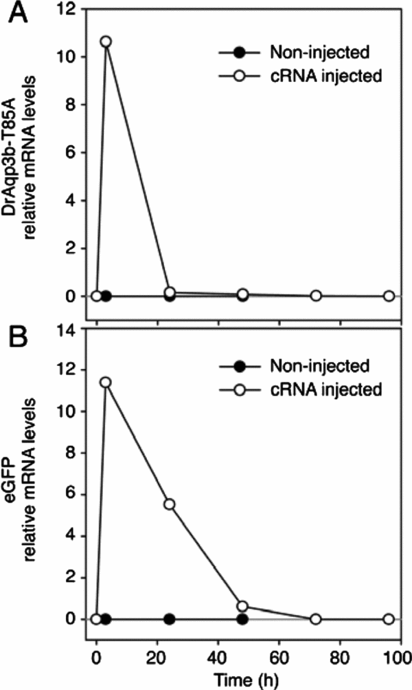
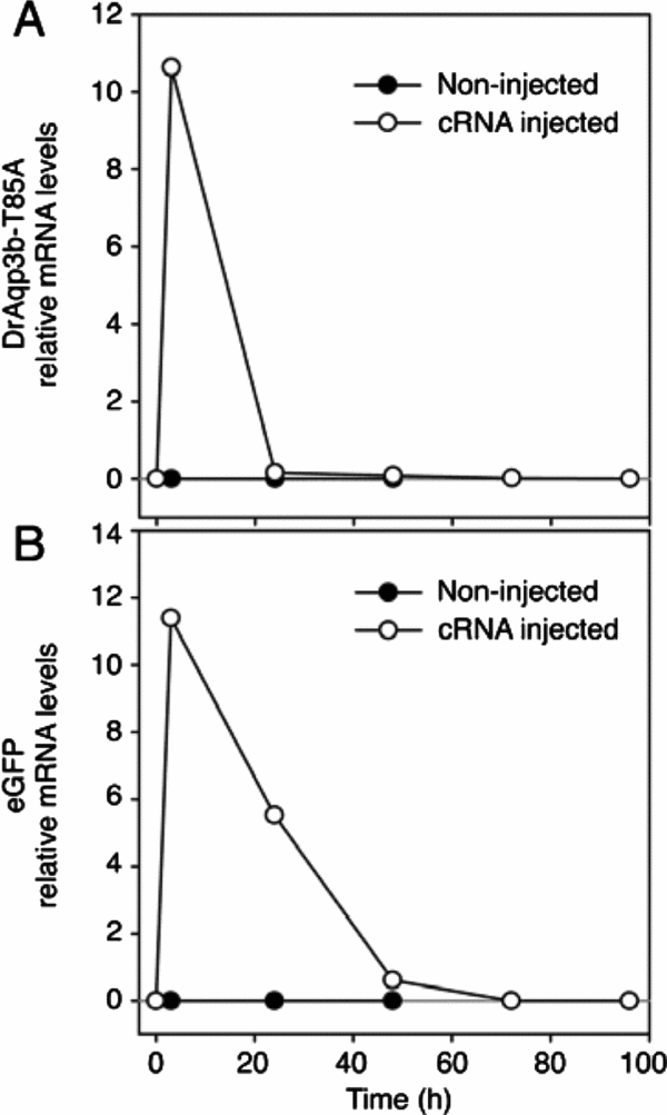
Given that the main objective of our study was precisely to test whether expression of an AQP highly selective for ethylene glycol would improve post-thaw survival of cryopreserved embryos, we decided to move forward with the lower concentration of the cryoprotectant for further experiments. Interestingly, when 1-celled embryos were microinjected with eGFP + DrAqp3b-T85A cRNA and frozen at the 2-cell stage in 1.5 M ethylene glycol-containing medium, post-thaw survival rates were almost three times higher than those observed in uninjected contemporary frozen–thawed controls (Table 2). Higher post-thaw survival rates were clearly due to expression of DrAqp3b-T85A and had no relationship to cRNA injection, given that those injected with eGFP alone displayed very low survival rates, similar to those in uninjected frozen–thawed control embryos. Hence, although not evaluated herein, the higher survival rates of DrAqp3b-T85A-expressing embryos likely stemmed from a higher permeability to ethylene glycol (Chauvigné et al., Reference Chauvigné, Lubzens and Cerdà2011; Morató et al., Reference Morató, Chauvigné, Novo, Bonet and Cerdà2014). Given a potential for future application of AQP expression as a means to improve the freezability of cells or tissues, it was particularly important to ensure that all foreign cRNA would be eventually used or degraded. Notably, all DrAqp3b-T85A and eGFP cRNA was undetectable by 72 h following injection (Fig. 2).
Embryo development rates for embryos injected with Aquaporin T85A and frozen in 1.5 M ethylene glycol

a Data expressed as mean±standard error of the mean (SEM); based upon five replicates.
b,c,d Different superscripts within column denote significant differences; P<0.05.
e Survival = Percentage of embryos surviving the freeze/thaw procedure.
For survival rates: FT control vs. FT T85A P = 0.01; FT eGFP vs. FT T85A P = 0.006.
Control, unfrozen embryos; FT control, uninjected frozen–thawed control embryos; eGFP, embryos microinjected with 2–4 pg eGFP cRNA and then frozen–thawed ~18–21 h later; FT T85A, embryos microinjected with 2–4 pg eGFP + T85A cRNA and then frozen–thawed ~18–21 h later; n1, initial number of oocytes; n2, number of oocytes surviving the freeze–thaw process (not applicable to control).
Persistence of expression of zebrafish aquaporin mutant (DrAqp3b-T85A; A) and enhanced green fluorescent protein (eGFP; B) cRNA at 3, 24, 48, 72 and 96 h following microinjection into mouse zygotes as determined by qRT-PCR. The analysis was performed in triplicate with RNA extracted from five injected embryos at each time point. Both for DrAqp3b-T85A and eGFP RNA was undetectable in embryos by 72 h following microinjection. Negative controls were contemporary uninjected embryos.
Overall, our data show that expression of a genetically modified fish AQP with high selectivity for ethylene glycol was able to rescue low survival rates of 2-cell mouse embryos frozen–thawed using a suboptimal cryopreservation protocol employing a low concentration of ethylene glycol. However, embryo development rates were lower than those observed for control unfrozen embryos, in particular for more advance stages (i.e. blastocysts, P < 0.05; Table 2). Likely, the freeze–thaw protocol used herein produced such a drastic damaging effect in embryo structures and/or DNA that could not be overcome by expression of the AQP. Improving the embryo development rates herein was beyond the scope of this study, in particular as better cryopreservation protocols are already used for mouse oocytes and embryos (Cha et al., Reference Cha, Kim, Kim, Kim, Lee, Yoon and Lee2011). Importantly, we were able to show that expression of DrAqp3b-T85A benefited cell cryosurvival in a suboptimal system probably by ameliorating the embryo permeability to the cryoprotectant (Kasai, Reference Kasai1996; Kasai & Mukaida, Reference Kasai and Mukaida2004; Vajta, Reference Vajta2013). Hence we propose that the expression of AQPs, in particular those that may be genetically modified to increase permeability to specific cryoprotectants, may provide a potential avenue to improve cryopreservation results of large cells and tissues in which current vitrification protocols have not yielded the desired success.
Acknowledgements
The authors would like to thank the CMRB core facilities for providing technical support.
Financial support
Dr Sylvia J. Bedford-Guaus was supported and this research was partially funded by the Subprograma Ramón y Cajal (RYC-2012–10053), Ministerio de Economía y Competitividad (MINECO), Spain. This work was also partially funded by the Instituto de Salud Carlos III, FEDER, Plan Estatal de Investigación Científica y Técnica y de Innovación (PEICTI) 2013–2016, PI14/02184 (to SJB-G) and the MINECO project AGL2013-41196-R (to JC).
Conflicts of interest
There are no conflicts of interest.