Introduction
The skin provides a protective barrier against the external environment. It also plays an important role in the defence against pathogenic microbes, through both acquired and innate immune responses.Reference Sirigu, Perra, Ferreli, Maxia and Turno1–Reference Nizet, Ohtake, Lauth, Trowbridge, Rudisill and Dorschner4
Antimicrobial peptides are an important component of the early innate defences against infection.Reference Boman2–Reference Braff, Bardan, Nizef and Gallo5 A large number of antimicrobial peptides have been identified in a wide variety of cells and tissues of different organs.Reference McCray and Bentley6–Reference Ali, Falconer, Ikram, Bissett, Cerio and Quinn11
The antimicrobial activity of skin probably depends upon the secretion and interaction of multiple factors.Reference Befus, Mowat, Gilchrist, Hu, Solomon and Bateman12 The glands in the skin of the external auditory canal release various lipids and proteins, which may play a role in protection against microbes.Reference Chai and Chai13–Reference Jankowski, Kapusta and Nowacka16 However, the control of the secretion of these substances is poorly defined.
Mast cells are known to infiltrate the skin and to degranulate in response to pathogenic insults. It has been suggested that mast cells play an important role in regulating gland secretion.Reference Yoon17
In the skin of the external auditory canal, the inter-relationship between antimicrobial peptides and mast cells could be of particular clinical significance, due to several significant intrinsic properties.
In the present study, we sought to characterise the distribution of mast cells and of the antimicrobial peptides human β-defensin-1 and -2 and LL-37, within the skin of the external auditory canal.
Materials and methods
Tissue specimens
Skin was collected from the cartilaginous external auditory canal of 12 individuals undergoing middle ear surgery with canaloplasty (for chronic otitis media). The skin samples collected were in a healthy state.
Immediately after surgical removal, the skin specimens were fixed overnight in freshly prepared 4 per cent paraformaldehyde in phosphate buffer at pH 7.4. All samples were then dehydrated in a graded series of ethanol to xylene and embedded in paraffin wax. Paraffin-embedded skin specimens were sectioned (to 4 µm thickness) and mounted on albumin-coated glass slides. The tissue sections were heated for a minimum of 30 minutes at 60°C.
The study was approved by the institutional review board of Chonbuk National University Hospital. Written, informed consent was obtained from all patients.
Immunohistochemistry
Tissue sections were de-waxed in xylene over three 5-minute periods, and then rehydrated with graded ethanol (at 100, 95, 80, 70 and 50 per cent) and water, each for a 3-minute period. Endogenous peroxidase activity was blocked by incubating the sections with 3 per cent hydrogen peroxide in ice-cold methanol for 30 minutes, followed by rinsing in phosphate-buffered saline. Non-specific binding was blocked by separately incubating sections with 10 per cent normal goat serum in phosphate-buffered saline for 10 minutes.
Immunoreactivity was detected using a standard avidin-biotin complex peroxidase method (Vectastain Universal Elite ABC kit; Vector Laboratories, Burlinger, Calif., USA). Localisation of immunoreactivity was carried out using goat anti-human polyclonal antibodies (human β-defensin-1 and human β-defensin-2, LL-37, tryptase and chymase). Sections were incubated with diluted primary antibodies overnight at 4°C. Control sections were incubated with pre-absorbed antiserum in place of the primary antibody. The next day, slides were washed twice in phosphate-buffered saline, and then the secondary biotinylated antibody (Vectastain; Vector Laboratories) was applied at room temperature for 45 minutes. Sections were again washed twice in phosphate-buffered saline, before application of the tertiary antibody for 30 minutes at room temperature. The reaction product was visualised using 0.03 per cent diaminobenzidine tetrahydrochloride, and sections were counterstained with Meyer's haematoxylin.
Results and analysis
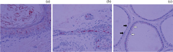
In normal external auditory canal skin, human β-defensin-1 was expressed mainly in the epidermis, hair follicles and glands (Figure 1).
Photomicrographs showing immunohistochemical localisation of human β-defensin-1 in (a) epithelium, (b) sebaceous gland and (c) ceruminous gland, within the external auditory canal skin. Human β-defensin-1 activity is present in the granular and prickle cell layers of the epidermis. Human β-defensin-1 staining is more intense in ceruminous than sebaceous glands. In active glandular cells, human β-defensin-1 is concentrated in a region directly below the apical protrusion (white arrows). In contrast, inactive gland cells are negative for human β-defensin-1 immunostaining (black arrows). (×200)

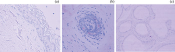
In the epidermis, human β-defensin-1 was present in the granular and prickle cell layers (Figure 1a). Human β-defensin-2 was not expressed in any epidermal layer (Figure 2a).
Photomicrographs showing immunohistochemical localisation of human β-defensin-2 in (a) epithelium (×200), (b) hair follicle (×200), and (c) sebaceous and ceruminous glands (×100), within the external auditory canal skin. Human β-defensin-2 is not expressed in any layer of the epithelium, or in the cytoplasm of secretory cells in the ceruminous glands. Only hair follicles display human β-defensin-2 expression.

In the dermis, human β-defensin-1 was localised to the glandular region and along the hair shaft, while human β-defensin-2 stained only weakly along the hair shaft, deep in the dermis (Figure 2b).
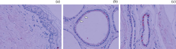
Regarding glands, human β-defensin-1 was expressed in both sebaceous (Figure 1b) and ceruminous gland cells (Figure 1c). The cytoplasm of secretory cells in the sebaceous glands stained weakly for human β-defensin-1, while the cytoplasm of secretory cells in the ceruminous glands stained positive for both human β-defensin-1 and LL-37 (Figure 3c). The nuclei of the secretory cells did not stain. The apical part of active glandular cells stained strongly positive for human β-defensin-1 and LL-37 (Figures 1c and 3c), while the inactive region was negative for all markers (Figures 1c and 3c). Human β-defensin-2 was not detected in the cytoplasm of secretory cells in either the ceruminous or the sebaceous glands (Figure 2).
Photomicrographs showing immunohistochemical localisation of LL-37 in epithelium (a; ×100), and ceruminous gland (b & c; ×200), within the external auditory canal skin. LL-37 expression is not present in the epidermis, but there is intense immunopositivity in the ceruminous glands. In highly active glandular cells (b & c), LL-37 is concentrated in a region directly below the apical protrusion (arrows).

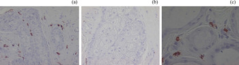
The majority of the mast cells were found in the subepithelial space and adjacent to glands. In the deep dermal layer, most mast cells were located next to ceruminous glands (Figure 4c), although some lay scattered around sebaceous glands (Figure 4b). Higher magnification revealed the typical granular appearance of mast cells (Figure 4c).
Photomicrographs showing immunohistochemical localisation of mast cells in (a) epithelium, (b) the subepithelial space, including a sebaceous gland, and (c) a ceruminous gland, within external auditory canal skin. Mast cells are not seen in the epithelium, but are abundant in the subepithelial space and around ceruminous glands (in the latter, preferentially located in interstitial areas), and show a typical granular appearance. Mast cells stained only slightly or not at all in sebaceous glands. (×200)

Discussion
In the skin of the external auditory canal, host defence may depend upon the physical barrier to lateral migration and the acidic pH (close to 5.0) presented by the skin. In the external auditory canal, the cerumen (ear wax) seems to have antimicrobial activity.Reference Chai and Chai13–Reference Jankowski, Kapusta and Nowacka16 The external auditory canal skin also produces a variety of antimicrobial factors that exhibit broad-spectrum activity against various pathogens, forming an innate epithelial biochemical protection complex. These antimicrobial peptides and proteins include human β-defensin, cathelicidin, dermicidin, lysozyme, lactoferrin, secretory leukocyte protease inhibitor and α1-antitrypsin. Antimicrobial peptides are found predominantly in cells and tissues involved in host defence, and have broad-spectrum activity against a wide range of micro-organisms including bacteria, viruses, fungi, yeasts and protozoa. Antimicrobial peptides are particularly important in the early host defence against microbes, and are a critical component of the innate defences of most organisms against invading pathogens.
Antimicrobial peptides and proteins have been identified in the human skin and mucosa.Reference McCray and Bentley6–Reference Harder, Bartels, Christophers and Schroder8 One of these compounds, human β-defensin-1, is produced by various epithelial tissues, including the urogenital and respiratory tracts. Expression of human β-defensin-1 is constitutive (i.e. continuous), while human β-defensin-2 expression increases following skin injury or inflammation. Another antimicrobial peptide, human cathelicidin (also known as LL-37) is produced by neutrophils, mast cells and keratinocytes in response to inflammatory processes. LL-37 also acts as a chemoattractant for neutrophils, monocytes, T cells and mast cells.
Antimicrobial peptides represent attractive examples of the potential therapeutic application of innate immune protection, and have therefore been the focus of research attention in recent years. Rising interest in natural antimicrobial factors has driven researchers to identify principal secretion sites. Antimicrobial peptides may be derived from epithelial and glandular cells. Human β-defensin-1 is constitutively produced by sweat glands and secreted into sweat, where it is proteolytically processed. Secretion from skin glands may be required for an effective cutaneous innate immune response.Reference Braff, Bardan, Nizef and Gallo5
Many types of glands are present in the dermis, particularly sebaceous and sweat glands. The external auditory canal skin contains both sebaceous and ceruminous glands, the latter being modified sweat glands closely linked to the formation of cerumen.Reference Main and Lim18 A number of reports have identified antimicrobial peptides in skin gland cells, including human β-defensins in apocrine sweat glands.Reference Harder, Bartels, Christophers and Schroder8–Reference Ali, Falconer, Ikram, Bissett, Cerio and Quinn11 Antimicrobial peptides are expressed in apocrine glands and transported via sweat to the epidermal surface.
Recently, antimicrobial factors (e.g. lysozyme, lactoferrin and α1-antitrypsin) have been detected in the external auditory canal skin, using immunohistochemical analysis, and their distribution at this site has been found to correlate positively with the location of ceruminous glands. The first author has previously examined the presence of human β-defensin-1 and -2 in cerumen, using Western blotting.Reference Yoon14
However, the exact source of antimicrobial peptide secretion has not been identified. Mast cells represent one likely candidate for the source of ceruminous gland peptide secretion.
• The antimicrobial activity of external auditory canal skin may depend upon the secretion and interaction of multiple factors
• This study identified mast cell markers (tryptase and chymase) and antimicrobial peptides (human β-defensin-1 and -2 and LL-37) in external auditory canal skin, using immunohistochemistry
• Immunoreactivity for mast cells, human β-defensin-1 and LL-37 was greater in ceruminous glands than sebaceous glands
• This suggests that mast cells are involved in ceruminous gland secretion of antimicrobial peptides
Using immunohistochemical analysis, we examined the distribution of mast cells and several antimicrobial peptides within the external auditory canal skin, specifically comparing ceruminous and sebaceous glands. Human β-defensin-1 activity was seen in ceruminous gland cells, but only weakly in sebaceous gland cells. This difference in distribution may be related to function, indicating a greater antimicrobial role for the ceruminous glands compared with the sebaceous glands. Mast cells were confined mainly to the subepithelial space just beneath the outer layer of keratinising stratified squamous epithelium, and to the gland regions. The ceruminous glands contained significantly more mast cells than the sebaceous glands, but most of those mast cells were actually located adjacent to the ceruminous glands. Some lay scattered in the region of the sebaceous glands. Overall, more mast cells and greater human β-defensin-1 expression were detected in ceruminous glands compared with sebaceous glands, within the skin of the external auditory canal.
Conclusion
There were significant differences in mast cell distribution and antimicrobial peptide expression, comparing the ceruminous and sebaceous glands of external auditory canal skin. Mast cell, human β-defensins and LL-37 immunoreactivity was conspicuously greater in the ceruminous glands compared with the sebaceous glands. We hypothesise that ceruminous glands influence the innate immune properties of external auditory canal skin, and that interactions between mast cells and antimicrobial peptides coordinate and promote innate immune pathways.
Acknowledgements
This work was supported by research funds from Chonbuk National University (2009) and Chonbuk National University Hospital Research Institute of Clinical Medicine.