Introduction
Dilated cardiomyopathy is a heterogeneous cardiomyopathy. Under the stimulation of infection, immunity, genetic, and other factors, the heart gradually expands, the myocardial tissue shows varying degrees of pallor, relaxation, and fibrosis, and the ventricular wall becomes thinner. Reference Japp, Gulati, Cook, Cowie and Prasad1 Due to structural changes in the heart, cardiac function gradually declines, and electrical conduction disorders can be accompanied by heart failure and arrhythmia. There is an urgent medical need to address the persistently high 5-year mortality rate. Reference Reichart, Magnussen, Zeller and Blankenberg2 Despite advances in therapeutic strategies, the exact mechanisms underlying the pathogenesis of dilated cardiomyopathy are still not fully understood, and further studies are needed to identify new therapeutic targets. Pathological changes influence the progression of dilated cardiomyopathy in inflammatory processes and autoimmune responses. However, specific mechanisms need to be further studied. Reference Wang, Zhou and Li3 The pathogenesis and progression of dilated cardiomyopathy are related to CD4+ T cells. Reference Zeng, Wang and Li4 In patients with dilated cardiomyopathy, CD4+ T cells have been identified as the primary cause of cardia-specific autoimmunity. Reference Wu, Sun and Chen5 Following tissue injury, cardiac autoantigens are released, and dendritic cells are activated to clear autoantigen presentation in draining lymph nodes. This may destroy cardiac-specific tolerance, triggering the production of cardiac-specific autoantibodies, expansion of autoreactive CD4+ T cells, and autoimmunity. Reference Van der Borght and Scott6 Activated by antigen-presenting cells, naive T cells rapidly proliferate and differentiate into effector T cells. When pathogens are controlled or eliminated, most of these effector T cells undergo apoptosis, and only 5 to 10% of memory T cells survive for a long time. CD4+T cells are activated by recognising major histocompatibility complex class II antigens. Driven by specific cytokines (IL-17, IL-25, IL-33, IL-6, TGF-β, ICOS), they differentiate into Th1, Th2, Th17, Treg, and TFH are involved in the regulation of immune function in dilated cardiomyopathy. Reference Van der Borght and Scott6
The IL-17 family is a complex and unique cytokine signalling system that shows high conservation in vertebrate evolution. The family has been confirmed to include six ligands (IL-17A to F) and five receptors (IL-17RA to RE). Among them, IL-17A is the most well-studied member. At first, it was thought that IL-17 was mainly produced by CD4+T cells, but with the deepening of research, it has been found that a variety of immune cells, such as NKT17, ILC3 subsets, and macrophages, can also secrete IL-17 under certain conditions. Dysregulation of such cytokines may lead to excessive inflammatory responses and chronic pathological states, which in turn trigger tissue damage and autoimmune problems. IL-17 plays a critical role in the pathogenesis of autoimmune diseases, including lupus nephritis and psoriasis, by inducing the expression of pro-inflammatory factors, chemokines, and other inflammation-related molecules. Reference Qian, Chen, Wu, Hu, Huang and Jin7–Reference Sundrud and Trivigno8 Due to the high homology of the gene between IL-17 and HVS13 herpesvirus, it was initially called CTLA-8 until it was officially renamed IL-17 in 1993. IL-17 is mainly secreted by Th17 cells, including IL-17A-E, in which IL-17A is important. Studies have shown that IL-17 expression is significantly increased in the serum of mice with viral myocarditis and patients with myocarditis. Reference Bousoik and Montazeri Aliabadi9 Yang et al. Reference Zhu and Paul10 found that blocking IL-17 could significantly improve myocardial inflammatory infiltration and cardiac function in viral myocarditis. In addition, some research has shown that IL-17 can work with other cytokines, IL-1β and IL-6, to promote the development of autoimmune myocarditis. Reference Di Napoli, Taccardi and Grilli11 Studies have shown that IL-17 (A) can recruit and activate neutrophils, improve T cell response and promote the release of inflammatory transmitters, eliminate microbial pathogens in the host body, induce inflammatory responses in tissues and organs, and thus cause the disorder of the body’s autoimmune function. Reference Van der Borght and Scott6–Reference Qian, Chen, Wu, Hu, Huang and Jin7 IL-17 (A) also has a synergistic effect with various cytokines, which can amplify the human body’s inflammatory response and exert a powerful pro-inflammatory effect.
The JAK/STAT signalling pathway has been a hot topic of research. Reference Sundrud and Trivigno8 Upon binding to transmembrane receptors, specific ligands, such as interleukins, can activate JAK, which in turn triggers the activation of downstream effectors. Reference Bousoik and Montazeri Aliabadi9 In CD4+T cell differentiation, multiple signalling pathways play key regulatory roles. These include the JAK2/STAT3 pathway, one of the primary pathways for CD4+ T cells to develop into Th17 cells. Reference Zhu and Paul10
Our study aimed to investigate how IL-17 mediates immunomodulatory mechanisms of CD4+ T cells in DCM. Understanding the interplay between IL-17, CD4+ T cells, and the JAK/STAT signalling cascade could provide valuable insights into the inflammatory processes of dilated cardiomyopathy and provide new avenues for therapeutic intervention.
Materials and methods
Selection of animals
Male BALB/c mice were selected and purchased from Vital River at 6 to 8 weeks. The mice were maintained in a controlled environment at 22 ± 1°C, with a relative humidity of 50 ± 1% and a 12-hour light/dark cycle. The animal studies were conducted in accordance with the regulations and guidelines of the animal regulatory authorities, the International Committee for the Evaluation and Accreditation of Laboratory Animal Feeding Management, and the guidelines of the Laboratory Animal Use and Management Committee. Intraperitoneal injection of adriamycin (Sigma, USA) at a dose of 2 mg/kg once a week for 8 weeks was used to establish the mouse model of dilated cardiomyopathy. The Second Hospital of Hebei Medical University approved the study.
T cell isolation and CD4+ T cell sorting
Two mice were killed by cervical bleaching. Spleens from mice were removed aseptically and placed in sterile RPMI1640 medium (Sigma) supplemented with human 10% fetal bovine serum penicillin (100 U/mL) and streptomycin (100 U/mL) (Sigma). Following the kit’s instructions, the lymphocytes were obtained using the Spleen Lymphocyte Isolation Kit (Solarbio Life Sciences, P.R. China). According to the manufacturer’s instructions, naive CD4+ T cells were isolated from mouse splenic lymphocytes using magnetic bead sorting and the Mouse Naive CD4+ T Cell Isolation Kit (Miltenyi Biotec, Germany). The purity of isolated cells was assessed by flow cytometry and found to be>90%. Naive CD4+ T cells were cultured in RPMI1640 medium at 37°C with 5% CO2, supplemented with 10% fetal bovine serum, penicillin, and streptomycin at 100 U/mL concentration.
Cell culture, lentiviral transduction, and generation of stable cell lines
IL-17 (NM_0021900.3) lentivirus, negative control, shRNA lentivirus targeting IL-17, and scrambled shRNA lentivirus was generated by cell culture. Lentivirus transduction and stable cell lines were purchased from Hanhang Biotech (Shanghai, China). All lentiviruses were labelled with green fluorescent protein. Sh IL-17 IL-17 target sequence of 5´- CCAGCGAGTTCAAGAAGAAGCTCTT - 3, Sh IL-17 - NC IL-17 target sequence of 5´ - TTCTCCGAACGTTGTCACGTAA - 3´. Lentiviral transduction and selection of stable cell lines were performed, as previously described. Briefly, murine splenic naive CD4+ T cells were seeded in 12-well plates (1 × 10⁵ cells/well) for adhesion, as described above. The cells were then transfected with a lentiviral vector at a multiplicity of infection of 30 in the presence of 5 μg/mL polybrene. Seventy-two hours after transduction, puromycin (8 μg/mL) was added, and stable cell lines were selected for 3 weeks.
Western blot analysis
The cells were treated with Radioimmunoprecipitation Assay lysis solution, which included protease inhibitors, and proteins were extracted using an extraction kit. Bradford tests were used to determine protein levels in the supernatant. Proteins were isolated using 10% SDS-PAGE electrophoresis and transferred to polyvinylidene fluoride membranes. Polyvinylidene fluoride membranes were blocked with 5% skim milk for 2 hours at room temperature before incubating with primary antibodies overnight at 4°C. After thoroughly washing with Tris-buffered saline with Tween 20 (TBST), which is a buffer used to remove unbound antibodies and reduce non-specific binding by the use of Tween 20 as a detergent, the polyvinylidene fluoride membrane was incubated with the appropriate horseradish peroxidase-labeled secondary antibody for 2 hours at 37°C. Chemiluminescence was used to see the protein bands, which were then quantified with ImageJ software.
Quantitative polymerase chain reaction analysis
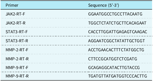
Total RNA was isolated from mouse spleen CD4T cells using Trizol reagent (Invitrogen), and reverse transcription-quantitative PCR was done using the TB GreenTM Advantage quantitative polymerase chain reaction Premix (Takara) kit, following the kit instructions. GAPDH served as an internal control for all mRNA expressions. The gene sequences are provided in Table 1. The Qrt-Pcr data were computed with the 2−ΔΔCt technique.
Sequences of the primers used in this study

Enzyme-linked immunosorbent assay analysis
According to the manufacturer’s directions, TNF-α, IL-1β, IL-4, and IFNγ levels were measured using ELISA kits (MultiSciences, Hangzhou, China). The detection antibody was added after the standard and sample were added to the wells. After incubation for 1.5 hours at 37°C, the plates were washed six times. Then, 100 µL of enzyme-labeled reagent and 100 µL of chromogen were pipetted into each well. After the mixture, the mixture was incubated at 37°C in the dark for about 15 min, and then the reaction was terminated by pipetting 100 μL of termination solution into each well. The optical density of each well was measured at a wavelength of 450 nm after zero adjustment. After plotting the standard curve, the optical density values of each sample were used to determine the concentrations of TNF-α, IL-1β, IL-4, and IFNγ from the standard curve. The units were pg/mL.
Flow cytometry
The cells were cultivated with protein transport inhibitors invitrogen for 6 hours. To examine the intracellular expression of NF-α, IL-1β, IL-4, and IFNγ in CD4+T cells, cells were fixed and membrane-broken using Cell fixation/disruption reagent (Invitrogen) as described in the instructions of the kit (cell concentration approximately 0.5 x 10%/well). Cells were stained with anti-CD4 (item 552051) and anti-CD3 antibodies (item 562286) and then fixed with Invitrogen’s fixation buffer in a 4°C dark room for 30 min. Cells were disrupted using Invitrogen’s disruption buffer (1x). The cells were stained with anti-Tnf-α, anti-Il-1β, anti-IL-4, and anti-IFNγ (all purchased from BD, NJ) labelled with fluorochrome. Stained cells were examined with a BD FACSCalibur flow cytometer, and the results were analysed with FowJo X software (Tree-star, Inc.).
Statistical methods
The results are reported as mean ± SEM. SPSS 16.0 software was used for one-way analysis of variance to compare statistical differences between multiple groups. The variance test was used for a one-way analysis, which was then compared to the post hoc and Student–Newman–Keuls tests. A P-value of <0.05 was judged statistically significant.
Results
Expression of inflammatory factors after overexpression or knockdown of IL-17 in a mouse model
In our mouse model, the main inflammatory factors in dilated cardiomyopathy are mainly TNF-α, IL-1β, IL-4, and IFNγ, Reference Japp, Gulati, Cook, Cowie and Prasad1 The expression levels of TNF-α, IL-1β, IL-4, and IFNγ were detected after overexpression or knockdown of IL-17. The flow cytometry experiments revealed that in the mouse model, overexpression of IL-17 led to an increase in the secretion ratio of TNF-α, IL-1β, IL-4, and IFNγ (compared with oe-IL-17-NC, P < 0.05) (Figure 1b). The ELISA showed that after IL-17 overexpression, the expression of TNF-α, IL-1β, IL-4, and IFNγ was also increased (compared with the oe-IL-17-NC group, P < 0.05) (Figure 1b). However, after knocking down IL-17, the expression of TNF-α, IL-1β, IL-4, and IFNγ decreased (compared with the sh-IL-17 group, P < 0.05) (Figure 1).
Expression of inflammatory factors after overexpression or knockdown of IL-17. ( a ) TNF-α, IL-1β, IL-4, IFNγ secretion ratio detected by flow cytometry. ( b ) ELISA for TNF-α, IL-1β, IL-4, and IFNγ expression.oe:overexpression.kd:knock down. NC = negative control. *P < 0.05 versus control.

Expression of JAK/STAT, matrix metalloproteinase-2, and MMP-9 after overexpression or knockdown of IL-17 in a mouse model
Western blot analysis in the mouse model showed that overexpression of IL-17 resulted in a significant increase (p < 0.05) in the levels of p-JAK2, p-STAT3, and target genes (MMP-2, MMP-9) compared to the control (Figure 2a). Knockdown of IL-17 had the opposite effect on the above proteins in the cells, and the levels of p-JAK2, p-STAT3, and target genes (MMP-2, MMP-9) were decreased after IL-17 compared with the control (p < 0.05) (Figure 2a). Additionally, quantitative polymerase chain reaction results in the mouse model showed that IL-17 overexpression significantly elevated the levels of MMP-2 and MMP-9 (p < 0.05) (Figure 2b). These findings suggest that IL-17 may modulate the expression of the JAK/STAT pathway and matrix metalloproteinases.
Expression of JAK/STAT, MMP-2, MMP-9 after overexpression or knockdown of IL-17. ( a ) WB detection of protein expression of JAK2, p-JAK2, STAT3, p-STAT3, MMP-2, MMP-9. ( b ) Quantitative polymerase chain reaction detection of mRNA expression of JAK2, p-JAK2, STAT3, p-STAT3, MMP-2, MMP-9.oe:overexpression.kd:knock down. NC = Negative control. *P < 0.05, ***P < 0.001 versus control. #P < 0.05, ##P < 0.01, ###P < 0.001vs oe-IL-17.

Effects of IL-17 overexpression or knockdown on apoptosis of CD4+ T cells in a mouse model
Flow cytometry analysis confirmed that IL-17 overexpression increased the apoptosis rate of CD4+ T cells in the mouse model compared to the control group (P < 0.05). Conversely, IL-17 knockdown decreased the apoptosis rate in CD4+ T cells in the mouse model compared to the control group (P < 0.05) (Figure 3).
Effect of overexpression or knockdown of IL-17 on apoptosis of CD4+T cells. Flow cytometry was used to detect the apoptosis rate of different groups.oe:overexpression.kd:knock down. NC = Negative control.***P < 0.001 versus control. ###P < 0.001vs oe-IL-17.

In our mouse model, treatment with E171 showed significant effects on colitis-associated colon tumorigenesis
Discussion
The aetiology of dilated cardiomyopathy is closely related to inflammatory responses, metabolism, genetics, and other factors. These factors can cause varying degrees of myocardial damage, leading to progressive expansion of the myocardium and eventually developing into chronic heart failure. Because of varying degrees of myocardial cell damage, myocardial fibrosis occurs, which affects the heart’s conduction system to a certain extent. Consequently, atrial fibrillation is a common type of arrhythmia in dilated cardiomyopathy. Atrial fibrillation aggravates the symptoms of heart failure, accelerates ventricular remodelling, and increases mortality in dilated cardiomyopathy.
The pathogenesis of dilated cardiomyopathy is very complex, and apoptosis is considered the leading cause of progressive cardiac dysfunction in dilated cardiomyopathy. Reference Di Napoli, Taccardi and Grilli11 Additionally, dilated cardiomyopathy may be linked to persistent viral infections, autoimmunity, and other immune responses, which manifest as cardiomegaly, heart failure, arrhythmia, embolism, and sudden death. Reference Ibe, Saraste and Lindemann12 Dilated cardiomyopathy has a poor prognosis and high mortality, which seriously threatens human health. Reference McKenna, Maron and Thiene13 Myocardial remodelling in dilated cardiomyopathy leads to cardiac enlargement and eventually progresses to heart failure. Intervention to inhibit myocardial cell apoptosis is a new therapeutic approach for dilated cardiomyopathy. Reference Moeinafshar, Yazdanpanah and Rezaei14
Effector CD4+T cells play a role by secreting effector cytokines such as IFN-γ from Th1 cells, IL-17A from Th17 cells, and IL-22 from Th22 cells. Reference Crawford, Sinha, Renavikar, Borcherding and Karandikar15 Studies have confirmed that effector Th1 and Th17 cells, along with their secreted pro-inflammatory cytokines like IFN-γ and IL-17A, have significant pathogenic effects in promoting the progression of acute myocarditis. Reference Nindl, Maier and Ratering16–Reference Huang, Wu and Wang17 However, following the peak of the immune response, the ability of effector CD4+T cells to release cytokines gradually declines, ultimately leading to exhaustion or apoptosis. Reference Kania, Siegert and Behnke18–Reference Thome, Yudanin and Ohmura19 .
Research shows that IL-17 has a key function in regulating inflammatory diseases. Many cell types, including fibroblasts, chondrocytes, macrophages, and synoviocytes, display enhanced anti-inflammatory responses to IL-17, alone or in combination with other pro-inflammatory cytokines like TNF or IFNγ. Reference Hu, Wang, Wu, Chen, Yuan and Chen20 In our mouse model, this study showed significantly higher levels of inflammatory factors in CD4+ T cells following IL-17 overexpression, including TNF-α, IL-1β, IL-4, and IFNγ. These inflammatory factors are significant markers in the immune response, and an increase in their levels may indicate an exacerbation of the immune response in CD4+T cells due to IL-17. This may, in turn, worsen the pathological process of dilated cardiomyopathy.
Studies have demonstrated a relationship between interleukin-17 (IL-17) levels in plasma and peripheral blood monocytes and the severity of dilated cardiomyopathy. Reference Zhang, Xu, Wang, Li and Zhao21 The mRNA level of IL-17 protein in peripheral blood mononuclear cells (PBMC) culture supernatant or PHA-stimulated PBMC was correlated with cardiac function in dilated cardiomyopathy patients, suggesting that IL-17 might play an essential role in dilated cardiomyopathy pathogenesis. Reference Zhang, Xu, Wang, Li and Zhao21 Liu et al. found that the IL-17 signalling pathway and acute inflammatory response were closely linked in the bioinformatics prediction of dilated cardiomyopathy mechanisms. Reference Antonov, Kozlov, Pal’tseva, Polyakova and Lin’kova22 In a related study in mouse models, IL-6 knockout inhibited myocardial cell apoptosis, improved cardiac function, and alleviated myocardial remodelling and fibrosis in adriamycin-induced dilated cardiomyopathy, possibly by suppressing STAT3 phosphorylation. Reference Sperandio, de Belle and Bredesen23
We also observed that, in our mouse model, IL-17 overexpression resulted in elevated phosphorylation of JAK2 and STAT3. The JAK/STAT system is essential for cellular signal transduction, and its activation is linked to inflammation, differentiation, and proliferation. In our mouse model, IL-17 may regulate the immune response of CD4+T cells through activation of the JAK/STAT pathway. Reference Kimura, Kishimoto and Kishi24 In disorders associated with the intervertebral disc, research has shown that IL-17 primarily utilises the JAK/STAT pathway for signalling and transcriptional activation of target genes. Reference Kimura, Kishimoto and Kishi24 The Th17/IL-17 axis’s activation via the JAK2/STAT3 pathway is associated with increased IL-6, TGF-β, RORγt, and other mediators. Reference Martinez-Moreno, Garcia-Castello and Ruiz-Llorente25
Moreover, this study revealed that altered IL-17 expression affected MMP-2 and MMP-9, matrix metalloproteinases involved in cardiovascular disease development and progression. In the mouse model, matrix metalloproteinases are crucial in extracellular matrix degradation and remodelling. MMP-2 and MMP-9 have been identified as markers of dilated cardiomyopathy in different age groups. Reference Wang, Li and Zhang26 Increased MMP activity during myocardial injury degrades collagen and elastin, leading to ventricular remodelling. Studies have shown that reducing MMP-2 protein expression in myocardial tissue can inhibit ventricular remodelling and improve cardiac function. Reference Wang, Li and Zhang26
Finally, our findings indicated that IL-17 influences the apoptosis levels of CD4+T cells. Apoptosis is a programed form of cell death necessary to maintain tissue homeostasis. In our mouse model, IL-17 overexpression increased apoptosis, which could further exacerbate the progression of dilated cardiomyopathy. Reference Sperandio, de Belle and Bredesen23
While our study utilised a mouse model to investigate the role of IL-17 in dilated cardiomyopathy, it is important to acknowledge that mice and humans can exhibit differences in disease pathology and progression. However, many key inflammatory pathways and mechanisms identified in mice are conserved in human dilated cardiomyopathy. For instance, both models and human studies have observed the IL-17 signalling pathway and its effects on inflammatory cytokine release and matrix metalloproteinase expression. These findings suggest that the mechanisms explored in our mouse model may be relevant for human dilated cardiomyopathy, although further validation in human clinical studies is necessary. Understanding these mechanisms in mice provides a foundational insight that can guide future research and potential therapeutic approaches in human patients.
In conclusion, this study, conducted in a mouse model, provides evidence for the critical role of IL-17 in the immunological modulation of CD4+T cells in dilated cardiomyopathy. Our findings indicate that IL-17 regulates the secretion of inflammatory cytokines, activates the JAK/STAT signalling pathway, influences matrix metalloproteinase (MMP-2 and MMP-9) expression, and promotes apoptosis in CD4+T cells. While these results offer valuable insights into the role of IL-17 in dilated cardiomyopathy, it is crucial to recognise that our findings are based on a murine model. Mice and humans can exhibit differences in disease pathology and progression, so the direct applicability of these results to human dilated cardiomyopathy remains to be validated. Future research should focus on translating these findings from mouse models to human studies to understand better how IL-17 contributes to the progression of dilated cardiomyopathy in humans. Investigating targeted therapeutic interventions to modulate IL-17 signalling could improve treatment outcomes by mitigating inflammation and myocardial damage associated with IL-17 dysregulation.
Availability of data and material
All data generated or analysed during this study are included. Further inquiries can be directed to the corresponding author.
Acknowledgements
None.
Financial support
This study was funded by the Hebei Provincial Natural Science Foundation Precision Medicine Joint Fund Cultivation Project; Funding Number (Project Number): H2020206332.
Competing interests
None.
Ethics standard
The Second Hospital of Hebei Medical University approved the study. The animal studies were conducted in accordance with the regulations and guidelines of the animal regulatory authorities, the International Committee for the Evaluation and Accreditation of Laboratory Animal Feeding Management, and the guidelines of the Laboratory Animal Use and Management Committee.
Author contribution
JH is responsible for the guarantor of integrity of the entire study, study concepts and design, the definition of intellectual content, and manuscript review; WWL is responsible for the study design, definition of intellectual content, literature research, experimental studies, data analysis, manuscript preparation, and editing; WJ is responsible for the clinical studies, literature research, experimental studies, data acquisition, statistical analysis, and manuscript preparation; FL is responsible for the clinical studies and data analysis; YNX is responsible for the data acquisition and statistical analysis. All authors read and approved the final manuscript.