INTRODUCTION
Barnacles are found in the oceans worldwide and in various habitats, from the intertidal zone to deep waters. They live on various substrata, including bodies of other animals. Although we sometimes find barnacles that live on deep-sea animals, little is known about their morphology, taxonomy and ecology.
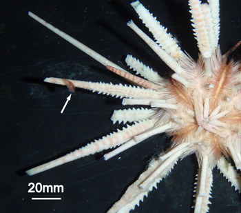
We were fortunate to obtain three rare pedunculate barnacle specimens from Okinawa, southern Japan that were attached to spines of the deep-sea urchin Prionocidaris sp. (Figure 1). These barnacles had beautiful red coloration that matched the colour of the sea urchin. They appeared to be Paralepas maculata Utinomi, Reference Utinomi1980. In his last paper, published posthumously, Dr Utinomi described this species based on specimens collected in Wakayama, south-western Japan. These barnacles were found on a sea urchin, Coelopleurus maculatus Agassiz & Clark, 1907. Because P. maculata has not otherwise been reported since the original description, our new material represents the second record of this species.
Paralepas maculata, rare pedunculate barnacles attached to spines of the deep-sea sea urchin Prionocidaris sp. in the Churaumi Aquarium, Okinawa. We can find only one barnacle in this photograph.

In this paper, the morphological features of these barnacles are figured and reported, and the type plus other material described by Utinomi (Reference Utinomi1980) is re-examined.
MATERIALS AND METHODS
Material examined
Three barnacle specimens were found attached to the spine of a sea urchin, Prionocidaris sp. in Okinawa Churaumi Aquarium. The urchin was caught with a gill net on 11 June 2010 near Minnajima Island (26°37′163″N 127°48′081″E), Okinawa, Japan, at a depth of 216 m. Since that time, the urchin has been reared in the Aquarium. On 10 October 2010, we removed the barnacles from the spines and fixed them with 100% ethanol (1 individual) and 10% formalin (2 individuals).
We also examined the type series of P. maculata Utinomi, Reference Utinomi1980. These specimens were found on the spines of a sea urchin collected off Minabe (33°46N135°19E). All these specimens are deposited in the Seto Marine Biological Laboratory as SMBL-Type 319 (although Utinomi mentioned it as SMBL-Type 290, it was renumbered (Harada, Reference Harada1991)). Utinomi designated ‘the largest one as the holotype’ and ‘five smaller young ones as the paratypes’, but we found 25 individuals in the same bottle and could not specify the five paratypes. The largest of these specimens is intact. Another was dissected, most likely by Utinomi.
Methods
We measured the capitulum length and the peduncle length and observed the body shape and surface of all individuals, including the type material. We then dissected two newly collected specimens (fixed with formalin) to examine the mouth parts, cirri, penises, caudal appendages and filamentary appendages and figured for the larger specimen. For comparison, the corresponding structures of the dissected type specimen were examined. Because a new specimen had eggs and nauplii just after hatching in its mantle cavity, we measured the length of five randomly chosen eggs and one larva (other larvae were damaged).
RESULTS
SYSTEMATICS
Subclass CIRRIPEDIA Burmeister, 1834
Order LEPADIFORMES Buckeridge & Newman, Reference Buckeridge and Newman2006
Family HETERALEPADIDAE Nilsson-Cantell, 1921
Genus Paralepas Pilsbry, 1907
Paralepas maculata Utinomi, Reference Utinomi1980
(Figures 1–4)
Paralepas maculata: Utinomi, Reference Utinomi1980.
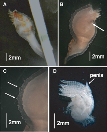
Newly collected specimens of Paralepas maculata: (A) living specimen. The barnacle showed beautiful red coloration; (B) specimen fixed in 10% formalin. The boundary between the peduncle and the capitulum is slightly narrow. The arrow indicates scutum; (C) small prominences on the capitulum's surface; (D) penis and cirri. The penis is very short. Cirri V and VI were similar in length. Scale bars are 2 mm.

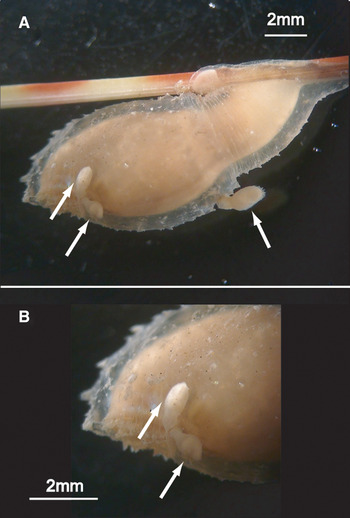
Holotype of Paralepas maculata, with smaller conspecific barnacles: (A) three smaller individuals attached to the holotype; (B) close-up of Figure 3A. Two smaller individuals attached just below the orifice of the holotype. Scale bars are 2 mm.

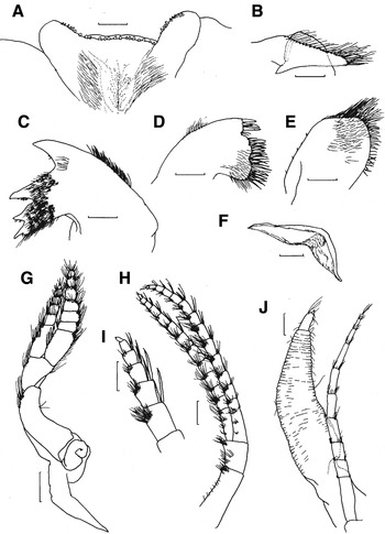
Newly collected specimen of Paralepas maculata: (A) labrum; (B) mandible palp; (C) mandible; (D) maxillule; (E) maxilla; (F) scutum; (G) cirrus I; (H) cirrus VI; (I) distal articles of cirrus VI; (J) penis and caudal appendage. Scale bars: A–E, G, I, J, 0.2 mm; F, H, 0.4 mm.

DESCRIPTION OF NEW SPECIMENS
The three newly collected specimens showed red coloration when alive (Figure 2A); however, the colour faded in alcohol. The globular capitulum is thick and hard, due to shrinkage by fixation process (Figure 2B) and has many small prominences on its surface (Figure 2B, C). A smaller number of prominences are observed on the peduncle. The boundary between the capitulum and the peduncle is slightly narrow (Figure 2B). The peduncle is shorter than the capitulum (capitulum = 5.53 ± 2.27 mm, N = 3; peduncle = 3.40 ± 1.67 mm, N = 3) and is cylindrical and hard (Figure 2B). The orifice is fringed by small prominences along the occludent margin. The scuta are small but present below the orifice, chitinous and implanted in capitulum skin (Figure 4F).
Mouth parts: the labrum (Figure 4A) bears a row of blunt denticles along the ventral margin. The mandible palp (Figure 4B) is tapering distally, fringed with setae along the ventral margin. The mandible (Figure 4C) has four teeth with small denticles along their lower edge. The maxillule (Figure 4D) bears strong spines along cutting edge which is divided by three notches; the strongest spine at the upper corner is broken off in this specimen. The maxilla (Figure 4E) is rounded in outline, setose marginally.
The cirrus I (Figure 4G) has unequal rami; inner ramus shorter than outer ramus. The filamentary appendage is digitiform and is situated at the base of cirrus I. The inner and outer rami of cirri V and VI are similar in length; articles of cirri longer than wide (Figures 2D & 4H); distal articles with strong spine-like setae along the posterior margin (Figure 4I). These characteristics are typically diagnostic of the genus Paralepas Pilsbry, 1907 (Newman, Reference Newman1960). The caudal appendage (Figure 4J) is uniformly narrow. It is 1/2 the length of cirri VI and 10-segmented (caudal appendage = 2.20 mm, 1.98 mm, N = 2, 10% formalin samples; cirri VI = 4.71 mm, 3.56 mm, N = 2, 10% formalin samples). The penis (Figure 4J) is annulated and tapered at the distal part: less than half as long as cirri VI (penis length = 1.56 mm, 1.21 mm; penis diameter = 0.41 mm, 0.25 mm, N = 2, 10 % formalin samples).
The larger specimen was fixed in 10% formalin and held a residual egg mass. On average, the eggs are 0.179 ± 0.017 mm long and 0.106 ± 0.009 mm wide (N = 5). A hatched nauplius is 0.235 mm long (N = 1).
DESCRIPTION OF UTINOMI'S TYPE SERIES
The capitulum length and peduncle length of the holotype are 8.56 mm and 4.34 mm, respectively. Therefore, the holotype's peduncle is shorter than its capitulum. Although Utinomi (Reference Utinomi1980) states that the scuta are ‘quite absent’, we found small, triangular scuta below the orifice in the type series, including the holotype. Utinomi (Reference Utinomi1980) reported that the annulation of the penis was indistinct. However, we observed in the dissected specimen that the penis showed annulation.
Three individuals are attached to the capitulum of the holotype (Figure 3A). Two are attached to the right side of the capitulum immediately below the orifice (perhaps on the scutum) (Figure 3B). The third is attached to the middle of the capitulum. We show the lengths of the peduncle (0.33–0.71 mm) and capitulum (0.82–1.50 mm) of these three small specimens attached to the holotype, along with those of the holotype (Table 1).
Capitulum and peduncle lengths of the holotype and three smaller individuals attached on the holotype.

DISCUSSION
The new specimens share several characteristics with Paralepas maculata as described by Utinomi (Reference Utinomi1980); e.g. small prominences on the body surface and the morphology of the cirri and the caudal appendages. Notably, our specimens showed a red colour pattern when alive (Figure 2A), consistent with the descriptions in Utinomi (figure 1 in Utinomi, Reference Utinomi1980). The characteristics of the peduncle in our specimens, e.g. shorter, cylindrical and hard, also coincide with Utinomi's description. Although certain traits, such as the existence of scuta and the annulation of the penis, do not agree with Utinomi's description, we confirmed that these traits are also shared by his specimens. Therefore, we conclude that our specimens should be identified as P. maculata.
Although P. maculata was clearly distinguished from other congeners in the original description (Utinomi, Reference Utinomi1980), this distinction among species would be ambiguous in view of the overlooked important characteristics cited above. For example, this species appears similar to Paralepas nodulosa (Broch, Reference Broch1922), reported by Liu & Ren (Reference Liu and Ren2007) from the East China Sea and the South China Sea. Indeed, we found no clear difference between our specimens and Liu & Ren's description. One difference is the coloration: Liu & Ren (Reference Liu and Ren2007) report that the colour is pale yellow or yellowish brown, not red. However, this statement may be based on preserved specimens and may therefore not represent the coloration during life. Paralepas nodulosa has been reported from quite different localities and from different host genera and/or species (Jones et al., Reference Jones, Hewitt and Sampey2000). At the present time, we cannot confirm the identity of various populations of P. nodulosa and P. maculata. Thus, the taxonomy of these species should be reconsidered in the future.
The original and only report on P. maculata is based on specimens collected in Wakayama, Japan, from a sea urchin, Coelopleurus maculatus. Therefore, this report represents the first record of this species from Okinawa and on Prionocidaris sp. It is interesting that specimens from different localities show the same red coloration, matching the colour of sea urchin spines. Red coloration is hypothesized to be cryptic in the deep-sea because red light penetrates less deeply in seawater.
The re-examination of the holotype revealed three small individuals attached to the capitulum. Although we did not investigate these individuals in detail, to avoid damaging the holotype, it is possible that the three individuals are dwarf males (we avoid the term ‘complemental male’ which means androdioecious males. See Høeg (Reference Høeg, Schram and Høeg1995) and Yusa et al. (Reference Yusa, Takemura, Miyazaki, Watanabe and Yamato2010)). If so, this species should be considered as androdioecious (showing the coexistence of hermaphrodites and males) because other individuals are hermaphrodites (one of our specimens has both eggs and a penis). Dwarf males on hermaphrodites are also reported in Paralepas klepalae Kolbasov & Zevina, Reference Kolbasov and Zevina1999 and Heteralepas vetula Pilsbry, Reference Pilsbry1911 (see Newman, Reference Newman and Forest1996) in the Heteralepadidae. However, the morphology of the dwarf individuals differs greatly among these species: the males in P. klepalae show a much reduced form without a peduncle, whereas the dwarf individuals in P. maculata and H. vetula have a form similar to conspecific non-dwarf individuals except for their small size. Such dwarf males are known in the pedunculate barnacle Octolasmis warwickii Gray, 1825 (Yusa et al., Reference Yusa, Takemura, Miyazaki, Watanabe and Yamato2010) and in the acorn barnacle Chelonibia patula (Ranzani, 1818) (Crisp, Reference Crisp1983). Dwarf males with diverse morphological characteristics are also found in thoracican barnacles in general (Klepal, Reference Klepal1987; Høeg, Reference Høeg, Schram and Høeg1995; Yusa et al., Reference Yusa, Takemura, Sawada and Yamaguchi2013) and may be important for the adaptive significance and life history strategy of dwarf males (Yamaguchi et al., Reference Yamaguchi, Ozaki, Yusa and Takahashi2007, Reference Yamaguchi, Sawada and Yusa2012). If dwarf individuals in P. maculata are males, they would provide an interesting opportunity for study, as the dwarf males in congeners are extremely different from these individuals. Detailed examination including dissection is required to determine whether the small individuals are males or not.
Paralepas maculata has not been reported for more than 30 years since its original description (Utinomi, Reference Utinomi1980), perhaps due to the difficulty of obtaining deep-sea animals. In this study, it was fortunate to find this species on an animal reared in an aquarium. Because many aquaria regularly collect and rear animals, some of which are very valuable for academic research, collaborations between scientists and aquaria have an unexplored potential for enhancing marine biodiversity studies.
ACKNOWLEDGEMENTS
We are extremely thankful to Mr Atsushi Kaneko, Ms Hiroko Takaoka and Ms Miyako Tanimoto at the Okinawa Churaumi Aquarium for supporting the collections and to Drs Yoichi Yusa and William A. Newman and two referees, Gregory A Kolbasov and John St J.S. Buckeridge, for their helpful comments. This work was performed under the support of a Grant-in-Aid for JSPS Fellow to Sachi Yamaguchi.