




No CrossRef data available.
Article contents
Characterization of an extracellular serine protease of Leishmania (Leishmania) amazonensis
Published online by Cambridge University Press: 28 February 2005
Abstract
A serine protease was purified 942-fold from culture supernatant of L. amazonensis promastigotes using (NH4)2SO4 precipitation followed by affinity chromatography on aprotinin-agarose and continuous elution electrophoresis by Prep Cell, yielding a total recovery of 61%. The molecular mass of the active enzyme estimated by SDS-PAGE under conditions of reduction was 56 kDa and 115 kDa under conditions of non-reduction, suggesting that the protease is a dimeric protein. Additionally, it was found to be a non-glycosylated enzyme, with a pI of 5·0. The optimal pH and temperature of the enzyme were 7·5 and 28 °C respectively, using α-N-ρ-tosyl-L-arginine-methyl ester (L-TAME) as substrate. Assays of thermal stability indicated that 61% of the enzyme activity was preserved after 1 h of pre-treatment at 42 °C. Haemoglobin, bovine serum albumin (BSA), ovalbumin, fibrinogen, collagen, gelatin and peptide substrates containing arginine in an ester bond and amide substrates containing hydrophobic residues at the P1 site were hydrolysed by this extracellular protease. The insulin β-chain was also hydrolysed by the enzyme and many peptidic bonds were susceptible to the protease action, and 4 of them (L11-V12, E13-A14, L15-Y16 and Y16-L17) were identified. Inhibition studies suggested that the enzyme belongs to the serine protease class inhibited by calcium and manganese and activated by zinc. These findings show that this enzyme of L. amazonensis is a novel serine protease, which differs from all known flagellate proteases characterized.
Information
- Type
- Research Article
- Information
- Copyright
- © 2005 Cambridge University Press
INTRODUCTION
Leishmania spp. belonging to the Trypanosomatidae family, are a group of protozoan pathogens that cause a spectrum of chronic diseases ranging from self-healing cutaneous lesions to lethal visceral disorders (Herwaldt, 1999). They are digenic parasites: the flagellated forms residing and multiplying within the alimentary tract of sandfly vectors, and the obligate intracellular amastigotes living and replicating in the mammalian mononuclear phagocyte system cells (Alexander & Russel, 1992). During their in-host development, Leishmania release antigens into the environment, especially proteins that have been considered as virulence factors (Chang et al. 2003). In order to understand the parasite physiology, the disease mechanisms and the host–parasite interaction, characterization of released leishmanial molecules has been the focus of extensive investigations. However, little is known about the identity and functions of secreted or released parasite antigens during the life-cycle and the infection. In addition to lipophosphoglycan and phosphoglycan, promastigotes of various Leishmania species have been shown to produce and release a variety of proteins during growth ‘in vitro’, and the best characterized soluble protein is the histidine acid phosphatase of Leishmania donovani (Joshi, Mallinson & Dwyer, 2004). More than 40 proteins, with molecular weights from 15 to >150 kDa, are released into the culture medium (Jaffe & Dwyer, 2003), many of which have been used in diagnostic ELISA for visceral leishmaniasis (Cibreleus et al. 1999) as well as being vaccine candidates (Webb et al. 1998).
Extracellular parasite proteases play crucial roles in parasite life-cycles and in the pathogenesis (McKerrow et al. 1993). They have been implicated in a wide variety of adaptation mechanisms for in-host parasite survival, which include modulation of the host immune system, invasion and destruction of host tissues, enabling parasites to migrate to specific sites for growth and development and/or acquire essential nutrients that guarantee survival and proliferation for infection maintenance (Rouggwille et al. 1996; Coombs & Mottram, 1997; Rosenthal, 1999; Burleigh & Woolsey, 2002). These extracellular proteases have been considered as determinants of pathogenicity (Rhoads & Fetterer, 1997). Serine proteases are among the most studied enzymes and have been identified in extracellular products of helminths, such as Schistosoma mansoni (Salter et al. 2000), Trichinella spiralis (Todora & Stoyanov, 2000) and Plagiorchis elegans (Moczon, 1996) and their functions in parasite–host tissue invasion and dissemination were confirmed. The blockage of parasite invasion and dissemination was observed through the use of specific protease inhibitors (Lim et al. 1999). Protozoa serine proteases play important functions in the host–parasite interaction, and the most well known enzyme is found in Plasmodium. This malarial enzyme digests proteins of red blood cell cytoplasmic membranes, thereby affording invasion and infection by the parasite and specific serine protease inhibitors prevent malaria parasite invasion (Rouggwille et al. 1996). Thermostable secreted serine proteases from Toxoplasma gondii (Conseil, Soête & Dubremetz, 1999) and Acantamoeba heayli (Kong, Kim & Chung, 2000) have been implicated in parasite invasion and pathogenesis of toxoplasmosis and amoebiasis respectively, because they are adapted to the inflammatory environment in which they act. Among Trypanosomatids, an extracellular serine peptidase of Trypanosoma cruzi was reported (Santana et al. 1997). This enzyme is supposed to be essential for parasite dissemination throughout host tissues, because it was highly active against connective tissue proteins such as human collagen types I and IV and consequently was identified as a good target for Chagas' disease chemotherapy (Santana et al. 1997).
In a previous study (Silva-Lopez et al. 2004) we identified an extracellular serine protease from the culture supernatant of L. amazonensis promastigotes and its subcellular location was studied. We demonstrated that the enzyme was located in the flagellar pocket and cytoplasmic vesicles of promastigote forms, whereas in amastigotes, besides the flagellar pocket, it was also found in electron-dense structures resembling megasomes. These results indicated that the extracellular serine protease was secreted to the extracellular milieu through the flagellar pocket, and its intracellular location suggested intracellular trafficking (Silva Lopez et al. 2004). In this study we report the purification and partial characterization of this extracellular serine protease of L. amazonensis promastigotes. The results obtained here will contribute to the elucidation of the function of the enzyme in the pathogenesis of leishmaniasis.
MATERIALS AND METHODS
Parasites and culture conditions
L. amazonensis promastigotes (IOC 575; IFLA/BR/67/PH8) were maintained at 28 °C in brain heart infusion medium (BHI Difco, Detroit, USA) supplemented with 10% (v/v) heat-inactivated fetal calf serum. For large-scale cultivation (2 L) the cultures were maintained at room temperature (25 °C) in Roller bottles using a Cel-Gro Rotator (Lab-Line Model, Thomas Scientific, New Jersey, USA). The cells (4th day of cultivation) were harvested by centrifugation (3000 g for 15 min at 4 °C), and the culture supernatant was collected for enzyme purification. Cell growth was estimated by counting the parasites in a Neubauer chamber, and the cell viability was assessed by trypan blue dye exclusion (Barankiewicz, Dosh & Cohen, 1988).
Ammonium sulfate precipitation and enzyme purification
Solid ammonium sulfate was added to the culture supernatant to 45% saturation. After gently stirring at 4 °C overnight, the suspension was centrifuged (12000 g for 60 min at 4 °C). The pellet was collected and resuspended in 10 mM Tris-HCl (pH 7·5) at one-tenth of the original volume and dialysed overnight against 200 volumes of the same buffer at 4 °C. After removal of the insoluble material by centrifugation (33000 g for 60 min at 4 °C), the clear supernatant was filtered through a 0·22 μm membrane and was loaded on to an aprotinin-agarose affinity column (2·5 ml, Sigma Co, St Louis, MO, USA), previously equilibrated with 10 mM Tris-HCl, pH 7·5 containing 5 mM CaCl2. After exhaustive washing (20 bed volumes) the active material was eluted (at flow rate of 15 ml/h) with the same buffer containing 1·5 M NaCl, without calcium. Fractions of 1 ml were collected on ice, and the Abs280nm of effluents was monitored to detect the protein peak. The enzymatic activity of the fractions was assayed with N-ρ-tosyl-L-arginine methyl ester (L-TAME) substrate. The active fractions were pooled and concentrated in Microcon (Amicon) concentrators (3kDa cut-off membrane) at 4 °C and submitted to a continuous elution electrophoresis preparation cell Model 491 (Bio-Rad), as described earlier (Ubeidat & Rutherford, 2002). The elution chamber contained 800 ml of elution buffer (50 mM Tris–HCl buffer, pH 7·5). After equilibrating the system to 4 °C the sample was mixed with SDS-sample buffer (62·5 mM Tris–HCl, pH 6·8, containing 10% glycerol, 0·05% bromophenol blue, 5% 2-mercaptoethanol, and 2% SDS) and was not heated. A 7·5% separating gel and a 4% stacking gel were prepared, as described previously (Laemmli, 1970), and poured in a 28 mM diameter electrophoresis tube. The sample was applied onto the top of the stacking gel. Electrophoresis was performed at 12 W (start: ca. 250 V/50 mA and end: ca. 340 V/35 mA) until the loading dye reached the bottom of the separating tube (approximately 4 h) and then continued for 1 h before the elution was initiated. The proteins were eluted at 1 ml/min; 1 ml fractions were collected for 5 h. Fractions containing the enzymatic activity were collected, concentrated and dialysed overnight against 100 mM Tris–HCl, pH 7·5 in order to determine the enzymatic activity, the protein content and analyse the profile of SDS-PAGE under non-reducing and reducing conditions.
Polyacrylamide gel electrophoresis and substrate gel electrophoresis
Sodium dodecyl sulfate-polyacrylamide gel electrophoresis (SDS-PAGE) was performed according to the method of Laemmli (Laemmli, 1970). The gels were stained with Coomassie Blue R-250 or silver stained (Sigma kit). Myosin (200 kDa), β-galactosidase (116 kDa), phosphorylase (97 kDa), bovine serum albumin (66 kDa), ovalbumin (45 kDa) and carbonic anhydrase (29 kDa) were used as molecular mass standards.
Gelatin substrate gel electrophoresis was carried out under non-reducing conditions as reported previously (Alves, Marzochi & De Simone, 1993). After electrophoresis, the gel was washed with 2·5% Triton X-100 for 1 h to remove SDS and incubated overnight at room temperature in 100 mM Tris-HCl pH 7·5 (Tris-buffer) to allow proteolysis. The next day, the gels were stained with 0·1% amide black and destained using methanol/ acetic acid/ distilled water (3[ratio ]1[ratio ]6).
Determination of optimal pH, temperature and heat stability
The assays for pH dependency were carried out incubating the enzyme (2 μg) for 30 min at room temperature with 0·25 mM L-TAME with different buffers. The buffers used were as follows: 100 mM sodium citrate (pH 5·0–6·0) and Tris-HCl (pH 7·0–9·0). To determine the effect of the temperature on enzyme activity, the reaction mixture was incubated in 100 mM Tris-HCl, pH 7·5 (Tris-buffer) for 30 min at different temperatures ranging from 25 to 50 °C. Absorbance was monitored at 247 nm and each assay performed in triplicate. Specific activity was expressed in μ moles of formed product/min/mg of protein. For thermal stability assays, the enzyme was previously incubated in Tris-buffer at 42 °C for up to 1 h. The reaction was triggered by addition of L-TAME (0·25 mM) at 28 °C. The residual activity was calculated by defining the protease activity, (at 28 °C) without previous incubation, as 100%.
Deglycosylation assay
The presence of carbohydrate was investigated using an enzymatic deglycosylation kit (Bio-Rad, CA, USA) according to the manufacturer's instructions. In this method, glycosidases are employed to cleave all N and most O-linked oligosacharides from glycoproteins or glycopeptides. Fetuin was used as an internal standard control and the reaction products (before and after deglycosylation) were analysed by SDS-PAGE.
Determination of isoelectric point
Isoelectric focusing was conducted on a Phast System (Bio-Rad, CA, USA) using PhastGel IEF 3–9 according to the manufacturer's recommendation. The isoelectric point (pI) was determined comparing the migration of the isolated protein with the markers of the calibration kit (pI 3·5–9·3, Bio-Rad, CA, USA).
Enzyme assays and determination of kinetic parameters
Enzymatic activity was quantitated by measuring the hydrolysis of chromogenic substrates, such as N-Benzoyl-L-arginyl-ρ-nitroanilide, N-Benzoyl-valinyl-glycinyl-arginyl-ρ-nitroanilide, N-Succinyl-alanyl-alanyl-prolyl-phenylalanyl-ρ-nitroanilide, N-Benzoyl-prolyl-phenylalanyl-arginyl-ρ-nitro-anilide, N-Benzoyl-L-tyrosyl-ρ-nitroanilide (BTPNA), N-Carboxy-benzoyl-phenylalanyl-ρ-nitro-anilide (CBPPNA), N-Benzoyl-arginyl-methyl-ester (L-BAME) and L-TAME (0·25 mM), all purchased from Sigma, in Tris-buffer at 28 °C, essentially as described by Erlanger, Kokowsky & Cohen (1961). For each assay 100 μl of enzyme (0·03 mM) was added to 400 μl of substrate solution and, after incubation (1–10 min), the digestion of substrate was followed by measuring the absorbance increase at 247 nm for L-TAME, 253 nm for L-BAME and 410 nm for all other substrates containing ρ-nitroanilide, and the enzymatic activity was expressed as μmol/min and specific activity as μmol/min/mg protein (Sousa et al. 2002).
The steady-state parameters Km and Kcat were determined from initial velocity measurements at substrate concentrations between 1·25 μM and 2·5 mM. Plots of velocity versus substrate concentration were used to determine Km values through non-linear regression (Prism, version 3.0 GraphPad Software, San Diego, CA, USA). In order to calculate Kcat the molecular mass of the enzyme was 115 kDa, with 1 catalytic site per enzyme molecule. Kcat was expressed as the number of formed products/sec/enzyme molecule (Sousa et al. 2002).
Effect of protease inhibitors
The type of protease was determined using specific inhibitors for the known protease classes, using L-TAME as substrate. Different concentrations of protease inhibitors dissolved in water (aprotinin, antipain, chymostatin, amastatin, 6-amino-caproic acid, canafistuline, α2-macroglobulin, soybean trypsin inhibitor (SBTI) and ethylenediaminetetraacetic acid (EDTA), dimethyl sulfoxide (leupeptin and N-α-Tosyl-L-Lysine chloromethyl ketone (TLCK), methanol (phenylmethysulfonyl fluoride (PMSF) and pepstatin) or ethanol N-ρ-Tosyl-L-Phenylalanine chloromethyl ketone (TPCK) and 1,10 phenanthroline) were incubated with 2 μg of enzyme for 15 min at room temperature. The reaction commenced upon addition of the substrate (0·25 mM L-TAME) in Tris-buffer at 28 °C for 30 min, and the activity was measured as described in the previous section. Appropriate controls were carried out in parallel using the same enzyme solutions free of inhibitors. Inhibition was expressed as the percentage of the appropriate control activity. All inhibitors were purchased from Sigma Chemical Co, except the 6-amino-caproic-acid (Dojinco, Japan). Canafistuline is a Kunitz type inhibitor purified from Peltophorum dubium seeds (Rodrigues Macedo et al. 2003) and was kindly provided by Dr M. L. Rodrigues Macedo from UFMGS, Mato Grosso do Sul, Brazil.
Effect of ions on protease activity
The protease (2 μg) was incubated for 15 min at room temperature with 10 mM of ions (calcium, zinc, manganese and magnesium) before the substrate was added. The reactions were performed as described in the previous section. The percentage of inhibition was calculated considering the protease activity in the absence of added ions as an initial activity (100%). Each assay was performed in triplicate.
Enzyme assays toward protein substrates
Protein substrates 0·1% (w/v) of high grade (haemoglobin, BSA and ovalbumin, all from Sigma, Chemical Co) were dissolved in Tris-buffer (400 μl) and incubated with gentle agitation for 30 min at room temperature with 100 μl (2 μg) of purified enzyme. The reactions were stopped by addition of 500 μl of 10% (v/v) trichloroacetic acid. The tubes were centrifuged (12000 g for 10 min at 4 °C) and the absorbance of the supernatants was measured at 280 nm (Hitachi U-2000 spectrophotometer). One unit of enzymatic activity was defined as the amount of enzyme required to cause an increase of 0·1 in the absorbance under standard conditions.
All proteins of high grade were obtained from Sigma, except collagen type II that was purified from bovine articular cartilage according Pieper et al. (2002).
Hydrolysis of oxidized insulin β-chain, purification and sequencing of peptides
The oxidized insulin β-chain (40 nmol, HPLC grade Sigma) was dissolved in 80 μl of Tris-buffer, and 0·5 pmol of the purified enzyme (enzyme/substrate, 1: 80000 w/w) was added to the solution. The mixture was incubated at 37 °C for 16 h. Five microlitres of trifluoroacetic [(TFA) Fluka Chemie AG, Switerzerland] were added and the hydrolysed material dried under vacuum. The separation and identification of peptides from insulin digestion were carried out by reverse phase HPLC using an octadesyl column (15 cm×6 mm, I. D., Shimadzu). Peptides were separated (7–40%) at a flow rate of 1 ml/min using buffers A (7% acetonitrile-water) and B (0·1% TFA in acetonitrile, Merck, Germany). About 20 nmol of lyophilized peptides were dissolved in 50% (v/v) acetonitrile containing 7% (w/v) sinapinic acid and mixed with a Pt probe. After removing the solvent in warm air, the Pt probe adsorbed peptides were applied to a vacuum chamber and analysed. Alternatively, the sequences of the purified peptides were determined as described below. The purity and molecular mass of the peptides were confirmed by matrix-associated laser desorption ionization (MALDI) mass spectroscopy (Brucker Reflex, Max-Plank Institute for Molecular Genetics, Berlin, Germany) under the recommended conditions (Kussman et al. 1997).
Amino acid sequencing
The sequencing of the peptides derived from the insulin β-chain digestion and the N-terminal sequence were developed using a gas-phase sequencer (PSQ-1 model, Shimadzu, Kyoto, Japan) under the described conditions (De Simone et al. 1997).
Extracellular serine protease enzymatic activity and cell growth
The activity of extracellular serine protease during the Leishmania growth was determined in culture supernatant using L-TAME as substrate. The Leishmania growth was estimated by counting the number of parasites in a Neubauer chamber during 10 consecutive days. After counting the cells the culture supernatant was collected by 2 steps of centrifugation: 3000 g for 60 min at 4 °C followed by 12000 g for 60 min at 4 °C. The specific activity against L-TAME (0·25 mM) was assayed in 100 μl of clear culture supernatant and expressed as μmol/min/mg protein.
Protein measurement
Protein content was measured by Lowry's method (Lowry et al. 1951) using bovine serum albumin as standard.
RESULTS
Purification of the protease and molecular mass estimation
The purification of the serine protease from culture supernatant of L. amazonensis promastigotes, using the procedure herein described, is summarized in Table 1. The overall purification of approximately 942-fold yielded 61% of serine protease obtained from 2 L of culture supernatant containing 8·0×1010 parasites. Fig. 1 shows the purification profile of the enzyme in the aprotinin-affinity column and the SDS-PAGE analysis (Fig. 1A and B). Under reducing conditions, SDS-PAGE analysis demonstrated that the step of affinity chromatography purification did not produce homogeneous material; however, the band of 56 kDa represented a significant protein that exhibited the principal gelatinolytic activity, as determined through gelatin-SDS-PAGE (Silva-Lopez et al. 2004). The SDS-PAGE analyses under non-reducing conditions revealed a different electrophoretic pattern, indicating that a reducing agent had an important influence in the structure of this protein because proteins of higher molecular masses were predominant (Fig. 1A) and only one single 115 kDa-protein demonstrated proteolytic activity (Fig. 1B). In order to isolate the 115 kDa protease, the pooled material from affinity chromatography was submitted to a continuous elution polyacrylamide gel electrophoresis procedure (Fig. 2), yielding a homogeneous protein that retained enzymatic activity after dialysis. The fraction that contained a 115 kDa protease was collected, concentrated and its purity established by SDS-PAGE after silver staining. A single protein of 115 kDa was observed by SDS-PAGE under non-reducing conditions (Fig. 2A) and under reducing conditions a protein of 56 kDa was obtained (Fig. 2B). These results suggest that the extracellular protease might exist as a dimer.

Fig. 1. Affinity chromatography purification profile and Leishmania amazonensis extracellular serine protease SDS–PAGE analysis. The supernatant obtained from supernatant culture (NH4)2SO4 precipitation was dialysed and applied to an aprotinin-agarose column (2·5 ml) previously equilibrated with 10 mM Tris-HCl buffer, pH 7·5, containing 5 mM CaCl2. The fractions (1 ml) were eluted with 10 mM Tris-HCl buffer, pH 7·5, containing 1·5 M NaCl and collected on ice. The Abs280 nm of effluents was monitored to detect the protein peak ([bull ]) and the enzymatic activity was assayed with N-ρ-tosyl-L-arginine methyl ester (L-TAME) (). The active fractions were pooled and analysed by 12% SDS-PAGE (A) and gelatin-SDS-PAGE (B) using non-reducing conditions. The values of standard molecular mass proteins (kDa) are located to the right side of the gels. Gel A was stained with Coomassie Blue R-250 and Gel B was stained using amido black.

Fig. 2. Continuous elution preparative electrophoresis profile of the active fractions from affinity chromatography purification of the culture supernatant of Leishmania amazonensis. The sample obtained by the affinity chromatography purification was concentrated and applied to a continuous elution preparative electrophoresis (5% stacking, 7·5% resolving). The proteins were eluted at 1 ml/ min, and the fractions (1 ml) were collected for 5 h. The active fractions (arrow headed) were pooled, concentrated and dialysed in order to analyse the enzymatic activity, the protein content and SDS–PAGE profile. In detail, SDS–PAGE (12%) of the pooled peak under non-reducing conditions (A) and in conditions of reduction (B). The gels were silver stained and the values of standard molecular mass proteins (kDa) are located to the left side of the gels.

Effects of the pH and temperature on the enzymatic activity
The pH dependence of activity using L-TAME as substrate is demonstrated in Fig. 3A. This figure shows that the maximum activity was obtained at pH 7·5. Furthermore, the enzyme preserved about 55% of activity at pH 5·0 but only 15% of activity at pH 9·0 (Fig. 3A). The pH optimum profile of aprotinin-agarose eluted material was similar to that of purified protease (data not shown). The effect of temperature on the enzyme activity was analysed using pH 7·5 at temperatures ranging from 25 to 50 °C. The enzyme displayed maximal activity at 28 °C and preserved 72% of its activity at 50 °C (Fig. 3B). When protease was assayed in order to determine its thermal stability, a residual activity of 61% at 42 °C was observed under pre-incubation of the enzyme for 1 h (Fig. 3C). The activity was preserved even when the enzyme was stored for 6 months at 0 °C.

Fig. 3. Effects of pH and temperature on the enzymatic activity of Leishmania amazonensis extracellular serine protease. In all the experiments 2 μg of HPLC-purified enzyme and substrate L-TAME (0·25 mM) were used. (A) To study the effect of pH in 100 mM Solutions, the following buffers used were: sodium citrate buffer (pH 5·0–6·5) and Tris-HCl (pH 7·0–9·0). (B) The effect of temperature was investigated by incubating the enzyme and L-TAME for 30 min at several temperatures, and the heat stability was verified by pre-incubating the enzyme at 42 °C for up to 1h and then assaying the activity at 28 °C (C). Each value is the mean±S.D. (<±5%) of 2 separate experiments carried out in triplicate.
Carbohydrate detection and pI
According to the deglycosylation assays, the L. amazonensis extracellular protease did not have carbohydrates in its molecule, since glycosidase activities (neuraminidase II, O-glycosidase DS and N-glycosidase F) were not observed (data not shown). In isoelectric focusing the protease migrated as a sharp band at pH 5·0.
Substrate specificity and kinetic parameters
L. amazonensis protease exhibited proteolytic activity as demonstrated by substrate-SDS-PAGE (Fig. 1B), as well as when it was assayed using haemoglobin, fibrinogen, BSA, collagen type II and ovalbumin as substrates. The specific activities were 7·65±0·50, 4·03±0·16, 2·72±0·25, 1·95±0·40 and 1·87±0·15, respectively (U/mg enzyme). The enzyme was not able to cleave myoglobin and fibronectin. Considering all peptide substrates, L-TAME and BAME were better substrates for the L. amazonensis extracellular protease than the substrates containing an amide bond (Table 2), because this enzyme has better values of Km and Kcat/Km for these substrates. In general, serine proteases have more activity against substrates containing ester bond than those containing amide bonds. However, the ρ-nitroanilide substrates are more suitable to study the specificity of proteases as they are more similar to a peptide bond than ester substrates, such as L-TAME and BAME. The enzyme had no activity against N-benzoyl-L-arginyl-ρ-nitroanilide, N-benzoyl-valinyl-glycinyl-arginyl-ρ-nitroanilide, N-succinyl-alanyl-alanyl-prolyl-phenylalanyl-ρ-nitroanilide and N-benzoyl-prolyl-phenylalanyl-arginyl-ρ-nitroanilide although showed important activity against N-benzoyl-L-tyrosyl-ρ-nitroanilide (BTPNA) and N-Carboxy-benzoyl-phenylalanyl-ρ-nitroanilide (CBPPNA), both of which having aromatic residues at the P1 site. The values of Km, Kcat and Kcat/Km calculated for L-TAME, BAME, CBPPNA and BTPNA are summarized in Table 2. The enzyme presented Michaelis-Menten kinetics within the substrate concentration range analysed.

Effect of protease inhibitors and divalent cations
Various molecules were examined, within their analysed concentration range for the ability to inhibit the L. amazonensis extracellular protease activity under L-TAME hydrolysis. As shown in Table 3, the enzyme activity was strongly inhibited only by aprotinin. Inhibitors such as benzamidine reduced 80% of the protease activity, and PMSF and TPCK (chymotrypsin site-specific reagent) both reduced 60% of the protease activity. The 6-amino-caproic acid (general serine protease inhibitor) showed an inhibitory effect of about 40%. On the other hand other serine protease inhibitors such as TLCK (trypsin site-specific reagent), amastatin, chymostatin (competitive inhibitor of chymotrypsin) and antipain decreased the enzymatic activity only 30%, 26% and 13% and 6% respectively. Canafistuline (Kunitz type trypsin inhibitor, isolated of Peltophorum dubium seeds) and α2-macroglobulin (general endoprotease inhibitor of human serum) did not inhibit the enzymatic activity. Likewise, pepstatin and the chelator's agents EDTA and 1,10-phenathroline had no effect on the protease. The thiol protease inhibitor DTT did not affect the enzymatic activity while the E-64 (10 μM) demonstrated a non-significant inhibitory effect (5%) (Table 3). Divalent cations such as Ca2+and Mn2+, added to the reaction mixture, reduced the enzymatic activity by 62% and 37% respectively, while Zn2+ positively modulated the enzyme (about 50% higher than the control), and Mg2+ did not show any effect on enzymatic activity (Fig. 4).

Fig. 4. Effects of cations on the enzymatic activity of Leishmania amazonensis extracellular serine protease. The purified enzyme was pre-incubated with each ion (10 mM) in Tris-buffer, for 15 min at room temperature. Values are the residual activity of serine protease as a percentage of the activity on L-TAME without ions and represent the mean±S.D. (<±5%) of 3 separate experiments carried out in triplicate.
Table 3. Effects of different types of protease inhibitors on Leishmania amazonensis extracellular serine protease activity (The percentage of the remaining activity of protease on TAME compared with a control reaction without pre-incubation. The values refer to the mean of 4 separate experiments carried out in duplicate.)

Digestion of the oxidized insulin β-chain
After 16 h incubation of the oxidized insulin with the L. amazonensis extracellular serine protease, 11 major peptides were yielded, 7 of which whose sequences were determined (Fig. 5). The cleavage sites were evaluated, matching the peptide masses with various fragments of insulin. Considering the sequences of peptides determined by MALDI, the peptide bonds hydrolysed by the enzyme were designated Leu11-Val12, Glu13-Ala14, Leu15-Tyr16 and Tyr16-Leu17 (Table 4). The sequences of peaks 1, 2, 9 and 10 were ambiguous with fragments presenting the same mass. Confirmation of the peptide sequence by gas-phase sequencing was not absolute.

Fig. 5. Reverse-phase chromatography on a CLC-ODS column of the β-chain insulin hydrolysed by Leishmania amazonensis extracellular serine protease for 16 h. The peptides from hydrolysis of β-chain insulin were submitted to reverse-phase chromatography, and their primary structures were identified and their molecular masses determined by MALDI mass spectroscopy. The sequences of the peptides are located in the upper portion of the figure.
Table 4. Cleavage sites of oxidized insulin β-chain by various serine proteases (Complete digestion indicates the sites cleaved after extended overnight reaction.)

Cell growth and extracellular serine protease enzymatic activity
In order to evaluate the enzymatic activity in accordance with parasite growth, the culture was collected during 10 consecutive days, and the parasites were counted, after which the extracellular serine protease activity was measured in the culture supernatant. Fig. 6 shows that maximum enzymatic activity, observed on the 5th day of the culture, and curiously, the decay of this activity do not closely correspond to the parasite growth. Certainly this serine peptidase activity found in the culture supernatant comes from the intracellular serine proteases due to the rupture of dead parasites. This hypothesis is supported because a serine oligopeptidase was characterized from a soluble intracellular extract of L. amazonensis promastigotes (Ribeiro de Andrade et al. 1998).

Fig. 6. Cell growth and extracellular serine protease enzymatic activity. The culture of Leishmania amazonensis promastigotes was collected over 10 consecutive days, the number of the parasites were counted ([bull ]) and the culture supernatant was obtained in order to determine the extracellular serine protease activity against L-TAME ().
Structural analysis
The partial NH2-terminal amino acid sequence of the 56 kDa serine protease was DAVIAAD. Although a short sequence was obtained, no homology was observed with the common Ile-Val-Gly-Gly N-terminal sequence of some mammalian serine proteases. The comparison of these short sequences with other sequences of proteins and genes deposited in data bank are reliable.
DISCUSSION
The utilization of salt precipitation and affinity chromatography procedures greatly improved the Leishmania extracellular serine protease yield when compared to other parasite serine peptidases purification protocols (Ribeiro de Andrade et al. 1998; Kong et al. 2000). The active protease presented a molecular mass of 115 kDa by SDS-PAGE under non-reducing conditions and 56 kDa under reducing conditions (Silva-Lopez et al. 2004), suggesting that the enzyme may exist as a dimer assembled by sulfydryl bridges. The enzyme exhibited maximal proteolytic activity at 28 °C and pH 7·5. The intracellular serine oligopeptidase of L. amazonensis promastigotes (Ribeiro de Andrade et al. 1998) showed the same dependence on temperature as the extracellular serine protease. It seems that the optimum temperature of the extracellular protease and the other peptidase of L. amazonensis is an adaptation because the promastigotes are found in the intestinal tract of the sandfly where the temperature never exceeds 28 °C. Another interesting observation concerns the thermal stability of this serine protease at high temperatures. This behaviour is unusual for the majority of enzymes but was also observed for a serine protease isolated from A. healyi (Kong et al. 2000). The good thermal stability of the Leishmania enzyme could be another type of adaptation, since the enzyme is released by the parasite into the extracellular environment. Optimum pH, molecular mass and pI value of extracellular serine protease differs from that of intracellular peptidase, reinforcing the theory that the Leishmania needs different enzymes working in different compartments, in order to execute distinct functions in both invertebrate and vertebrate hosts. Furthermore, the presence of carbohydrates in the protein protects it against the attack of proteolytic enzymes and could give us clues about its possible cellular location and intracellular function. Cathepsins are good examples of glycosylated proteases that are located in lysosomes (Seglen & Bohley, 1992). On the other hand, the present protease did not have carbohydrate, although in amastigotes this enzyme is located in megasomes (Silva-Lopez et al. 2004) that display lysosome-like properties and are found only in species belonging to the mexicana complex (Pupkins, Tetley & Coombs, 1986).
The present enzyme showed hydrolytic activity against peptides and proteins, unlike the serine oligopeptidase purified from the soluble extract of the same parasite, which did not cleave proteins. The specific activity on haemoglobin was very significant. It is known that Leishmania parasites require haemin for their growth, but they are able to evolve on blood agar medium without addition of haemin, suggesting an ability to acquire this nutrient from haemoglobin (Sengputa et al. 1999). In fact, the Leishmania haemoglobin catabolism is not fully understood. The haemoprotein is internalized by promastigotes in vesicles at the level of the flagellar pocket membrane and subsequently degraded by fusion with other vesicles (Sengputa et al. 1999). However, it is uncertain whether or not the cell membrane attachment of the haemoprotein requires a modification from secreted parasite proteases, like the present serine protease. The extracellular serine protease was able to hydrolyse fibrinogen and collagen. However, the proteolytic activity observed on fibrinogen and collagen was not comparable to the cleavage observed for their specific enzymes, such as plasmin (Zhao et al. 2003) and collagenases (Chung et al. 2004) respectively. With respect to the synthetic peptide substrates, L-TAME and BAME were hydrolysed more readily by the Leishmania serine protease than the amide substrates. It is known that the esterolytic activity of serine proteases is always more efficient than the aminolytic activity, although esterolysis is much less specific. On the other hand, the enzyme hydrolysed amide substrates that contain hydrophobic residues (phenylanine and tyrosine respectively) at the P1 site, such as CBPPNA and BTPNA, indicating the tendency of protease to be a chymotrypsin-like serine protease that prefers hydrophobic residues at the P1 site. Although the enzyme did not hydrolyse N-succinyl-alanyl-alanyl-prolyl-phenylalanyl-ρ-nitroanilide which has phenylanine at the P1 site, its specificity depends on the nature of the amino acids other than in P1 position.
Inhibitors belonging to the serine proteases class specifically acted on the L. amazonensis extracellular serine protease activity, and the aprotinin, benzamidine and TPCK simultaneous sensitivity puts the L. amazonensis serine protease in a select class of serine protease, because aprotinin and benzamidine are inhibitors of trypsin-like serine proteases, while TPCK has a phenylalanine bond to the cloromethyl ketone group and is very specific for chymotrypsin-like serine proteases. This kind of inhibition profile was not observed for the intracellular serine peptidase of the same parasite (Ribeiro de Andrade et al. 1998) nor for serine peptidases of the other trypanosomatids, thus distinguishing it from the enzymes described so far (Burleigh & Andrews, 1995; Troeberg et al. 1996). Calcium ions had a negative effect on the activity of the extracellular serine protease and also on the activity of intracellular serine oligopeptidase of L. amazonensis promastigotes (Ribeiro de Andrade et al. 1998). On the other hand, pancreatic mammalian serine proteases trypsin and chymotrypsin require calcium for their activity (Berezin & Martinek, 1970) and this difference between Leishmania serine peptidase and mammalian enzymes is paramount. In Leishmania Ca2+ participates in signalling in the transformation of promastigotes to amastigotes in response to the temperature changes (Prasad et al. 2001) and in the Leishmania-macrophage attachment (Misra et al. 1991). It is possible that the activities of Leishmania serine peptidases are negatively modulated by calcium during the differentiation and the attachment to the macrophages. So these enzymes must be involved in different functions in the parasite physiology or in another pathological processes.
The specificity of extracellular Leishmania serine protease for insulin β-chain indicated that the peptide bonds between Leu11-Val12, Glu13-Ala14 and Leu15-Tyr16 were susceptible to hydrolysis by protease. These peptide bonds are the primary sites for protease K and other serine proteases (Abraham, Chow & Breuil, 1995; Clark et al. 2000). The extracellular serine protease also hydrolysed the bond between Tyr16-Leu17 (an unusual site for other serine proteases) but this was not the primary hydrolysis site for protease K and subtilisin BNP', enzymes displaying a broad peptide bond specificity (Abraham et al. 1995) nor for other enzymes, such as chymotrypsin of Eugraulis japonic (Heu, Kim & Pyeun, 1995) and a subtilisin of Thermoanaerobacter yonseiensis (Jang et al. 2002). No evidence was obtained in our study for cleavage between Phe25 and Tyr26, a common cleavage site for bovine chymotrypsin as well as several other serine proteases (Berezin & Martinek, 1970; Abraham et al. 1995). In addition, bonds involving aromatic amino acids, leucyl, methionyl, asparaginyl, and glutamyl residues are hydrolysed at a high rate by chymotrypsin (Berezin & Martinek, 1970). Cleavage after Leu11 was unusual (except for protease K), when compared to other digestion patterns and may be a specific target site for the extracellular L. amazonensis serine protease. No hydrolysis was obtained on arginine of the insulin chain, although the enzyme cleaved the same residue at the COOR side on the short peptide L-TAME and BAME substrates. Furthermore, the enzyme was able to hydrolyse other peptide bonds, but unfortunately it was not possible to determine the primary sequences of the peptides from these cleaved bonds, although peptides 9 and 4 seemed to be the primary sites for the Leishmania extracellular serine protease. Considering all these specificity studies, the results may suggest that the extracellular serine protease from L. amazonensis appears to prefer hydrophobic residues, such as Leu, Tyr and Phe, at the P1 site. The hydrolysis of a peptide bond involving a glutamic residue could be a secondary hydrolysis site. This preference has also been established for several serine proteases, such as chymotrypsin (Berezin & Martinek, 1970). Although insulin has been used as the polypeptide substrate of choice, in order to investigate the initial specificity of proteases, shorter peptide substrates have also been employed due to the simplicity of the procedure and the analysis of the results (Clark et al. 2000; Kong et al. 2000; Sousa et al. 2002).
Biochemically, the best-characterized enzymes that are released by Leishmania to the extracellular environment are histidine acid phosphatase, a filamentous proteophosphoglycan-modified phosphomonoesterase (Joshi et al. 2004), and surface metalloprotease gp63 (Jaffe & Dwyer, 2003). These enzymes employ different mechanisms in order to reach the extracellular milieu. The histidine acid phosphatase is a secreted enzyme, and like all secreted compounds, it is liberated from the flagellar pocket, an invagination of the plasma membrane at the base of the flagellum (Overath, Stierhof & Wiese, 1997). The flagellar pocket is the only site for the endo- and exocytosis in Leishmania and other trypanosomatids (Overath et al. 1997). We demonstrated that Leishmania extracellular serine protease follows the same route of secretion employing the flagellar pocket, because the enzyme was detected in internal structures as well as in the flagellar pocket of promastigotes and amastigotes (Silva-Lopez et al. 2004).
The functions of L. amazonensis extracellular serine protease are still unclear. It may be important for the survival of the promastigotes inside their hosts, since the enzyme is released into the extracellular environment, hydrolyses specific proteins and preserves excellent activity at high temperatures. Furthermore, the enzyme exhibits different biochemical properties from those mammalian enzymes and, therefore, it may be crucial for the pathogenesis of the disease, subsequently being a suitable target for the development of drugs against Leishmania.
This work was supported with grants from Fundação de Amparo a Pesquisa do Estado do Rio de Janeiro (FAPERJ), Fundação Oswaldo Cruz (FIOCRUZ) and Conselho Nacional de Desenvolvimento Científico e Tecnológico (CNPq). We are gratefully indebted to Mr G. A. de Souza for technical assistance on Leishmania cultures, Dr M. L. Rodrigues Macedo for the generous gift of an amount of canafistuline and Mr A. L. Antelo for computer assistance.
References
REFERENCES
ABRAHAM, L. D.,
CHOW, D. T. &
BREUIL, C. (
1995).
Characterization of the cleavage specificity of a subtilisin-like serine proteases from Ophistoma piceae by liquid chromatography/mass spectrometry and tandem MS.
FEBS Letters
374,
208–210.CrossRefGoogle Scholar
ALEXANDER, J. &
RUSSEL, D. G. (
1992).
The interaction of Leishmania species and macrophages.
Advances in Parasitology
31,
175–254.CrossRefGoogle Scholar
ALVES, C. R.,
MARZOCHI, M. C. A. &
De SIMONE, S. G. (
1993).
Heterogeneity of cysteine protease in Leishmania braziliensis and Leishmania major.
Brazilian Journal of Medical Biology Research
26,
167–171.Google Scholar
BARANKIEWICZ, J.,
DOSH, H. M. &
COHEN, A. (
1988).
Extracellular nucleotide catabolism in human B and T lymphocytes.
Journal of Biological Chemistry
263,
7094–7098.Google Scholar
BEREZEIN, I. &
MARTINEK, K. (
1970).
Specificity of [alfa]-chymotrypsin.
FEBS Letters
8,
261–267.CrossRefGoogle Scholar
BURLEIGH, B. A. &
ANDREWS, N. A. (
1995).
120-kDa alkaline peptidase from Trypanosoma cruzi is involved in the generation of a novel calcium-signaling factor for mammalian cells.
Journal of Biological Chemistry
270,
5172–5180.CrossRefGoogle Scholar
BURLEIGH, B. A. &
WOOLSEY, A. M. (
2002).
Cell signaling and Trypanosoma cruzi invasion.
Cellular Microbiology
4,
701–711.CrossRefGoogle Scholar
CHANG, K. P.,
REED, S. G.,
McGWIRE, B. S. &
SOONG, L. (
2003).
Leishmania model for microbial virulence: the relevance of parasite multiplication and pathoantigenicity.
Acta Tropica
85,
375–390.CrossRefGoogle Scholar
CHUNG, L.,
DINAKARPANDIAN, D.,
YOSHIDA, N.,
LAUER-FIELDS, J. L.,
FIELDS, G. B.,
VISSE, R. &
NAGASE, H. (
2004).
Collagenase unwinds triple-helical collagen prior to peptide bond hydrolysis.
EMBO Journal
23,
3020–3030.CrossRefGoogle Scholar
CIBRELEUS, P.,
PRECIGOUT, E.,
SERENO, D.,
CARCY, B.,
LEMERSE, J. L. &
GORENFLOT, A. (
1999).
Secreted antigens of the amastigote and promastigote forms of Leishmania infantum inducing a humoral response in human and dogs.
Parasite
6,
121–129.CrossRefGoogle Scholar
CLARK, D. J.,
HAWRYLIK, S. J.,
KAVANAGH, E. &
OPHEIN, D. J. (
2000).
Purification and characterization of a unique alkaline elastase from Micrococcus luteus.
Protein Experimental Purification
18,
46–55.CrossRefGoogle Scholar
CONSEIL, V.,
SOÊTE, M. &
DUBREMETZ, J. F. (
1999).
Serine protease inhibitors block invasion of host cells by Toxoplasma gondii.
Antimicrobial agents and Chemotherapy
46,
1358–1361.Google Scholar
COOMBS, G. H. &
MOTTRAM, J. C. (
1997).
Proteases in trypanosomatids. In
Trypanosomiasis and Leishmaniasis
(ed. Hide, G., Mottram, J. C., Coombs, G. H. & Holmes, P. H.), pp.
176–197.
CAB International,
London.
ERLANGER, B. F.,
KOKOWSKY, N. &
COHEN, W. (
1961).
The preparation and properties of two new chromogenic substrates of trypsin.
Archives of Biochemistry and Biophysics
95,
271–278.CrossRefGoogle Scholar
De SIMONE, S. G.,
AGUIAR, A. S.,
GIMENEZ, A. R.,
NOVELINO, K. A. &
MOURA, R. S. (
1997).
Purification, properties and N-terminal amino acid sequence of a kalikrein-like enzyme from the venom of Lachesis Muta rhombeata (Bushmaster).
Journal of Protein Chemistry
16,
809–818.CrossRefGoogle Scholar
HEU, M. S.,
KIM, H. R. &
PYEUN, J. H. (
1995).
Comparison of trypsin and chymotrypsin from the viscera of anchovy, Engraulis japonic.
Comparative Biochemical Physiology B
112,
557–567.CrossRefGoogle Scholar
JAFFE, C. L. &
DWYER, D. M. (
2003).
Extracellular releases of the metalloproteases, gp63, from Leishmania and insect trypanosomatids.
Parasitology Research
91,
229–237.CrossRefGoogle Scholar
JANG, H. J.,
KIM, B. C.,
PYU, Y. R. &
KIM, Y. S. (
2002).
A novel subtilisin-like serine protease from Thermoanaerobacter yonseiensis KB-1: its cloning, expression, and biochemical properties.
Extremophiles
6,
233–243.CrossRefGoogle Scholar
JOSHI, M. B.,
MALLINSON, D. J. &
DWYER, D. M. (
2004).
The human pathogen Leishmania donovani secretes a histidine acid phosphatase activity that is resistant to proteolytic degradation.
Journal Eukaryotic Microbiology
51,
108–112.CrossRefGoogle Scholar
KONG, H. H.,
KIM, T. H. &
CHUNG, D-II. (
2000).
Purification and characterization of a secretory serine protease of Acanthamoeba healyi isolated from GAE.
Journal of Parasitology
86,
12–17.CrossRefGoogle Scholar
KUSSMAN, M.,
NORDHOFF, E.,
RAHBEK-NIELSEN, H.,
HAEBEL, S.,
LARSEN, M. R.,
JAKBOSEN, L.,
GOBOM, J.,
MIRGORODSKAYA, E.,
KROLL-KRISTENSEN, A.,
PALM, L. &
ROEPSTORFF, P. (
1997).
Matrix-associated laser desorption/ionization mass spectrometry sample preparation techniques designed for various peptide and protein analytes.
Journal of Mass Spectrometry
32,
593–601.3.0.CO;2-D>CrossRefGoogle Scholar
LAEMMLI, U. K. (
1970).
Cleavage of structural proteins during the assembly of the head bacteriophage T4.
Nature, London
227,
680–685.CrossRefGoogle Scholar
LIM, K. C.,
BAHGAT, M.,
BUCKS, D.,
GUY, R.,
HINZ, R. S.,
CULLANDER, C. &
Mc KERROW, J. H. (
1999).
Blockage of skin invasion by schistosome cercariae by serine protease inhibitors.
American Journal of Tropical Medicine and Hygiene
60,
487–492.CrossRefGoogle Scholar
LOWRY, O. H.,
ROSEBROUGH, N. J.,
FARR, A. L. &
RANDALL, R. J. (
1951).
Protein measurement with the Folin reagent.
Journal of Biological Chemistry
193,
264–275.Google Scholar
McKERROW, J. H.,
SUN, E.,
ROSENTHAL, P. J. &
BOUVIER, J. (
1993).
The proteases and the pathogenicity of parasitic protozoa.
Annual Review Microbiology
47,
821–853.CrossRefGoogle Scholar
MISRA, S.,
NASKAR, K.,
SARKAR, D. &
GHOSH, D. K. (
1991).
Role of Ca2+ ion on Leishmania-macrophage attachment.
Molecular Cellular Biochemistry
27,
13–18.CrossRefGoogle Scholar
MOCZON, T. (
1996).
A serine protease in the penetration glands of the cercariae of Plagiorchis elegans (Trematoda, Plagioochiidae).
Parasitology Research
82,
72–76.CrossRefGoogle Scholar
OVERATH, P.,
STIERHOF, Y. D. &
WIESE, M. (
1997).
Endocytosis and secretion in trypanosomatid parasites-tumultuous traffic in a pocket.
Trends in Cell Biology
7,
27–33.CrossRefGoogle Scholar
PIEPER, J. S,
VAN DER KRAAN, P. M.,
HAFMANS, T.,
KAMP, J.,
BUMA, P.,
VAN SUSANTE, J. L.,
VAN DER BERG, W. B,
VEERKAMP, J. H. &
VAN KUPPEVELT, T. H. (
2002).
Crosslinked type II collagen matrices: preparation, characterization, and potential for cartilage engineering.
Biomaterials
23,
3183–3192.CrossRefGoogle Scholar
PRASAD, A.,
KAUR, S.,
MALLA, N.,
GANGULY, N. K. &
MAHAJAN, R. C. (
2001).
Ca++ signaling in the transformation of promastigotes to axenic amastigotes of Leishmania donovani.
Molecular Cellular Biochemistry
224,
39–44.CrossRefGoogle Scholar
PUPKINS, M. E.,
TETLEY, L. &
COOMBS, G. H. (
1986).
Leishmania mexicana: Amastigote hydrolases in unusual lysosomes.
Experimental Parasitology
62,
29–39.CrossRefGoogle Scholar
RHOADS, M. L. &
FETTERER, R. H. (
1997).
Extracellular matrix: a tool for defining the extracorporeal function of parasite proteases.
Parasitology Today
13,
119–122.CrossRefGoogle Scholar
RIBEIRO de ANDRADE, A.,
SANTORO, M. M.,
NORMA de MELO, M. &
MARES-GUIA, M. (
1998).
Leishmania (Leishmania) amazonensis: purification and enzyme characterization of a soluble serine oligopeptidase from promastigotes.
Experimental Parasitology
89,
153–160.CrossRefGoogle Scholar
RODRIGUES MACEDO, M. L.,
MACHADO FREIRE, M. G.,
CABRINI, E. C.,
TOYAMA, M. H.,
NOVELLO, J. C. &
MARANGONI, S. (
2003).
A trypsin inhibitor from Peltophorum dubium seeds active against pest proteases and its effect on the survival of Anagasta kuehniella (Lepidoptera: Pyralidae).
Biochimica et Biophysica Acta
2,
170–182.CrossRefGoogle Scholar
ROSENTHAL, P. J. (
1999).
Proteases of protozoan parasites.
Advances in Parasitology
43,
105–159.CrossRefGoogle Scholar
ROUGGWILLE, E.,
BÉTOULLE, M. E. M,
BLISNICK, T. &
BRAUN-BRETON, C. (
1996).
A role for erythrocyte band 3 by the parasite gp76 serine protease in the formation of the parasitophorous vacuole during the invasion of erythrocytes by Plasmodium falciparum.
Molecular Biochemical Parasitology
82,
13–24.CrossRefGoogle Scholar
SALTER, J. P.,
KEE-CHONG, L.,
HANSELL, E.,
HSIEH, I. &
McKERROW, J. H. (
2000).
Schistosoma invasion of human skin and degradation of dermal elastin are mediated by a single serine protease.
Journal of Cellular Biology
275,
38667–38673.Google Scholar
SANTANA, J. M.,
GRELLIER, P.,
SCHERÉVEL, J. &
TEIXEIRA, A. R. (
1997).
A Trypanosoma cruzi-secreted 80 kDa protease with specificity for human collagen types I and IV.
Biochemical Journal
324,
129–137.CrossRefGoogle Scholar
SEGLEN, P. O. &
BOHLEY, P. (
1992).
Autophagy and other vacuolar protein degradation mechanisms.
Experientia
48,
158–172.CrossRefGoogle Scholar
SENGPUTA, S.,
TRIPATHI, T.,
TANDON, R.,
RAJE, M.,
ROY, R. P.,
BASU, S. K. &
MUKHOPADHYAY, A. (
1999).
Hemoglobin endocytosis in Leishmania is mediated through a 46-kDa protein located in the flagellar pocket.
Journal of Biological Chemistry
274,
2758–2765.CrossRefGoogle Scholar
SILVA-LOPEZ, R. E.,
MORGADO-DÍAZ, J. A.,
ALVES, C. R.,
CÔRTE-REAL, S. &
GIOVANNI De SIMONE, S. (
2004).
Subcellular localization of an extracellular serine protease in Leishmania (Leishmania) amazonensis.
Parasitology Research
93,
328–331.CrossRefGoogle Scholar
SOUSA, M. O.,
MIRANDA, T. S. L.,
MAIA, C. N.,
BITTAR, E. R.,
SANTORO, M. M. &
FIGUEIREDO, A. F. S. (
2002).
Kinetic peculiarities of human tissue kallikrein: 1-Substrate activation in the catalyzed hydrolysis of H-D-valyl-leucyl-L-arginine 4-nitroanilide and H-D-valyl-leucyl-L-lysine 4-nitroanilide; 2-Substrate inhibition in the catalysed hydrolysis of N-ρ-tosyl-L-arginine methyl ester.
Archives of Biochemistry and Biophysics
400,
7–14.CrossRefGoogle Scholar
TODORA, V. K. &
STOYANOV, D. I. (
2000).
Partial characterization of a serine protease secreted by adult Trichinella spiralis.
Parasitology Research
86,
684–687.CrossRefGoogle Scholar
TROEBERG, L.,
PIKE, R. N.,
MORTY, R. E.,
BERRY, R. K.,
COETHER, T. H. T. &
LONSDALE-ECCLES, J. D. (
1996).
Proteases from Trypanosoma brucei brucei.
European Journal of Biochemistry
238,
728–736.CrossRefGoogle Scholar
UBEIDAT, M. &
RUTHERFORD, C. L. (
2002).
Expression and one-step purification of a developmentally regulated protein from Dictyostelium discoideum.
Protein Expression and Purification
25,
472–480.CrossRefGoogle Scholar
WEBB, J. R.,
CAMPOS-NETO, A.,
OVENDALE, P. J.,
MARTIN, T. I.,
STROMBERG, E. J.,
BADARÓ, R. &
REED, S. G. (
1998).
Human and murine immune responses to a novel Leishmania major recombinant protein encoded by members of a multicopy gene family.
Infection and Immunity
66,
3279–3289.Google Scholar
ZHAO, J.,
LI, L.,
WU, C. &
HE, R. Q. (
2003).
Hydrolysis of fibrinogen and plasminogen by immobilized earthworm fibrinolytic enzyme II from Eisenia fetida.
International Journal of Biological Macromolecules
32,
165–171.CrossRefGoogle Scholar
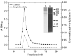
Fig. 1. Affinity chromatography purification profile and Leishmania amazonensis extracellular serine protease SDS–PAGE analysis. The supernatant obtained from supernatant culture (NH4)2SO4 precipitation was dialysed and applied to an aprotinin-agarose column (2·5 ml) previously equilibrated with 10 mM Tris-HCl buffer, pH 7·5, containing 5 mM CaCl2. The fractions (1 ml) were eluted with 10 mM Tris-HCl buffer, pH 7·5, containing 1·5 M NaCl and collected on ice. The Abs280 nm of effluents was monitored to detect the protein peak ([bull ]) and the enzymatic activity was assayed with N-ρ-tosyl-L-arginine methyl ester (L-TAME) (). The active fractions were pooled and analysed by 12% SDS-PAGE (A) and gelatin-SDS-PAGE (B) using non-reducing conditions. The values of standard molecular mass proteins (kDa) are located to the right side of the gels. Gel A was stained with Coomassie Blue R-250 and Gel B was stained using amido black.
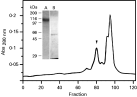
Fig. 2. Continuous elution preparative electrophoresis profile of the active fractions from affinity chromatography purification of the culture supernatant of Leishmania amazonensis. The sample obtained by the affinity chromatography purification was concentrated and applied to a continuous elution preparative electrophoresis (5% stacking, 7·5% resolving). The proteins were eluted at 1 ml/ min, and the fractions (1 ml) were collected for 5 h. The active fractions (arrow headed) were pooled, concentrated and dialysed in order to analyse the enzymatic activity, the protein content and SDS–PAGE profile. In detail, SDS–PAGE (12%) of the pooled peak under non-reducing conditions (A) and in conditions of reduction (B). The gels were silver stained and the values of standard molecular mass proteins (kDa) are located to the left side of the gels.
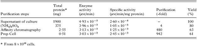
Table 1. Purification of Leishmania amazonensis extracellular serine protease
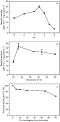
Fig. 3. Effects of pH and temperature on the enzymatic activity of Leishmania amazonensis extracellular serine protease. In all the experiments 2 μg of HPLC-purified enzyme and substrate L-TAME (0·25 mM) were used. (A) To study the effect of pH in 100 mM Solutions, the following buffers used were: sodium citrate buffer (pH 5·0–6·5) and Tris-HCl (pH 7·0–9·0). (B) The effect of temperature was investigated by incubating the enzyme and L-TAME for 30 min at several temperatures, and the heat stability was verified by pre-incubating the enzyme at 42 °C for up to 1h and then assaying the activity at 28 °C (C). Each value is the mean±S.D. (<±5%) of 2 separate experiments carried out in triplicate.
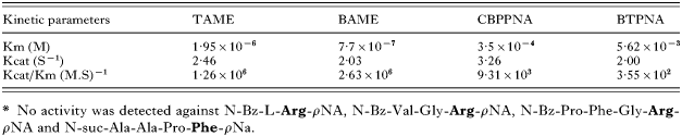
Table 2. Substrate specificity of Leishmania amazonensis extracellular serine protease on synthetic peptide substrates*
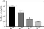
Fig. 4. Effects of cations on the enzymatic activity of Leishmania amazonensis extracellular serine protease. The purified enzyme was pre-incubated with each ion (10 mM) in Tris-buffer, for 15 min at room temperature. Values are the residual activity of serine protease as a percentage of the activity on L-TAME without ions and represent the mean±S.D. (<±5%) of 3 separate experiments carried out in triplicate.
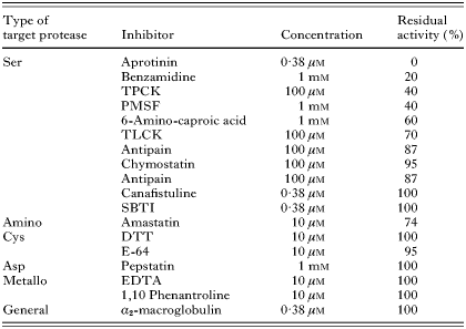
Table 3. Effects of different types of protease inhibitors on Leishmania amazonensis extracellular serine protease activity
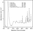
Fig. 5. Reverse-phase chromatography on a CLC-ODS column of the β-chain insulin hydrolysed by Leishmania amazonensis extracellular serine protease for 16 h. The peptides from hydrolysis of β-chain insulin were submitted to reverse-phase chromatography, and their primary structures were identified and their molecular masses determined by MALDI mass spectroscopy. The sequences of the peptides are located in the upper portion of the figure.
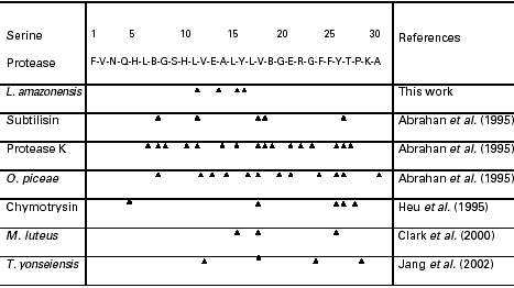
Table 4. Cleavage sites of oxidized insulin β-chain by various serine proteases
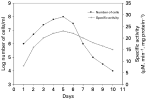
Fig. 6. Cell growth and extracellular serine protease enzymatic activity. The culture of Leishmania amazonensis promastigotes was collected over 10 consecutive days, the number of the parasites were counted ([bull ]) and the culture supernatant was obtained in order to determine the extracellular serine protease activity against L-TAME ().
 You have
Access
- 32
- Cited by