Introduction
Inflammatory myofibroblastic tumour was first described by Brunn in 1939.Reference Brunn1 Bahadori and Liebow published the first case series of inflammatory myofibroblastic tumour in the lung, in 1973, and coined the term ‘plasma cell granuloma’.Reference Bahdori and Liebow2 In 1994, the term ‘inflammatory myofibroblastic tumour’ was formalised by the World Health Organization to describe a soft tissue neoplasm of ‘intermediate’ malignancy which has the potential to recur but with a low metastatic risk. The lesion is histologically benign but locally destructive. Histologically, lesions are composed of differentiated myofibroblastic spindle cells accompanied by numerous plasma cells and/or lymphocytes.Reference Weiss, Weiss, Sobin and Enzinger3
In the head and neck region, inflammatory myofibroblastic tumour mainly involves the orbits, followed by the upper aerodigestive tract (nasopharynx, oropharynx, larynx and trachea), nasal cavity, paranasal sinuses, temporal bone and soft tissues of the neck.Reference Narla, Newman, Spottswood, Narla and Kolli4 Over the years, articles published on inflammatory myofibroblastic tumour in the temporal bone have borne other synonyms, such as inflammatory pseudotumor, plasma cell pseudotumor, pseudosarcomas myofibroblastic proliferation, myofibroblastoma, xanthomatous pseudotumor and atypical fibromyxoid nodule.Reference Cho, Lee, Jung and Kim5 However, literature on metachronous inflammatory myofibroblastic tumour is scarce. An online medical literature search for publications posted up to July 2008 revealed only five articles on metachronous inflammatory myofibroblastic tumour, which all involved regions other than the temporal bone.Reference Chan, White and Brash6–Reference Chen, Lai, Huang, Chen, Lu and Pan10 To our knowledge, the current paper represents the first reported case of metachronous inflammatory myofibroblastic tumour in the temporal bone.
Case report
A 27-year-old woman initially presented in 2003 with intermittent, right-sided otalgia and progressive hearing loss of 10 months' duration. She reported a history of an inflammatory myofibroblastic tumour in the paranasal sinuses three years prior to this presentation, which had been treated by surgical removal followed by oral steroids. She had been asymptomatic until her current problems.
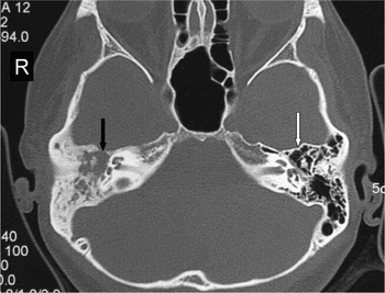
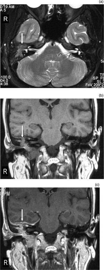
Otoscopic examination revealed a mass in the right middle ear. Nasoendoscopy showed a wide middle meatal antrostomy bilaterally as a result of previous surgery, with no evidence of recurrence. Hearing assessment revealed right-sided, profound, sensorineural hearing loss (SNHL). Hearing on the left was normal. High resolution computed tomography (CT) of the temporal bones showed an erosive mass in the right middle-ear cavity and mastoid air cells (Figure 1). The left temporal bone was normal. This lesion was isointense to brain on both T2- and T1-weighted images (Figures 2a and 2b, respectively), with homogeneous enhancement post-contrast (Figure 2c).
Axial, high resolution computed tomography scan of the right temporal bone at initial presentation, showing an erosive mass in the right middle-ear cavity extending into the mastoid air cells (black arrow). Note the normal left middle-ear cavity (white arrow).

(a) Axial, T2-weighted magnetic resonance imaging (MRI) scan of the temporal bone, showing a lesion isointense to brain parenchyma in the right middle-ear cavity (arrow). (b) Coronal, T1-weighted MRI of the temporal bone, showing that the right middle-ear cavity lesion is isointense to brain parenchyma (arrow). (c) Coronal, post-contrast, T1-weighted MRI of the temporal bone showing homogeneous enhancement of the right middle-ear cavity lesion with associated enhancement of the adjacent meninges of the floor of the right middle cranial fossa (arrow).

The patient underwent right canal wall down mastoidectomy and tumour excision.
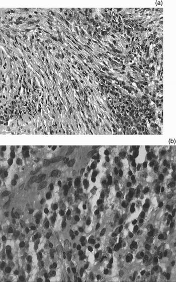
Histological examination of the tumour tissue showed multiple areas of intense, predominantly plasmacytic infiltrate surrounded by spindle cells within a fibro-collagenous stroma. Several lymphoid aggregates with germinal centres were noted. No cellular atypia or necrotic areas were seen. Immunohistochemical staining showed that the spindle cells were positive for smooth muscle actin but negative for CD34, cytokeratin and S-100 protein. These features were consistent with a diagnosis of inflammatory myofibroblastic tumour (Figure 3).
(a) Photomicrograph showing predominantly plasma cell infiltrate and spindle cells within a fibro-collagenous stroma (H&E; ×200). (b) Infiltrate of plasma cells within the tumour (H&E; ×400).

The patient was commenced on high dose steroids tapered down over six weeks, followed by a low maintenance dose for four months.
Following this treatment, the patient was asymptomatic until a year later, when she complained of intermittent otalgia and reduced hearing in the left ear, of two weeks' duration. There was no history of trauma and nothing to suggest infection. Otoscopic examination showed a middle-ear mass protruding through the tympanic membrane.
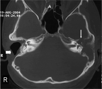
Hearing assessment showed profound, right-sided SNHL and mixed left-sided hearing loss. High resolution CT of the temporal bone this time showed an erosive mass in the left middle-ear cavity and mastoid air cells (Figure 4).
Axial, high resolution computed tomography scan of the temporal bone one year after initial presentation, showing an erosive mass in the left middle-ear cavity, extending into the mastoid air cells and consistent with a metachronous tumour (thin arrow). Note also the right mastoidectomy (thick arrow).

Due to these findings, the patient underwent left canal wall down mastoidectomy and excision of the new tumour.
Histological examination of the surgical specimen confirmed the diagnosis of metachronous inflammatory myofibroblastic tumour.
At follow up three years after the patient's second surgical procedure, she remained asymptomatic with no local recurrence of tumour. However, her hearing loss had progressively worsened.
As a result, the patient underwent a right cochlear implantation in 2006, and regained good hearing and speech.
Discussion
The clinical features of inflammatory myofibroblastic tumour depend on the site of tumour involvement. If the tumour involves the temporal bone, the patient can present with common ear symptoms such as otalgia, hearing loss, vertigo and even cranial nerve involvement if the tumour is extensive. On high resolution CT, inflammatory myofibroblastic tumour presents as a locally destructive, soft tissue mass in the middle ear which is indistinguishable from a malignant tumour. The magnetic resonance imaging (MRI) features of inflammatory myofibroblastic tumour are non-specific. Inflammatory myofibroblastic tumour has been described as isointense to brain on both T1- and T2-weighted MRI sequences, with homogeneous contrast enhancement.Reference Gasparotti, Zanetti, Bolzoni, Gamba, Morassi and Ungari11 Imaging is useful in determining the anatomical limits of these lesions, but is not sufficient to make the diagnosis of inflammatory myofibroblastic tumour.
The aetiology of inflammatory myofibroblastic tumour remains unknown but some theories have been proposed, including infection, post-surgical or foreign body reaction, and even an immunological cause.Reference Dehner12
• Inflammatory myofibroblastic tumour in the temporal bone is a benign but locally destructive lesion
• This paper presents a patient with bilateral, metachronous inflammatory myoblastic tumours
• Complete surgical excision is the treatment of choice for local disease control
The management of inflammatory myofibroblastic tumour also remains a controversial issue. There are three modalities described in the literature: surgery, steroids and radiotherapy. In general, surgery is advocated in surgically accessible locations and has the highest response rate. Steroids have been shown to be highly effective and have been recommended as first line treatment by the authors with the largest case series.Reference Olsen, DeSanto, Wold and Weiland13 High doses of steroids have been used for initial treatment, tapered over the ensuing months. However, the ideal duration of long-term therapy is unclear. Radiation has been used for patients who cannot continue oral steroids due to side effects, or for patients with tumours in poorly accessible areas, but it appears to be the least effective treatment modality.Reference Olsen, DeSanto, Wold and Weiland13
Our patient was successfully treated with complete surgical tumour excision and post-operative, long-term steroid therapy, and had no evidence of recurrence at three-year follow up.