Introduction
Carcinoma of the external auditory canal is a rare disease, with an incidence of 1 per 1 000 000 population.Reference Arriaga, Curtin, Takahashi, Hirsch and Kamerer1–Reference Nakagawa, Kumamoto, Natori, Shiratsuchi, Toh and Kakazu3 Owing to the tumour's rarity, it has been difficult to formulate a treatment strategy. With the development of skull base surgery and plastic surgery, surgical resection has become the main treatment for external auditory canal carcinoma.
Between 1981 and 2006, 33 patients were treated for carcinoma of the external auditory canal at Kurume University Hospital. According to Arriaga and colleagues' staging system,Reference Arriaga, Curtin, Takahashi, Hirsch and Kamerer1 this group contained 11 T1, six T2, eight T3 and eight T4 tumours. Patients' survival rates were calculated by the Kaplan–Meier method. The cause-specific, five-year survival rates were 100, 80, 38 and 50 per cent for stage T1, T2, T3 and T4 tumours, respectively. Although the five-year survival rate for T1 plus T2 patients reached 93 per cent, surgical resection for early cancer severely compromised patients' quality of life, due to the sacrifice of organ function. On the other hand, the five-year survival rate for T3 plus T4 cancer was 43 per cent, with a high rate of recurrence due to incomplete tumour resection.
In the treatment of external auditory canal carcinoma, local control of the primary tumour is of great importance, because neck and distant metastases are rare and most treatment failures result from local recurrence. In order to enable organ preservation in this severe disease, and to obtain cancer-free surgical margins, we introduced the use of superselective, intra-arterial, rapid infusion chemotherapy combined with radiotherapy.
Since 2003, we have treated four patients with superselective, intra-arterial, rapid infusion chemotherapy combined with radiotherapy, for carcinoma of the external auditory canal. The aim of this study was to evaluate the efficacy of intra-arterial chemotherapy administered concurrently with radiotherapy, for carcinoma of the external auditory canal.
Patients and methods
The study included four patients with primary squamous cell carcinoma of the external auditory canal (Table I). We retrospectively reviewed the records of all four patients, treated at our hospital for this condition between July 2003 to February 2006. The following information was collected: age, sex, tumour–node–metastasis (TNM) status, therapeutic method (i.e. feeder artery, frequency of intra-arterial chemotherapy and radiation dose), outcome and complications.
Four patients with external auditory canal carcinoma: summary

No = number; yrs = years; RT = radiotherapy; mths = months; F = female; M = male; T = tumour; N = node; M = metastasis; PPA = posterior auricular artery; STA = superficial temporal artery; AAW = alive and well.
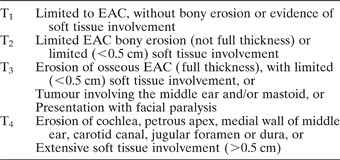
Radiography-based TNM staging was performed according to the Pittsburgh staging system. This system is based on improved radiographic methods for assessing temporal bone carcinoma, and is outlined in Table II.
University of pittsburgh staging system for external auditory canal carcinoma: tumour status

EAC = external auditory canal.
Chemotherapy was administered intra-arterially in the angiography suite via transfemoral catheterisation of the feeding arteries. In principle, patients received a cisplatin infusion (100 mg/patient) for up to one week with simultaneous intravenous administration of sodium thiosulphate, a neutralising agent. Patients received external beam radiation simultaneously with the cisplatin infusion chemotherapy. Radiotherapy was administered at 2.0 Gy per fraction, once daily, five days a week, with a photon beam energy of 6 MV. Two laterally angled, pair-wedged fields were used in all patients.
After two or three intra-arterial infusion chemotherapy sessions, the response to the therapy was evaluated using computed tomography (CT) or magnetic resonance imaging (MRI). Patients who appeared to have a complete response were continuously treated with chemo-radiation therapy for up to four infusions and a total dose of 60 Gy. On the other hand, patients who appeared to have a partial response or no change were treated by surgery, lateral temporal bone resection or subtotal temporal bone resection. The response of the disease to treatment was determined by either CT or MRI, six months after completion of therapy. The criteria for complete response, partial response and no change were based on the standard definitions established by the World Health Organization. We recorded any complications, such as osteoradionecrosis, sensorineural hearing loss and thromboembolic events.
All patients underwent audiological testing before commencing therapy and again 20 months afterwards. Assessment of patients' bone condition was used as an indicator of the stability of their cochlear function. Patients' sensorineural hearing level was calculated as the mean of bone conduction values for 250, 500, 1000, 2000 and 4000 Hz. Significant hearing loss was defined as greater than 15 dB sensorineural hearing loss.
Results
The four patients comprised two men and two women, with a median age of 68 years (range 46–79 years). All had squamous cell carcinoma. According to the Pittsburgh staging system, two patients had stage T2 tumour, one had stage T3 and one had stage T4. All patients were free of neck or distant metastases. The median follow-up period was 30 months, ranging from 19 to 50 months. The mean dose of radiation was 62.5 Gy (range 60–70 Gy). Cisplatin was delivered via the posterior auricular artery in three patients and via the superficial temporal artery in one. The posterior auricular artery is an important feeding artery. Intra-arterial cisplatin infusions were delivered as follows: two infusions in two patients, three infusions in one patient and four infusions in one patient. All patients were evaluated in order to assess their response. A complete reponse was obtained in all four patients. Following treatment, all patients were well with no signs of recurrence.
Overall, toxic side effects were modest. Osteoradionecrosis of the temporal bone and thromboembolic events were not observed. The median follow-up period for hearing assessment was 27 months, ranging from 22 to 36 month. The average hearing loss in the five frequency areas tested was 5.25 dB (range −17 to 18 dB). A hearing loss of more than 15 dB occurred in only one patient.
Case reports
Case one
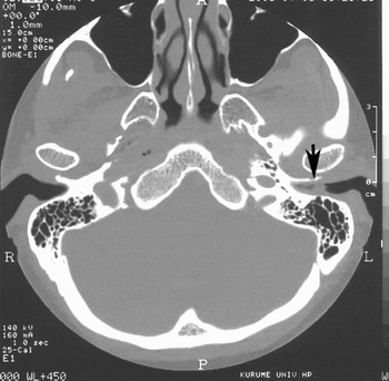
In June 2003, a 46-year-old woman was referred to our hospital after presenting with a one-year history of otorrhoea. The tumour was located on the posteroinferior wall of the left external auditory canal. A CT showed the tumour to involve the left external auditory canal with bony erosion, but not to invade the soft tissue (Figure 1). A MRI showed that the mass measured 10 × 5 × 5 mm and was enhanced by gadolinium administration. A biopsy specimen suggested a diagnosis of squamous cell carcinoma. The patient was diagnosed with a T2 N0 M0 external auditory canal squamous cell carcinoma.
Axial computed tomography scan for case one. The tumour (arrow) involved the left external auditory canal. Bony erosion was present.

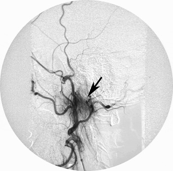
We selected intra-arterial infusion chemotherapy combined with radiotherapy, because the patient was eager for therapy which would preserve her hearing function. Cisplatin was delivered via the posterior auricular artery (Figure 2), and radiotherapy (60 Gy) was performed.
Angiographic scan for case one. A rapid infusion of cisplatin was delivered twice through the posterior auricular artery. Arrow indicates the posterior auricular artery (the feeding artery).

After this treatment, the tumour disappeared. The follow-up examination revealed no local recurrence or distant metastasis.
Case two
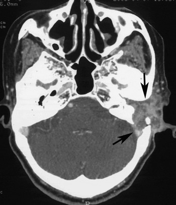
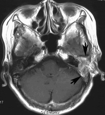
In March 2005, a 79-year-old woman was referred to our hospital with a one-year history of otorrhoea and otalgia. Her left external auditory canal was filled with tumour. A CT showed that the tumour had destroyed the bony wall of the external auditory canal and was invading into the tympanic cavity and mastoid. In addition, it had eroded into the basilar skull, including the posterior and middle fossae (Figure 3). A MRI demonstrated the presence of dural invasion, and the tumour was well enhanced by gadolinium administration (Figure 4). The histological diagnosis from a biopsy specimen was squamous cell carcinoma. The patient was diagnosed with stage T4 N0 M0 external auditory canal squamous cell carcinoma.
Axial computed tomography scan for case two. The tumour (arrows) had destroyed the bony wall of the external auditory canal and eroded into the basilar skull, including the posterior and middle fossae.

Axial magnetic resonance imaging scan with gadolinium enhancement, for case two. The tumour (arrows) was well enhanced by gadolinium administration, and was invading the dura.

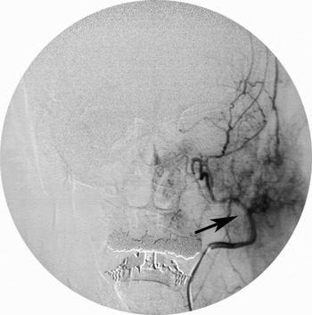
Because of the patient's advanced age and the fact that her tumour was not deemed to be resectable, she underwent intra-arterial infusion chemotherapy combined with radiotherapy. Cisplatin was delivered through the posterior auricular artery three times (Figure 5), and radiotherapy (70 Gy) was performed.
Angiographic scan for case two. Rapid infusion of cisplatin was delivered three times through the posterior auricular artery. Arrow indicates the posterior auricular artery (the feeding artery).

After the treatment, the tumour disappeared. The follow-up examination revealed no local recurrence or distant metastasis.
Discussion
Treatment principles for external auditory canal carcinoma
Surgery plays a central role in the treatment of squamous cell carcinoma arising from the external auditory canal, and most patients are considered to benefit from a combination of surgery and radiation therapy. The role of chemotherapy remains uncertain. Because distant metastases are not commonly reported and local control of the primary tumour is important, systemic chemotherapy is not routinely used. It is widely accepted that radiotherapy or radiotherapy combined with surgery is a reasonable treatment for patients with T1 disease.Reference Ogawa, Nakamura, Hatano, Uno, Fuwa and Itami4–Reference Hashi, Shirato, Omatsu, Kagei, Nishioka and Hashimoto7 On the other hand, the prognosis of patients with T2 or more advanced disease treated by radiotherapy alone is poor, and radiotherapy combined with surgery is recommended as the standard treatment for these patients.Reference Ogawa, Nakamura, Hatano, Uno, Fuwa and Itami4, Reference Hashi, Shirato, Omatsu, Kagei, Nishioka and Hashimoto7–Reference Korzeniowski and Pszon10 Surgical resection for external auditory canal SCC is often followed by local recurrence, due to frequent invasion of the adjacent temporal bone.Reference Wang6 Most investigators agree that a wide en bloc resection of the tumour, with clear surgical margins, is the optimal treatment.Reference Nakagawa, Kumamoto, Natori, Shiratsuchi, Toh and Kakazu3, Reference Ogawa, Nakamura, Hatano, Uno, Fuwa and Itami4, Reference Yin, Ishikawa, Honda, Arakawa, Harabuchi and Nagabashi11 Nevertheless, a pathologically positive margin was mainly observed in patients with stage III and IV tumours.Reference Yin, Ishikawa, Honda, Arakawa, Harabuchi and Nagabashi11 Some investigators have found the five-year disease-free survival rate to be significantly higher in patients with a negative surgical margin than in patients with a positive surgical margin.Reference Nakagawa, Kumamoto, Natori, Shiratsuchi, Toh and Kakazu3, Reference Yin, Ishikawa, Honda, Arakawa, Harabuchi and Nagabashi11, Reference Nyrop and Grontved12 Several investigators have indicated incomplete resection to be the major cause of recurrence, and have found post-operative radiotherapy not to be beneficial.Reference Nakagawa, Kumamoto, Natori, Shiratsuchi, Toh and Kakazu3, Reference Goodwin and Jesse13 Patients with advanced cancer who undergo an incomplete tumour resection should be treated with neoadjuvant therapy. Therefore, some investigators have recommended pre-operative radiotherapy.Reference Nakagawa, Kumamoto, Natori, Shiratsuchi, Toh and Kakazu3 Nakagawa et al. Reference Nakagawa, Kumamoto, Natori, Shiratsuchi, Toh and Kakazu3 stated that a complete en bloc resection with tumour-free surgical margins plus pre-operative chemoradiotherapy effectively improved the estimated survival of patients with advanced SCC of the temporal bone. Comparing concurrent chemo-radiotherapy with radiotherapy alone, several investigators have found that chemo-radiotherapy improved local control, while often resulting in tumour absence at the surgical margin.Reference Pignon, Bourhis, Domenge and Designe14, Reference Al-Sarrf, Marts, Herskovic, Leichman, Brindle and Vaitkevicius15
Intra-arterial infusion chemotherapy combined with radiotherapy
No previous reports have indicated that intra-arterial infusion chemotherapy combined with radiotherapy is equally effective for the treatment of external auditory canal carcinoma, compared with surgical resection. However, Robbins et al. Reference Robbins, Staniolo, Kerber, Seagren, Berson and Howell16 developed a delivery system for administration of high dose cisplatin to patients with head and neck cancer. Intra-arterial chemotherapy has a theoretical advantage over standard intravenous chemotherapy because of the higher concentration of chemotherapeutic agent delivered directly to the tumour bed, and the lower concentration delivered to other organs.
Intra-arterial infusion is an especially relevant delivery system for cisplatin, because this agent's tumour cell killing effect is known to be dose-dependent.Reference Los, Blommaert, Barton, Heath, den Engelse and Hanchett17 Intra-arterial, high dose, cisplatin chemotherapy combined with concurrent radiation therapy showed high complete response rates and good local control rates in patients with paranasal sinus, pharyngeal and laryngeal cancer.Reference Robbins, Vicario, Seagren, Weisman, Pellitteri and Kerber18–Reference Samant, Robbins, Vang, Wan and Robertson21 Furthermore, intra-arterial infusion chemotherapy combined with radiotherapy has been effectively used in the treatment of maxillary sinus carcinoma, and it has been proven as an effective organ preservation treatment with a high cure rate.Reference Samant, Robbins, Vang, Wan and Robertson21–Reference Shiga, Yokoyama, Hashimoto, Saijo, Tateda and Ogawa23
It is our opinion that intra-arterial infusion chemotherapy combined with radiotherapy is also effective for the treatment of external auditory canal carcinoma. The following three points are thought to support this belief. (Reference Arriaga, Curtin, Takahashi, Hirsch and Kamerer1) Anatomically speaking, it is difficult to remove external auditory canal carcinoma by en bloc resection with tumour-free surgical margins, because the external auditory canal is enclosed by a bony wall and important organs are located close by. (Reference Arena and Keen2) Functionally speaking, wide surgical resection severely compromises the patient's quality of life, due to sacrifice of organ function. (Reference Nakagawa, Kumamoto, Natori, Shiratsuchi, Toh and Kakazu3) Regarding the availability of a feeding artery, the external auditory canal is supplied by branches of the external carotid artery.
In our study, cisplatin was delivered via the postauricular artery in three patients (75 per cent); this artery was identified as an important feeding artery of external auditory canal carcinoma. A complete response was obtained in all four patients; these results indicate that intra-arterial infusion chemotherapy may be an effective organ preservation treatment with a high cure rate. In addition, these results suggest this treatment modality to be an effective therapy for advanced external auditory canal carcinoma.
Complications
Osteoradionecrosis
Osteoradionecrosis of the temporal bone is a major and not infrequent complication of radiotherapy; its reported incidence ranges from 5 to 30 per cent.Reference Wang, Tu and Liu24–Reference Nadol and Schuknecht27 In a series reported by Nadol and Schuknecht,Reference Nadol and Schuknecht27 of 27 patients with carcinoma of the ear treated with a combination of surgery and post-operative radiation therapy, osteoradionecrosis developed in eight (30 per cent). These authors reported that osteoradionecrosis occurred in 42 per cent of cases (eight of 19) treated with surgery leaving an open cavity plus post-operative radiotherapy of average 5840 rad. On the other hand, they reported no osteoradionecrosis in eight patients treated by surgery with cavity obliteration plus post-operative radiotherapy of average 5700 rad. As a result, Nadol and Schuknecht concluded that cavity obliteration significantly decreased the incidence of osteoradionecrosis. The latent period between radiotherapy and the onset of osteoradionecrosis has been reported to range from eight months to 13 years.Reference Pemberton, Swindell and Sykes26, Reference Nadol and Schuknecht27 Further investigations of osteoradionecrosis in such cases should therefore be conducted.
Sensorineural hearing loss
The effects of radiation on the auditory apparatus have long been a topic of interest. In such circumstances, hearing loss is due to the direct effect of ionising radiation on postmitotic cells and/or altered vascular physiology which interferes with oxygen supply to the cochlea.Reference Moretti28 The latent period between radiotherapy and the onset of sensorineural hearing loss has been reported to range from six month to four years.Reference Grau, Moller, Overgaard, Overgaard and Elbrond29–Reference Ho, Wei and Kwong32 In some studies, mainly those involving nasopharyngeal carcinoma and parotid tumours, the incidence of sensorineural hearing loss following radiotherapy ranged from approximately 30 to 50 per cent.Reference Grau, Moller, Overgaard, Overgaard and Elbrond29, Reference Schot, Hilgers, Keus, Schouwenburg and Dreschler33 In the current study, the average hearing loss over five frequencies was 5.25 dB, and significant hearing loss occurred in only one patient; no deafness was found to occur.
Thromboembolic events
We did not observe any thromboembolic events in our series. However, we recognise that catheter-related thromboembolic events remain a potential complication associated with intra-arterial infusion chemotherapy.
Conclusions
With the development of skull base surgery and plastic surgery, the survival rate for external auditory canal carcinoma has improved. However, surgical treatment for this tumour can often cause facial paresis, cosmetic damage (due to deformation of the auricle) and hearing loss. Considering such quality of life issues, a less aggressive treatment is therefore desirable for patients with external auditory canal cancer. Our results indicate that superselective intra-arterial infusion chemotherapy may be a safe and effective organ preservation treatment, with a high cure rate.