Introduction
Teak (Tectona grandis L. (Verbenaceae)) is a highly valued tropical deciduous timber. Two seasonal defoliator pests Hyblaea puera Cramer (Lepidoptera: Hyblaeidae), commonly known as the teak defoliator, and Paliga machoeralis Walker (Lepidoptera: Pyralidae), popularly called the teak skeletonizer, attack teak plants and affect the growth of the trees considerably (Nair et al., Reference Nair, Sudheendrakumar, Varma and Chacko1985, Reference Nair, Sudheendrakumar, Varma, Chacko and Jayaraman1996). The infestation of H. puera begins with the first monsoonal showers when new sprouts appear on the trees. At times infestation can be very severe with caterpillars completely defoliating the trees within a short period of time. The management of these defoliators in large plantations is often difficult and no effective methods of control have been developed. Currently, naturally occurring native entomopathogens are being explored as possible biological control agents against this pest. A potential baculovirus, tentatively designated as H. puera nucleopolyhedrovirus has been recently isolated from H. puera and a method for mass production of the virus for management of this pest has been developed (Sajeev and Sudheendrakumar, Reference Sajeev and Sudheendrakumar2005).
Many species of forest Lepidoptera are affected by microsporidia that are considered important naturally occurring regulatory factors of the populations of these insects. Many potentially pathogenic species belonging to the genus Nosema have been isolated from forest Lepidoptera. Nosema thomsoni from the tortricid moth Choristoneura conflictana (Walker) (Lepidoptera: Tortricidae) (Wilson and Burke, Reference Wilson and Burke1971), Nosema disstriae from the forest tent caterpillar Malacosoma disstriae Hübner (Lepidoptera: Lasiocampidae) (Thomson, Reference Thomson1959; Wilson, Reference Wilson1977), a microsporidium from the fall webworm Hyphantria cunea Drury (Lepidoptera: Arctiidae) (Nordin and Maddox, Reference Nordin and Maddox1974), Nosema fumiferanae from the spruce bud worm Choristoneura fumiferana (Clemens) (Lepidoptera: Tortricidae) (Wilson, Reference Wilson1980), Nosema portugal from populations of Lymantria dispar L. (Lepidoptera: Lymantriidae) in Portugal (Maddox et al., Reference Maddox, Baker, Jeffords, Kuras, Linde, Solter, McManus, Vavra and Vossbrinck1999) and many others have been the subjects of careful study by various workers. A new microsporidian parasite N. chrysorrhoeae was recently isolated from the browntail moth Euproctis chrysorrhoea L. (Lepidoptera: Lymantriidae) and the authors have indicated the parasite's potential for control of this pest (Hylis et al., Reference Hylis, Pilarska, Obornik, Vavra, Solter, Weiser, Linde and McManus2006). Furthermore, recently, Kyei-Poku et al. (Reference Kyei-Poku, Gauthier and Frankenhuyzen2008) have published a detailed account of the several Nosema species infecting lepidopteran forest defoliators in the genera Choristoneura and Malacosoma.
Members of the other genera particularly Vairimorpha and Endoreticulatus are also commonly recorded from forest Lepidoptera (Maddox et al., Reference Maddox, Brooks, Fuxa and Burges1981, Reference Maddox, McManus, Solter, McManus and Liebhold1998; Wang et al., Reference Wang, Solter, T'sui and Wang2005). Microsporidians of the gypsy moth are perhaps the most extensively studied among those infecting forest Lepidoptera. Many isolates are reported as important pathogens of L. dispar in Europe. Their distribution and significance in biological control have also been discussed by a number of researchers (McManus et al., Reference McManus, Maddox, Jeffords and Webb1989).
Microsporidia are undoubtedly prevalent in the insects inhabiting the tropical forests too, but only very few studies have gone into documenting their occurrence, distribution and role in many insect epizootics. Barring some of the studies on Nosema disease of silkworm (Ananthalakshmi et al., Reference Ananthalakshmi, Samson, Baig and Datta1994; Sasidharan et al., Reference Sasidharan, Singh, Samson, Manjula, Santha and Shekhariah1994), there has been very little work on microsporidia in India, especially on those infecting forest Lepidoptera (Johny et al., Reference Johny, Kanginakudru, Muralirangan and Nagaraju2006). It was the severity of defoliation and damage caused by the teak moth as a defoliator that prompted our search for enzootic microsporidia that could be exploited for biocontrol purposes.
Materials and methods
We conducted a survey of teak plantations during the pest outbreak seasons (usually from May to September after the first monsoonal showers). Weak and diseased H. puera larvae were collected, brought to the laboratory and screened for microsporidian pathogens. An unidentified microsporidium was recovered from H. puera larvae collected from a teak nursery in Kanakpura, Bangalore district, Karnataka as previously reported (Sasidharan et al., Reference Sasidharan, Remadevi, Anitha Rani, Priyadarsanan and Bhattacharya2008).
The spores of the microsporidium were purified from homogenized infected larvae by filtration and repeated centrifugation as described earlier (Sasidharan et al., Reference Sasidharan, Singh, Samson, Manjula, Santha and Shekhariah1994). They were further purified by the method of Sato and Watanabe (Reference Sato and Watanabe1980) using a gradient of neutralized Percoll (Sigma, St Louis, MO, USA). After centrifugation at 73,000 g for 30 min, a band of purified spores was collected from the gradient. The spores were washed twice in distilled water and stored in fresh sterilized distilled water at 4 °C.
Infectivity
Infectivity of the microsporidium was assessed by oral inoculation of spores to 3rd instar larvae of H. puera. The spore suspension (0.1 ml) with a concentration of 105 spores/ml was smeared on a small quantity of artificial diet placed in small glass vials and an individual 3rd instar larva was transferred to each vial. Each larva received a dose of 104 spores. Fresh diet was provided after ensuring complete consumption of the contaminated diet. The larvae were reared in an incubator at 26 ± 1 °C and 70–80% relative humidity (RH) under a light regime of 14 h light-10 h dark and allowed to pupate. Five replicates of 30 larvae were maintained for the trial. Faecal pellets discharged by the larvae and the larvae that succumbed to infection before pupation were examined for the presence of mature spores of the microsporidium. Adult emergence and infection in the adults were also recorded to assess the infectivity potential of the parasite. Body weight of at least ten infected larvae was also simultaneously recorded on alternate days till the fifth day after pupation, to assess the effect of infection on growth and development of the insect.
Sequence of infection and multiplication in different organs
A group of H. puera larvae on the first day of the 3rd instar were separately inoculated as per the method described above for studying the sequence of infection of the various organs. Midgut, fat body, tracheal membrane and Malpighian tubules were carefully dissected out, washed in a small quantity of sterile distilled water and examined every day from the late 4th instar till eclosion of the adult moths. The reproductive organs were examined from the late 5th instar onwards as they were not well developed before that time. Organs from at least five individuals were examined on each day to confirm the sequence of infection.
Besides studying the sequence of infection, concentration of spores in the organs was also assessed in pupae on successive days of development. In some cases, each specific organ from a group of pupae was pooled together because of the very small quantity of tissue obtained from an individual pupa. The dissected organs were washed carefully in a small quantity of sterile distilled water before macerating to determine the concentration of spores. For each organ, five replicates were maintained for the subsequent spore number assay. The weight of each tissue sample was recorded before homogenizing and recovering the spores. The samples were homogenized in a small quantity of 0.5% K2CO3 solution and the homogenate filtered through a single layer of thin cheesecloth. K2CO3 was used as the degrading solution for homogenizing the tissues because it facilitates effective release of the spores from the tissues (Sasidharan et al., Reference Sasidharan, Singh, Samson, Manjula, Santha and Shekhariah1994). The tissue debris collected in the cloth was washed again into a small quantity of K2CO3 solution, vortexed and filtered to collect any trapped spore in the debris. The filtrate was centrifuged at 3500 rpm for 5 min to sediment the spores. The sediment was dispersed in a known volume of sterile distilled water over a vortex and the spores were counted using an improved Neubaur haemocytometer. The quantity of spores was expressed per unit wet-weight of tissue. The data were analysed by performing a two-factor ANOVA.
Horizontal transmission
To check the horizontal transmission potential of the parasite, a single infected 4th instar larva, inoculated on the first day in the 2nd instar with 103 spores as per the procedure described above, was introduced into a group of 20 healthy larvae just after the 3rd ecdysis. The excretion of spores by the introduced larva was confirmed by examination of the faecal pellets discharged by the larva. These larvae generally excreted the parasite spores by the middle of the 4th instar (earlier observation). The larvae (20+1 infected larva) were then reared together on artificial diet in polypropylene plates placed in an incubator at 26 ± 1 °C and 70–80% RH under a light regime of 14 h light-10 h dark, until pupation. The introduced larva was allowed to remain together with the group until all the experimental larvae pupated. Any introduced larva dying during the experiment was identified by regular observation and replaced by an infected one from the inoculated group maintained separately. All the larvae pupated within 6 days of the experiment, and thus the chances of the larvae transmitting the infection to each other through contaminated faeces were presumed to be remote or negligible. Further, the faecal pellets discharged by the experimental larvae were separately collected and periodically examined. In most cases, spores could not be observed in their faeces until just before they pupated. Therefore, the test larvae transmitting the disease could be further ruled out and thus the source of infection in the test larvae was only from the introduced infected larva. Five replicates were run, each with 20 larvae. The moths emerging from the pupae were immediately examined and the percentage of infection was assessed. The experiment was repeated three times following the same procedure to verify the results.
Vertical transmission
To assess the potential and degree of vertical transmission, 3rd instar H. puera larvae were inoculated individually through artificial diet, each with 0.1 ml of a suspension of spores having a concentration of 105 spores/ml. Each larva received a spore load of 104. Larvae were reared on artificial diet until pupation. The male and female moths that emerged were separated and allowed to selectively mate as indicated below:
Group 1: Infected female × infected male
Group 2: Infected female × healthy male
Group 3: Healthy female × infected male
Group 4: Healthy female × healthy male
The uninfected moths used for the experiment were obtained from a healthy batch of larvae reared separately (Fig. 1). Five sets of parents (five replicates) were used in each treatment group. The eggs from each set of parents from all the four treatment groups were collected, rinsed in a dilute solution of sodium hypochlorite and incubated separately. The hatched larvae from each set of parents (replicate) in the four treatment groups were reared separately until the 3rd instar and 30 larvae from each replicate were individually homogenized and examined for assessing the infection. The percentages of infection in the larvae from the four treatment groups were compared to assess the degree of vertical transmission.
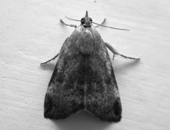
Teak defoliator moth, Hyblaea puera

DNA sequencing
The standard procedures were followed for spore isolation and DNA sequencing. Infected moths were homogenized in water, filtered through 50 μm nylon cloth and centrifuged through a 50% Percoll cushion in a 1.5 ml micro-centrifuge tube. The spores were then washed twice in STE buffer. DNA was liberated by bead beating in STE buffer. The solution was heated to 95 °C for 5 min and 4 μl of this solution was added to a standard polymerase chain reaction mixture using cycling parameters and amplification primers (18f 5′ CACCAGGTTGATTCTGCC 3′, 1492r 5′ GGTTACCTTGTTACGACTT 3′) as described previously (Vossbrinck et al., Reference Vossbrinck, Andreadis and Debrunner-Vossbrinck1998).
Results and Discussion
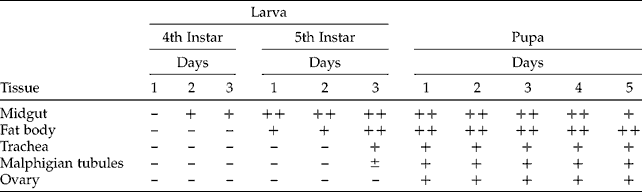
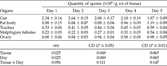
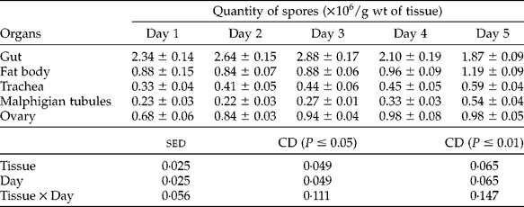
The spores of the microsporidium were ovo-cylindrical and had a mean size of 5.1 × 2.8 μm. It was highly infective to H. puera larvae at the dose (104 spores) tested and only about 39% developed into adults. All the emerged moths were also infected. Infection produced a marked negative effect on the growth and development of larvae. The weight of infected larvae increased only about 12 times up to pupation while the increase was about 22 times in the case of healthy larvae, indicating the strong debilitating effect of the parasite on growth and development of the insect. The midgut and fat body of larvae dosed with the microsporidium were heavily infected with spores. During later stages, infection was observed in the tracheal epithelium, Malphigian tubules and gonads. The parasite sequentially infected the different organs in the order: midgut – fat body – tracheal membrane – Malpighian tubules – gonad (Table 1). In general, the spore production in the different organs of pupae increased with age of pupae, except in the gut where it declined after an initial increase, possibly because of the gradual atrophy of the gut as the pupae became older (Table 2). This may suggest the tendency of the parasite to transform into mature spores for ensuring their survival as the nutrients of the host body are gradually depleted with advancing age. Microsporidian development has been reported to be closely related to the host body nutritional state (Sasidharan et al., 1994), besides a number of environmental factors (Becnel and Undeen, Reference Becnel and Undeen1992).
Sequence of infection in different organs of Hyblaea puera
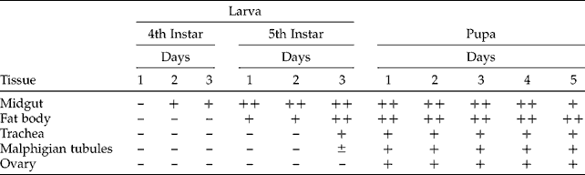
± , low infection; +, moderate infection; ++, high infection.
Spore number counted in different organs of Hyblaea puera pupa
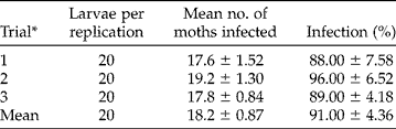
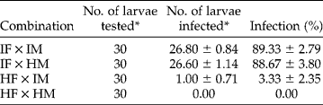
Studies on horizontal transmission in the laboratory showed that a single infected larva introduced into a healthy brood of larvae could result in >90% infection among the individuals of the brood developing together (Table 3). This suggested the potential of the parasite to cause efficient horizontal transmission in the field populations of the insect during outbreaks. Infection of the ovaries in females caused a high degree of vertical transmission of the microsporidium to the progeny. The transovarian transmission was 88.5% in the progeny larvae of infected females (Table 4). The mild infection (3.3%) in the progeny larvae from healthy females mated with infected males probably suggests the role of venereal transmission of the microsporidium via spores on the egg surface. Some of the spores sticking to the surface of the eggs could have escaped from the surface sterilization treatment. These characteristics tend to indicate the efficiency of the microsporidium to cause disease epidemic in the field, suggesting its prospects for biological control of the defoliator if exploited appropriately. However, the relevance and validity of these results need to be tested in the field through carefully designed long-term field experiments. Under experimental conditions, this microsporidium has been observed to infect another highly damaging teak pest, the teak skeletoniser P. machoeralis also (O.K. Remadevi, personal observation), further broadening its possible usefulness as a candidate organism for management of both the teak pests.
Horizontal transmission of the microsporidium in Hyblaea puera (mean±SE)
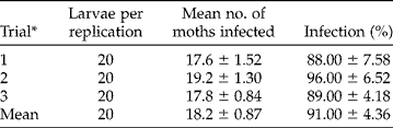
* Each trial replicated five times with 20 larvae each.
Vertical transmission of the microsporidium in Hyblaea puera (mean±SE)
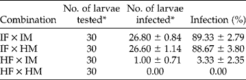
IF, infected female; IM, infected male; HM, healthy male; HF, healthy female. *Mean of five replicates.
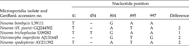
Comparative small subunit (SSU) rDNA sequence analysis showed that the H. puera microsporidium (accession number GQ244502) had a high sequence similarity to Nosema bombycis. There was only a two-nucleotide difference in the SSU rDNA between N. bombycis and the teak moth microsporidium. Table 5 shows a few other microsporidia isolates that have high similarity to N. bombycis. The teak moth isolate, like N. bombycis, is also transovarially transmitted. The teak moth and the silk moth are not as closely related as these two parasites appear to be, implying the likelihood of host switching.
Nucleotide difference from Nosema bombycis in five closely related microsporidian isolates
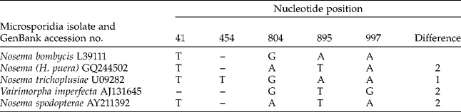
The teak microsporidium (from Hyblaea puera) shows two nucleotide differences: at position 804 (A versus G) and at position 895 (T versus A).
Conclusion
This study demonstrated some of the pathological effects and transmission potential of a newly isolated microsporidium (Nosema sp.) in the lepidopteran teak defoliator H. puera. The microsporidium was observed to be very efficiently transmitted in the teak defoliator, both horizontally and vertically and therefore holds good prospects for biological control of the pest. SSU rDNA analysis showed that this microsporidium was a close relative of N. bombycis of silk moth from which it differed only by two nucleotides. The teak moths and the silk moths are not as closely related as these two parasites appear to be, and therefore the occurrence of these closely related microsporidia in these two moths might imply the likelihood of host switching by the parasite.
Acknowledgements
This work was supported by a grant from the Department of Science and Technology, Government of India, New Delhi. The authors thank the Directors of ATREE and IWST, Bangalore for providing facilities to undertake the study. Permission granted by the PCCFs of Karnataka and Andhra Pradesh State Forest Departments to undertake survey for collection of the teak pests is thankfully acknowledged.