Case report
A 2-month-old boy with a body weight of 5.7 kg was admitted in ICU in our institution for irritation and poor feeding. Electrocardiography confirmed the diagnosis of supraventricular tachycardia with a heart rate of 260 beats/minute. Chest X-ray showed increased cardiothoracic ratio and lung congestion. Transthoracic echocardiography found increased left ventricular size with decreased left ventricular ejection fraction of 46% and moderate mitral regurgitation. Laboratory data revealed significant elevated plasma N-terminal prohormone level over 35,000 ng/ml. Electrical cardioversion could revert the rhythm to sinus, but sinus rhythm could not be maintained despite of combinations of intravenous amiodarone, oral digoxin, and propafenone for 16 days.
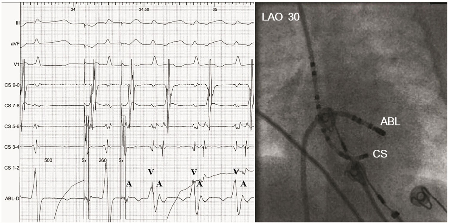
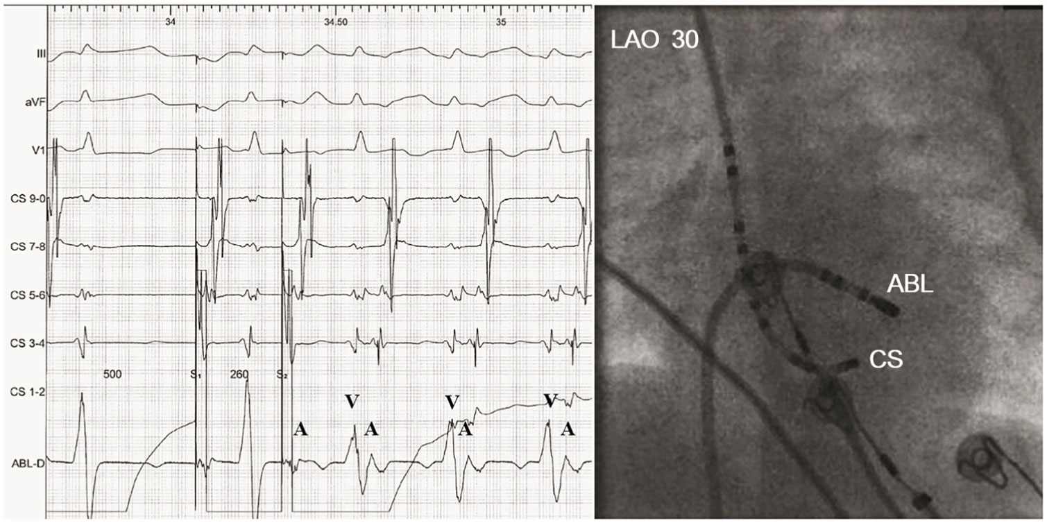
After informed consent was obtained, electrophysiology study was performed for the unstable and drug-refractory supraventricular tachycardia. In brief, a decapolar catheter (5Fr, Bard Electrophysiology) and a quadpolar catheter (6Fr, Bard Electrophysiology) were inserted at the coronary sinus and right ventricular apex, respectively, for standard pacing and extrastimulation protocols. Then, the catheter in right ventricle was removed and a 4-mm tip ablation catheter (7Fr, A curve, Bard Electrophysiology) was introduced via the right femoral vein and patent foramen ovale for mapping and subsequent ablation. Tachycardia was easily induced by atrial extrastimulation and the site of earliest A wave along mitral annulus was targeted for ablation (Fig 1). Radiofrequency energy was delivered during sinus rhythm, starting with the temperature of 50°C and the power of 15 W. The maximum power setting was gradually increased up to 40 W in order to reach the target temperature. After the first application of a total duration time of 90 seconds, tachycardia could not be induced by the same atrial protocol. Two other applications were given at the same site with the same power and duration to avoid recurrence, considering the impaired catheter stability. A successful elimination of a left posterior-lateral accessory pathway was achieved with the total fluoroscopy time and a radiation dose of 5.4 minutes and 11.4 µ Gy/m2, respectively.
Radiofrequency ablation was performed to eliminate a left posterior-lateral accessory pathway. The fluoroscopic image (right panel) depicted the position of the decapolar and ablation catheters. The decapolar catheter was introduced via the right internal jugular vein with the tip of the catheter (CS1-2) positioned in the proximal coronary sinus. The ablation catheter was positioned at the posterior-lateral mitral annulus by transseptal approach. Tachycardia was easily induced by atrial extrastimulation with an earliest A wave on the ablation catheter (left panel).
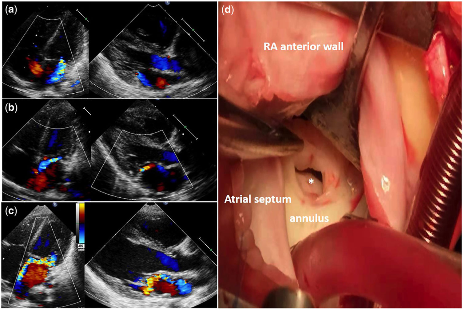
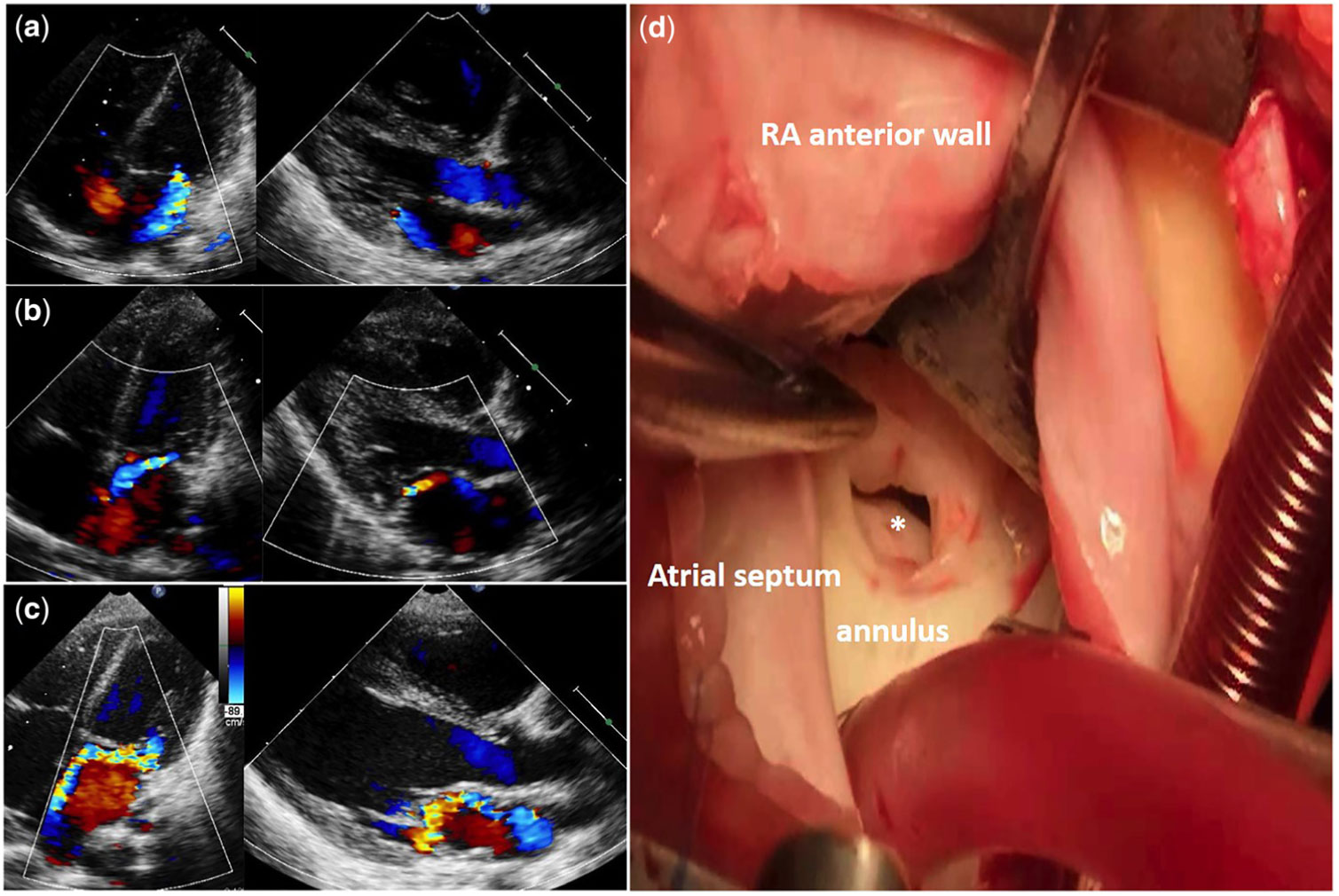
The patient was weaned off all antiarrhythmic medications and discharged 3 days after the procedure. Echocardiography on the same day of discharge showed a normal left ventricular ejection fraction of 62% and central moderate mitral regurgitation similar to pre-procedural imaging (Fig 2a). Three weeks after ablation, a repeat echocardiography discovered mild eccentric mitral regurgitation towards the septum (Fig 2b). The eccentric mild mitral regurgitation was found to have progressed to moderate degree at 3-month follow-up (Fig 2c). The location and direction of the regurgitation jet suggested that a small hole was made in the posterior leaflet of mitral valve. During follow-up period, the patient remained free of symptoms and arrhythmia, and stable of severity of mitral regurgitation. An uneventful successful surgical repair was performed when the patient was 2.5 years old, and a 5-mm hole adjacent to the annulus in the posterior leaflet of mitral valve was identified (Fig 2d).
A progressive mitral valve perforation was identified by post-procedural serial echocardiography studies and observation during open-heart surgery. ( a ) Echocardiography 3 days after ablation showed moderate central mitral regurgitation. ( b ) Echocardiography on 3-week follow-up discovered mild eccentric mitral regurgitation towards the septum. ( c ) Echocardiography on 3-month follow-up revealed mild eccentric mitral regurgitation progressing to moderate degree. The location and direction of regurgitation jet suggested that a small hole was made in the posterior leaflet of mitral valve. ( d ) A 5-mm hole (*) adjacent to the annulus in posterior leaflet of mitral valve was identified by surgical approach of right atrial anterior wall and atrial septum incision.
Discussion
Animal studies have demonstrated enlargement of the ablation lesions of both radiofrequency ablation and cryoablation in immature myocardium, although the underlying mechanism is unclear.Reference Khairy, Guerra and Rivard 1 , Reference Saul, Hulse, Papagiannis, Van Praagh and Walsh 2 The late effects related to lesion growth in immature myocardium of small children remain uncertain and are certainly of great concern. To our knowledge, this is the first report of clinically significant complication related to late effects of radiofrequency ablation in small children. In our case, serial echocardiography studies indicate that radiofrequency ablation lesions can expand over time and result in significant complication in infant. The eccentric mitral regurgitation caused by posterior leaflet perforation was not found 3 days after radiofrequency ablation. The subsequent echocardiographic follow-up revealed the perforation occurred and continued to enlarge until 3 months after ablation. Therefore, both timing of occurrence and progression of severity of mitral regurgitation suggest that perforation is resulted from late effects of radiofrequency ablation lesions rather than acute damage of improper manipulation or radiofrequency ablation lesions. We speculate that radiofrequency current was delivered directly on or close to the valve tissue during procedure, resulting in a damaged region within the posterior leaflet. The lesion continued to grow over time, subsequently became a hole and was getting larger.
Radiofrequency ablation has been reported to perform successfully and safely in infants and neonates without modification to standard procedure.Reference Akdeniz, Ergul, Kiplapinar and Tuzcu 3 – Reference Ozaki, Nakamura and Suzuki 5 However, in controlled animal studies, radiofrequency ablation lesion size is related to catheter tip size, temperature, ablation power, and application duration.Reference Avitall, Helms, Huang and Wilber 6 Furthermore, Blaufox et al showed that complications of radiofrequency ablation in small children appeared to be related to the number of applications over 20 seconds indexed to patients’ weight.Reference Blaufox, Paul and Saul 7 Thus, theoretical risk of injuring cardiac structures in small children might depend specifically on ablation parameters that influence lesion size.Reference Blaufox, Paul and Saul 7 In our case, the ablation parameters used were similar to those for older children, which might be responsible for the occurrence of complication. Therefore, despite the lack of evidence obtained from clinical studies, procedural modifications in small children, including the use of smaller ablation catheter, reducing power and temperature setting, and minimising application duration and number, have been advocated in the expert consensus statementsReference Philip Saul, Kanter and Writing 8 , Reference Brugada, Blom and Sarquella-Brugada 9 and should be considered in clinical practice to potentially lower the risk of cardiac complications.
Conclusion
In the present case, we demonstrate the growth of radiofrequency ablation lesion in infant. Catheter ablation in small children should be reserved for haemodynamically significant arrhythmia refractory to conventional medical management, and procedural modifications should be considered.
Acknowledgements
We thank Haosheng Yang (College of Natural Sciences, The University of Texas at Austin, USA, 78705) for his English polishing.
Financial Support
This study was funded by the National Key Research and Development Program (2016YFC1000500) and Innovation Project for Clinical Science and Technology Development (SHDC12016125).
Conflicts of Interest
None.
Ethical Standards
The authors assert that all procedures contributing to this work comply with the Helsinki Declaration of 1975, as revised in 2008, and have been approved by the institutional ethical committee of Children’s Hospital of Fudan University (2017-162).