Implications
The supplementation of CLA and essential fatty acids (EFAs) in bovine mammary epithelial cell line exposed to severe inflammatory conditions, reduced the gene expression of pro-inflammatory cytokines. This study suggests and strengthen the utilization of these fatty acids as a useful management tool for dairy cattle, their use on high-yielding dairy cow at the time of calving, could act by reducing the development of inflammatory processes related to parturition.
Introduction
The chronic inflammatory reactions are important in the pathogenesis of several cow disorder, particularly during the transition period of dairy cow (Bertoni et al., Reference Bertoni, Trevisi, Han and Bionaz2008). During this period dairy cow cannot adapt to growing demand of energy, which is necessary for the fetal growth, calving and onset of lactation (Abuelo et al., Reference Abuelo, Hernández, Benedito and Castillo2015). As a consequence, non-esterified fatty acids (NEFA) increase in bloodstream and impact both inflammatory responses and immune function of transition cows (Scalia et al., Reference Scalia, Lacetera, Bernabucci, Demeyere, Duchateau, Burvenich, Aoki, Etoh, Hashimoto, Naruse, Sano, Utsumi and Nawata2006; Lacetera et al., Reference Lacetera, Scalia, Mashek, Bernabucci and Grummer2007; Sordillo and Raphael, Reference Sordillo and Raphael2013). Moreover, plasma NEFA composition is modified: saturated (palmitate and stearate) and monounsaturated fatty acids (oleic acid) are increased, in contrast polyunsaturated fatty acids (especially n-3 fatty acids) are reduced (Sordillo and Raphael, Reference Sordillo and Raphael2013). Lipid changes can be responsible for the changes of the composition of the immune and endothelial cells membranes, and so may affect their functions.
Some studies have observed that T-cells treated with fatty acids modulated the secretion of various cytokines, such as tumor necrosis factor-α (TNF-α), interleukin-1β (IL-1β), interleukin-6 (IL-6), interleukin-8, interleukin-2, interleukin-10 (IL-10) and interferon-γ (de Jong et al., Reference Hontecillas, Wannemeulher, Zimmerman, Hutto, Wilson, Ahn and Bassaganya-Riera2014), inducing a pro-inflammatory cascade by activating toll-like receptors and modulating the production of adipokines (de Heredia et al., Reference Hammond, Regna, Chafin and Reilly2012).
Inflammatory state of the transition cow is also induced by oxidative stress which frequently occurs in this physiological phase (Bernabucci et al., Reference Bernabucci, Ronchi, Lacetera and Nardone2005) due to the excessive lipolysis that accentuates the production of reactive oxygen species (ROS) through the process of β-oxidation in overconditioned cows (Bernabucci et al., Reference Bernabucci, Ronchi, Lacetera and Nardone2005). The excessive amount of ROS would act by activating the nuclear factor κ-light-chain-enhancer of activated B cells (NF-Kβ) transcription factor that increases the expression of pro-inflammatory mediators, including cytokines (Sordillo and Raphael, Reference Sordillo and Raphael2013).
Fatty acids are important modulators of inflammatory responses; in particular, n-3 and n-6 EFAs and CLA isomers have received particular attention. Important active and short-lived hormones termed ‘eicosanoids’ are produced from EFAs. The eicosanoids originated from n-3 are known to have anti-inflammatory properties, whereas those derived from n-6 have pro-inflammatory properties (Teng et al., Reference Teng, Chang, Chang and Nesaretnam2014). Also CLA play an important protective role in the immune and inflammatory responses (Reynolds and Roche Reference Reynolds and Roche2010; Hammond et al., Reference Erdinest, Shmueli, Grossman, Ovadia and Solomon2014). Hontecillas et al. (Reference Gonzalez, Rimondi, Perez Aguirreburualde, Mozgovoj, Bellido, Wigdorovitz and Dus Santos2002) observed that dietary CLA supplementation suppressed colon inflammation in pigs with bacterial-induced colitis.
Some authors (Boudjellab et al., Reference Boudjellab, Chan-Tang and Zhao2000; McClenahan et al., Reference McClenahan, Sotos and Czuprynski2005; Medina-Estrada et al., Reference Medina-Estrada, López-Meza, Ochoa-Zarzosa and Ochoa-Zarzosa2016) observed that cultured bovine mammary gland epithelial cells could secrete IL-1α, IL-6 and TNF-α in response to inflammation induced by lipopolysaccharides (LPS).
Therefore, the aim of the current study was to evaluate and to compare the protective efficacy of CLA isomers (c9,t11 and t10,c12 CLA) and EFAs (aLnA, gLnA and LA) on inflammatory status of mammary gland after LPS stimulation by an in vitro model on bovine mammary epithelial cells (BME-UV1).
Material and methods
Cell culture conditions
The BME-UV1 cell line was created at the University of Vermont from primary bovine mammary epithelial cells in culture by stable transfection with SV-40 large T-antigen. Bovine mammary epithelial cells were kindly provided by Prof. Antonella Baldi (Department of Health, Animal Science and Food Safety, University of Milan, Italy). Cells were routinely cultivated into 75 cm2 tissue culture flasks (Costar, Corning, NY, USA), in a mixture of 50% DMEM-F12, 30% RPMI-1640 and 20% NCTC-135 (Sigma-Aldrich, St. Louis, MO, USA), supplemented with 10% fetal bovine serum, 0.1% lactose, 0.1% lactalbumin hydrolysate, 1.2 Mm glutathione, 1 μg/ml insulin, 5 μg/ml transferrin, 1 μg/ml hydrocortisone, 0.5 μg/ml progesterone, 10 μg/ml l-ascorbic acid and antibiotics (penicillin 100 IU/ml; streptomycin 100 μg/ml). All medium supplements were from Sigma-Aldrich (Milano, Italy). The cells were maintained at 37°C in a humidified 5% CO2 incubator until confluence. Cells used in the present work were at passage number between 39 and 41.
Experimental design
In order to test the effects of fatty acids on inflammatory status of bovine mammary gland induced by LPS stimulation (from Escherichia coli 055:B5, Sigma-Aldrich, Milano, Italy), Bovine mammary epithelial cells were re-suspended in complete culture medium to a concentration of 5×10⁵ cells/ml and dispensed into cells seeded in culture flasks and 96-wells tissue culture plates. After 24 h of incubation at 37°C in an atmosphere of 5% CO2, medium was removed and cells were treated for 48 h with complete medium containing either 50 μM of cis-9,trans-11 (c9,t11) CLA, or trans-10,cis-12 (t10,c12) CLA, or (α)-linolenic acid, (aLnA; 18:3n-3), or (γ)-linolenic acid (gLnA; 18:3n-6) or linoleic acid (LA; 18:2n-6). After this period cells were incubated at 37°C for 3 h with LPS. Control cells were treated for 48 h with complete medium and then treated with LPS at 37°C for 3 h. Gene expression of pro- and anti-inflammatory cytokines, peroxisome proliferator-activated receptor-γ and α (PPARγ/α) was determined.
Also cell viability and ROS production after LPS addition was measured. CLA isomers and EFAs were pure (CLA isomers >98%; EFA >99%), free molecules and purchased from Larodan (Malmo, Sweden). The fatty acids were first dissolved in ethanol 95%, and the dilutions of the stock solution were made into aqueous buffers (culture medium) before performing biological experiments. To ensure that the residual amount of organic solvent was insignificant, as organic solvents may have physiological effects at low concentrations, a control test of cell viability was performed and no cytotoxic effects or biological differences were observed (data not shown). The experiments included at least three replicates per treatment and were repeated at least twice.
Cell viability
Bovine mammary epithelial cells were seeded into 96-wells microplates at an optimal density (5×10⁵ cells/ml) and were grown with different fatty acids for 48 h. Then cells were incubated at 37°C for 3 h with LPS (20 μM). Cell viability after incubation with LPS assay was determined using Cell Proliferation kit II (XTT: sodium 30-[1-(phenylaminocarbonyl) 4-tetrazolium]-bis (4-methoxy-6-nitro) benzene sulfonic acid hydrate; Roche Applied Science, Indianapolis, IN, USA) according to the manufacturer’s instructions. For each treatment, after 3 h of exposure to LPS, 50 μl of XTT labeling mixture was added to each well. After 24 h incubation at 37°C, absorbance was measured at 450 nm. Background absorbance was subtracted from each value. Results were expressed as optical density.
Measurement of reactive oxygen species production
To determine ROS concentration, cells were washed twice with PBS (Lonza, Swiss) and incubated with 20 μM 2',7'-dichlorodihydrofluorescin diacetate probe (Sigma-Aldrich) in PBS at 37°C for 40 m. Fluorescence was measured at 485 nm (excitation) and 535 nm (emission) wavelengths on a microplate reader (Multimode Detector DTX 880; Beckman Coulter Inc., Indianapolis, IN, USA).
RNA isolation and real-time polymerase chain reaction
The messenger RNA (mRNA) abundance of bovine pro and anti-inflammatory interleukins (TNF-α, IL-1β, IL-6 and IL-10) and PPARγ/α were assayed in BME-UV1 cultured under the conditions described above and were carried out by real-time PCR. All primers and probes sequences used, were previously reported by Bionaz et al. (Reference Bionaz, Baumrucker, Shirk, Vanden Heuvel, Block and Varga2008), Gonzalez et al. (Reference de Jong, Kloppenburg, Toes and Ioan-Facsinay2013) and Liu et al. (Reference Liu, Biarnés Costa and Gerhardinger2012).
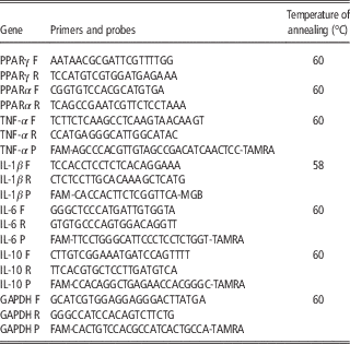
In order to isolate total RNA, BME-UV1 were seeded in cell culture flasks at the concentration of 5×10⁵ cells/ml in complete medium containing 10% FBS (Thermo Fisher Scientific, Waltham, MA, USA) and treated as described above. Total RNA was extracted by QIAzol Lysis Reagent (Qiagen, Hilden, Germany), according to the manufacturer’s instructions and stored at −80°C. RNA was quantified using Quant-iT RNA assay Kit (Invitrogen, Carlsbad, CA, USA) and fluorescence was measured at excitation/emission of 644/673 nm. Total RNA of 1 µg was reverse transcribed using Quantitect reverse transcription kit (Qiagen) in a total volume of 20 μl on a PCR Express thermal cycler (Hybaid, Ashford, UK). Quantitative SYBR Green and probes real-time PCR were performed following the manufacturer’s recommendations using LightCycler® 2.0 (Roche; Roche Applied Science, Indianapolis, IN, USA). To account for possible variation related to complementary DNA input or the presence of PCR inhibitors, the endogenous reference gene glyceraldehyde 3-phosphate dehydrogenase was simultaneously quantified for each sample, and data were normalized accordingly. In Table 1 are shown the specific characteristics of primers used for the real-time PCR.
DNA sequences of bovine sense and antisense primers and probes used for real-time PCR-analysis

PPARγ=peroxisome proliferator-activated receptorα-γ/α; TNF-α=tumor necrosis factor-α; IL-1β=interleukin-1β; IL-6=interleukin-6; IL-10=interleukin-10; GAPDH= glyceraldehyde 3-phosphate dehydrogenase; F=Forward; R=reverse; P=probe.
PCR products were subjected to a melting curve analysis on the LightCycler. To allow relative quantification after PCR, standard curves were constructed from the standard reactions for each target and housekeeping genes by plotting crossing point values, that is, the cycle number at which the fluorescence signal exceeds background v. log complementary DNA dilution. The crossing point readings for each of the unknown samples where then used to calculate the amount of either the target or housekeeping relative to the standard, using the second derivative maximum method with the LyghtCycler analysis software 3.5 (Roche Applied Science).
Statistical analysis
All data of the experiment are presented as least-squares means and SEM. The data were analyzed by ANOVA using Statistica-7 Software package (Stat Soft Inc., USA). The model included fatty acid as fixed effect, replicates as random effect and the error term. The significance of the differences was assessed by the Fisher’s LSD test. Significance was declared at P<0.05.
Results
Viability of bovine mammary epithelial cells
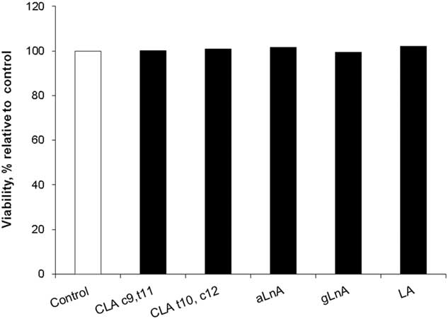
Cell viability (Figure 1) was carried out using the Cell Proliferation kit II and was evaluated after 3 h of LPS exposure. As showed in Figure 1, LPS treatments did not modify cell viability. These results indicate that exposure to LPS, does not affect the metabolic activity of BME-UV1 through a decrease or an increase in cells viability and that the treatment did not have a cytotoxic effect.
Cell viability of bovine mammary epithelial cells (BME-UV1) observed, after 48 h of fatty acids supplementation, and after 3 h of lipopolysaccharide treatment (20 μM). Absorbance was measured at 450 nm, and data are reported as least square means±SEM (n=6). CLA c9,t11=cis9, trans 11 conjugated linoleic acid; CLA t10,c12=conjugated linoleic acid; aLnA=α-linolenic acid; gLnA=γ-linolenic acid; LA=linoleic acid.

Quantification of reactive oxygen species production
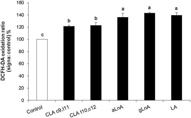
Intracellular ROS production by 2',7'-dichlorodihydrofluorescin diacetate probe fluorescence measurement was assessed in cells supplemented with fatty acids and stimulated with LPS (Figure 2). Cells treated with fatty acids showed an increased ROS production (P<0.05) compared with the control. Among treatments, cells treated with c9,t11 CLA and t10,c12 CLA revealed significant lower levels of ROS production compared with other fatty acids.
Intracellular production of reacting oxygen species by 2', 7'-dichlorodihydrofluorescin diacetate probe (DCFH-DA) in bovine mammary epithelial cells (BME-UV1) after 48 h of fatty acids supplementation, and after 3 h of lipopolysaccharide treatment (20 μM). Data are reported as least square means±SEM (n=6). Significant differences among control and treatments are represented by different letters (P<0.05). CLA c9,t11=cis9, trans 11 conjugated linoleic acid; CLA t10,c12=conjugated linoleic acid; aLnA=α-linolenic acid; gLnA=γ-linolenic acid; LA=linoleic acid.

Quantification of messenger RNA expression of pro and anti-inflammatory cytokines
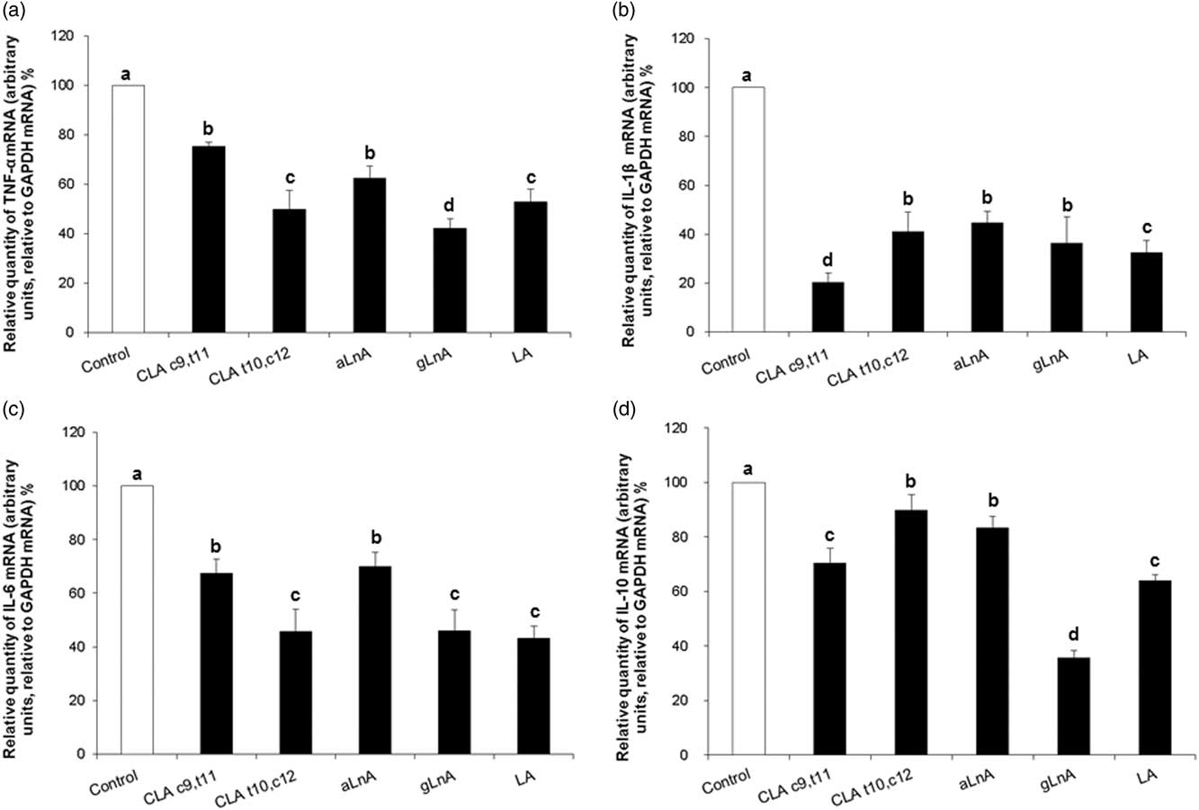
The mRNA expression of TNF-α, IL-1β, IL-6 and IL-10 are shown in Figure 3a, b, c and d, respectively. The gene expression of pro-inflammatory cytokines was lower (P<0.05) in cells treated with different fatty acids compared with the control. However, some differences were observed between the different fatty acids.
Messenger RNA abundance of tumor necrosis factor-α (TNF-α) (a), interleukin-1β (IL-1β) (b), interleukin-6 (IL-6) (c), and interleukin-10 (IL-10) (d) in bovine mammary epithelial cells (BME-UV1) after 48 h of fatty acids supplementation, and after 3 h of lipopolysaccharide treatment (20 μM). Data are reported as least square means±SEM (n=6). Significant differences between control and treatments and treatments between them are represented by different letters (P<0.05). CLA c9,t11=cis9, trans 11 conjugated linoleic acid; CLA t10,c12 = conjugated linoleic acid; aLnA=α-linolenic acid; gLnA=γ-linolenic acid; LA=linoleic acid.
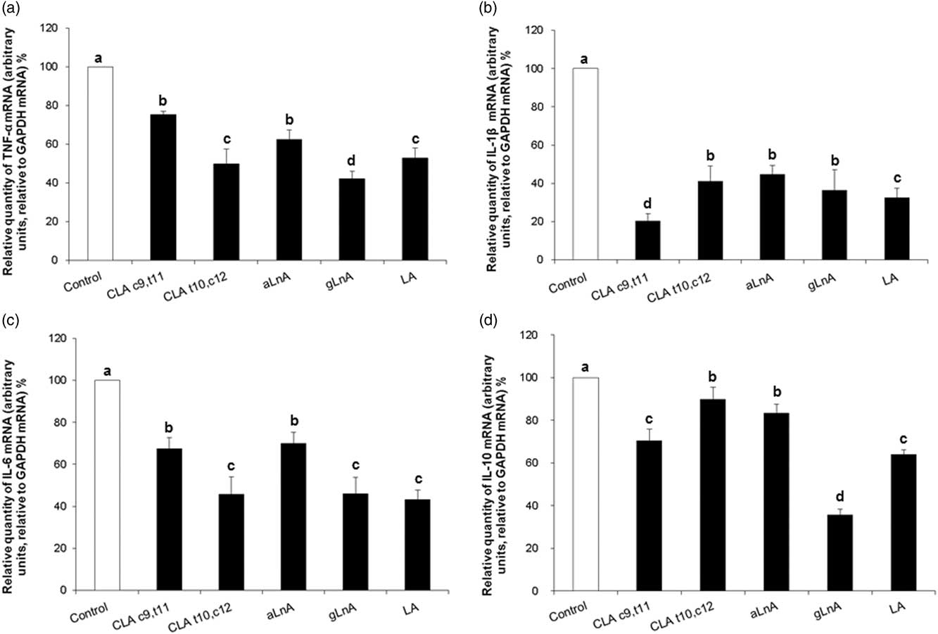
The lowest level of TNF-α gene expression (Figure 3a) was observed in cells treated with gLnA, compared with the other fatty acids (P<0.05). TNF-α gene expression was not different between cells treated with aLnA and c9,t11 CLA and between t10,c12 CLA and LA. aLnA and c9,t11 CLA showed greater (P<0.05) TNF-α mRNA when compared with t10,c12 CLA and LA.
The lowest level (P<0.05) of IL-1β gene expression (Figure 3b) was observed in cells treated with c9,t11 CLA compared with other FA. LA treatment showed greater (P<0.05) IL-1β gene expression compared with c9,t11 CLA and lower when compared with t10,c12 CLA, aLnA and gLnA. No differences were observed between t10,c12 CLA, aLnA and gLnA.
The lowest (P<0.05) level of IL-6 gene expression (Figure 3c) was observed in cells treated with t10,c12 CLA, gLnA and LA, compared with c9,t11 CLA and aLnA. No differences were observed between t10,c12 CLA, gLnA and LA, and between c9,t11 CLA and aLnA.
The gene expression of IL-10 (Figure 3d) was lower (P<0.05) in cells treated with different fatty acids compared with the control. Among fatty acids, the lowest (P<0.05) level of IL-10 gene expression was observed in cells treated with gLnA compared with other FA. IL-10 gene expression was not different between cells treated with t10,c12 CLA and aLnA, and between c9,t11 CLA and LA. t10,c12 CLA and aLnA showed greater (P<0.05) IL-10 mRNA when compared with c9,t11 CLA and LA.
Quantification of messenger RNA expression of peroxisome proliferator-activated receptor-γ and -α
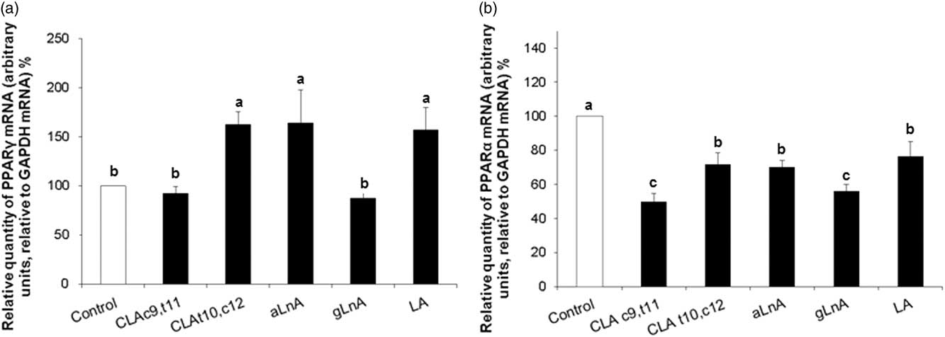
The quantification of PPARγ and PPARα mRNA expression is showed in Figure 4a and b, respectively. PPARγ gene expression was greater (P<0.05) in cells treated with t10,c12 CLA, aLnA and LA compared with control and with c9,t11 CLA and gLnA. No differences were observed between cells treated with t10,c12 CLA, aLnA and LA, and between control, c9,t11 CLA and gLnA.
Messenger RNA abundance of peroxisome proliferator-activated receptorα- γ/α [PPARγ (a) and PPARα (b), respectively] in bovine mammary epithelial cells (BME-UV1) after 48 h of fatty acids supplementation, and after 3 h of lipopolysaccharide treatment (20 μM). Data are reported as least square means±SEM (n=6). Significant differences between control and treatments and treatments between them are represented by different letters (P<0.05). CLA c9,t11=cis9, trans 11 conjugated linoleic acid; CLA t10,c12=conjugated linoleic acid; aLnA=α-linolenic acid; gLnA=γ-linolenic acid; LA=linoleic acid.

The gene expression of PPARα was lower (P<0.05) in cells treated with different FA when compared with control cells. Between FA treatments, PPARα gene expression was lower (P<0.05) in cells treated with c9,t11 CLA and gLnA compared with LA, aLnA and t10,c12 CLA. No differences were observed between c9,t11 CLA and gLnA and between LA, aLnA and t10,c12 CLA.
Discussion
In the present study, for the first time, the effect of several fatty acids (c9,t11 CLA, t10,c12 CLA, aLnA, gLnA and LA) on the inflammatory response of BME-UV1 cells was investigated. The results showed that bovine cell line had different susceptibility to the different EFAs and CLA.
Under the experimental conditions, the exposure to 50 µM of fatty acids for 48 h has protected the cells limiting the triggering of the inflammatory process induced by LPS, known as a potent endotoxin responsible to stimulate the inflammation response and inflammatory cytokine synthesis. The results of the present study showed that cell supplemented with fatty acids has reduced the gene expression of the main pro-inflammatory cytokines and of the anti-inflammatory cytokine IL-10, matched by increased gene expression of PPARγ. The data presented here are consistent with other findings indicating an improvement of the inflammatory status in cells treated with CLA and EFAs (Changhua et al., Reference Changhua, Jindong, Defa, Lidan, Shiyan and Jianjun2005; Zhao et al., Reference Wojdasiewicz, Poniatowski and Szukiewicz2005; Erdinest et al., Reference de Heredia, Gómez-Martínez and Marcos2012). Erdinest et al. (Reference de Heredia, Gómez-Martínez and Marcos2012), on human corneal epithelial cells culture incubated for 2 h with different concentrations of aLnA, gLna and LA, showed a reduced gene expression of TNF-α, IL-1β and IL-6 in cells treated with aLnA. Those authors stated that inhibitory effect of aLnA was mediated through NF-Kβ signal transduction. Also Zhao et al. (Reference Wojdasiewicz, Poniatowski and Szukiewicz2005) in a study on monocytic THP-1 cells pre-incubated with aLnA, LA and DHA observed that fatty acids decreased TNF-α, IL-1β and IL-6 gene expression and the NF-Kβ DNA-binding activity, whereas PPARγ activity was increased and the anti-inflammatory properties of fatty acids were attributed, at least in part, to PPARγ-dependent mechanism. Changhua et al. (Reference Changhua, Jindong, Defa, Lidan, Shiyan and Jianjun2005) in an in vitro study on the anti-inflammatory effect of c9,t11 CLA and t10,c12 CLA on cultured peripheral blood mononuclear cells, noted that CLA isomers, in particular t10,c12 CLA, suppressed the production and expression of TNF-α, IL-1 and IL-6. A recent study (Cao et al., Reference Cao, Luo, Zang, Chen, Xu, Shi and Jing2016) on primary goat mammary gland epithelial cells treated with gLnA and LA showed that gLnA but not LA reduced inflammation induced by LPS, through the inhibition of the activity of NF-Kβ transcription factor and therefore the transcription of IL-1, IL-6 and TNF-α. Also in vivo studies revealed similar results (Lin et al., Reference Lin, Hou, Xiang, Zhang, Che, Lin, Xu, Tian, Zeng, Yu, Zhang, Chen, Wu and Fang2013). Song et al. (Reference Song, Grant, Rotondo, Mohede, Sattar, Heys and Wahle2005), in humans, observed that dietary CLA supplementation reduced levels of TNF-α and IL-1β while increased the anti-inflammatory cytokine IL-10. Lin et al. (Reference Lin, Hou, Xiang, Zhang, Che, Lin, Xu, Tian, Zeng, Yu, Zhang, Chen, Wu and Fang2013) showed that rats, supplemented with diet rich in n-3 (fish oil to 5%), had lower mammary mRNA abundance of xanthine oxidoreductase, protein level of TNF-α and greater levels of IL-10 and PPAR-γ. Renner et al. (Reference Renner, Kersten, Duevel, Schuberth and Dänicke2013) showed that there were no differences in IL-10 expression in bovine peripheral blood mononuclear cells treated with c9,t11, t10,c12 CLA isomers and LA. In contrast, Verlengia et al. (Reference Verlengia, Gorjão, Kanunfre, Verlengia, Gorjão, Kanunfre, Bordin, de Lima, Martins, Newsholme and Curi2004) reported that the B-lymphocyte cell line treated with eicosapentanoic acid and docosahexaenoic acid reduced the production of IL-10. This suppression of cytokine production was also observed in spleen lymphocytes from mice fed fish oil-rich diets (Wallace et al., 2001). The effects of fatty acids on the gene expression of the anti-inflammatory IL-10 are currently ambiguous and contradictory. On the other hand, Tamayo et al. (Reference Tak and Firestein2011) in a human study on the relationship between the responses of cytokines in septic shock, observed that the secretion of pro- and anti-inflammatory cytokines occurs in a simultaneous manner by the moment which begins the immuno-inflammatory response. Results of the present study and findings from others (Wojdasiewicz et al., Reference Zhao, Etherton, Martin, Vanden Heuvel, Gillies, West and Kris-Etherton2014) suggest the hypothesis that the reduced anti-inflammatory cytokines gene transcription could be positively correlated with reduced transcription levels of pro-inflammatory cytokines in view of an harmonized inflammatory response.
The PPARγ transcription factor, through its activation or gene up-regulation, plays an important role in the control of inflammation (Mandard and Patsouris, Reference Mandard and Patsouris2013). PPARγ inhibits the production of inflammatory mediators such as TNF-α, IL-1 and IL-6 interacting physically with NF-Kβ and preventing NF-Kβ translocation into the nucleus (Tak and Firestein, Reference Tamayo, Fernández, Almansa, Carrasco, Heredia, Lajo, Goncalves, Gómez-Herreras, de Lejarazu and Bermejo-Martin2001). NF-Kβ is the main transcription factor that activate the expression of pro-inflammatory cytokines (Lawrence, Reference Lawrence2009).
In this study it was observed an up-regulation of PPARγ in cells treated with t10,c12 CLA, aLnA and LA, but no differences on the expression of PPARγ were observed in cells treated with c9,t11 CLA and gLna. Our results suggest that t10,c12 CLA, aLna and LA inhibited the transcription of pro-inflammatory cytokines through the upregulation of PPARγ expression, whereas c9,t11 CLA and gLnA probably acted stimulating the activity of the constitutive PPARγ, as also observed and reported by Cao et al. (Reference Cao, Luo, Zang, Chen, Xu, Shi and Jing2016).
The level of expression of the PPARα gene, contrarily to PPARγ, was lowest in all cells treated with the different fatty acids and stimulated with LPS. Probably this phenomenon is linked to higher production of ROS observed in BME-UV1 cells. Cabrero et al. (Reference Cabrero, Alegret, Sanchez, Adzet, Laguna and Carrera2002) observed that skeletal muscle cells treated with low concentration of etomoxir, a substance which contains a fatty acid-derived structure, showed a downregulation of PPARα mRNA expression, through increased production of ROS and NF-Kβ activation. It is important to remark that generation of ROS have not solely negative effects but within certain concentrations ROS are useful signaling molecules regulating physiological processes. The beneficial or detrimental effects depends on many variables such as the site of ROS production, the persistence of ROS flow or the antioxidant status of target cells (Barbieri and Sestili, Reference Barbieri and Sestili2012).
Conclusions
All fatty acids reduced the gene expression of pro-inflammatory cytokines, among fatty acids, t10,c12 CLA, LA and gLna showed an homogeneous reduction of the three cytokines and this may correspond to more balanced and efficient physiological activity and may trigger a better protective effect. Our results suggest that fatty acids inhibited the transcription of pro-inflammatory cytokines by the upregulation of PPARγ expression.
Acknowledgements
None.
Declaration of interest
There are no conflict of interests.
Ethics statement
None.
Software and data repository resources
Data is not deposited in an official repository.