Introduction
Lesions of the orbital apex pose both a diagnostic and surgical challenge. The diversity of normal tissue types encountered in this complex compact anatomical regionReference Abuzayed, Tanriover, Gazioglu, Eraslan and Akar1,Reference Yeh and Foroozan2 gives rise to a spectrum of pathological entities. These may present with a variety of ocular symptoms, including visual loss, pain, ophthalmoplegia, sensory loss in the ophthalmic nerve (V1) distribution and the associated orbital apex, and cavernous sinus syndromes.
Traditional, transcranial open approaches to the orbital apex have been associated with significant cosmetic morbidity. Transconjunctival and other orbitotomy techniques remain surgical options. However, in appropriately selected cases, and certainly for lesions presenting medially to the optic nerve, transnasal endoscopic surgery with image guidance provides superior anatomical definition and exposure of the orbital apex.Reference Sethi and Lau3,Reference Stokken, Gumber, Antisdel and Sindwani4 Facial incisions and scarring may be avoided.
Endoscopic surgery is predominantly utilised for inferomedially placed intraconal and extraconal lesions. However, with increasing combined surgical expertise, better imaging and high-definition camera optics, with caution, the remit of endoscopic surgery may be extended to include posterolaterally based lesions.Reference Stokken, Gumber, Antisdel and Sindwani4 Additionally, endoscopic surgery permits a magnified view to the optic nerve, thereby facilitating concurrent decompressions of the optic canal and nerve and medial orbital wall.Reference Sia, Chan, Wormald, Davis and Selva5
Given the narrow anatomical space, transnasal endoscopic surgery is invariably carried out with image guidance. Magnetic resonance imaging (MRI) remains the imaging modality of choice, and is often used in conjunction with computed tomography (CT) and magnetic resonance angiography. Current image fusion and segmentation techniques permit the pre-operative identification of the smallest of lesions, limiting non-diagnostic samples and delineating key neurovascular structures, thereby reducing complications.
The present study was prompted by the perceived reluctance of clinicians involved in the initial management of these patients, who have positive but frequently small volume mass lesions in this site, to pursue a tissue diagnosis on account of the potential risk to key neurovascular structures. Whilst it is often tempting to avoid the perceived potential surgical morbidity, the delay in referral to a multidisciplinary skull base unit with endoscopic expertise, coupled with previous semi-empirical treatment, has the potential for equally unfavourable outcomes. This study aimed to evaluate the spectrum of lesions at this site, and to evaluate the success and morbidity of endoscopic transnasal approaches to lesions presenting medially at the orbital apex.
Despite the improvement in imaging and surgical approaches, there remains much controversy regarding the timing of initiating treatment, particularly with corticosteroids. It may be argued that steroid treatment ought to be initiated prior to obtaining a definitive histological diagnosis. In inflammatory causes, such as orbital pseudotumours, this would be optimal. However, the differential diagnosis of lesions of the orbital apex is considerable,Reference Yeh and Foroozan2,Reference Stokken, Gumber, Antisdel and Sindwani4,Reference Vohra, Escott, Stevens and Branstetter6 and includes primary and metastatic tumours of the head and neck, lymphoma, and invasive fungal disease, where steroid treatment might adversely affect the prognosis. Clinically and radiologically, these lesions may mimic one another,Reference Vohra, Escott, Stevens and Branstetter6 and radiological features are often non-specific. Ultimately, without definitive histology, improper treatment may lead to adverse outcomes and catastrophic complications for the patients.
This paper reports on a prospective series of 21 consecutive patients who presented over an 11-year period, and examines the post-operative outcomes, wide differential diagnoses and potential complications following an exclusively transnasal endoscopic approach to orbital apex lesions.
Materials and methods
Prospective data were collected on 21 consecutive patients, presenting at a tertiary referral unit, to a single surgeon working as part of a multidisciplinary team, over an 11-year period between 2006 and 2017. Presenting symptoms, pre-operative imaging findings, final histopathology findings, ocular symptoms, post-operative visual changes and surgical complications were all recorded.
Patients presenting with dysthyroid eye disease, or those with known or obvious chronic rhinosinusitis, were excluded from the study. Patients with an eventual diagnosis of fungal disease, in whom the diagnosis was initially unclear radiologically but confirmed subsequently on histopathology, were included in the study.
Surgical technique
All surgical procedures were performed endoscopically via a transnasal approach, with the assistance of BrainLab (Munich, Germany) image guidance system. Each patient underwent pre-operative imaging with CT, MRI, and either CT angiography or magnetic resonance angiography. Images were fused to precisely delineate the lesion, and its relation to the optic nerve and canal, and related bony and vascular structures including the variable path of the ophthalmic artery.
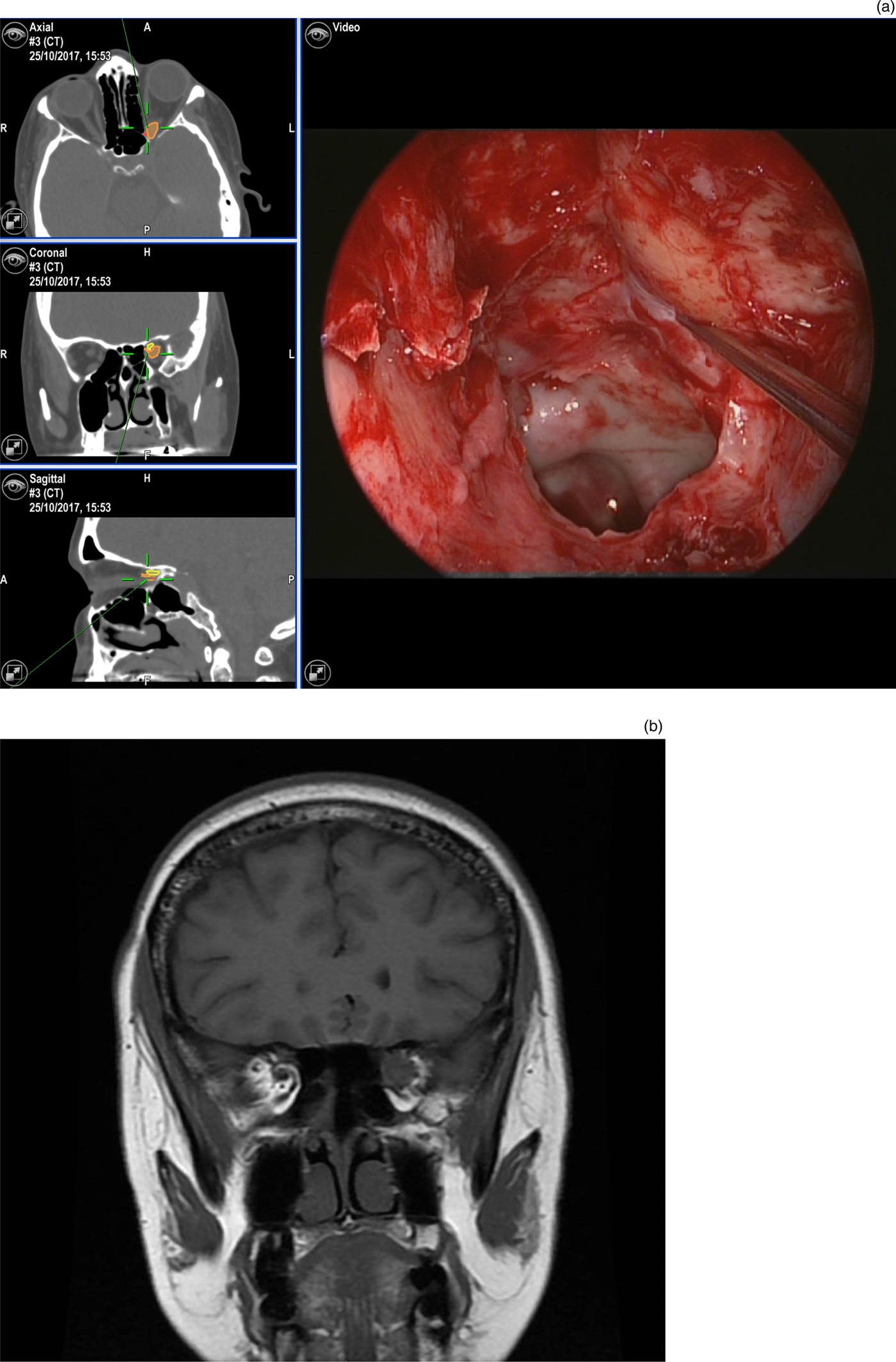
For most lesions presenting in the medial orbital apex or optic canal, the approach involved an initial middle meatal antrostomy and uncinectomy, and posterior ethmoidectomy with a wide sphenoidotomy, with exposure of the lamina papyracea as required. The middle turbinate was resected only if necessary to optimise access. Image guidance was used to precisely locate the lesion (Figure 1a).
Images of patient one (World Health Organization grade 1 meningioma), showing: (a) computed tomography (CT) fused image guidance pictures (axial, coronal and sagittal views; left) of the lesion (orange) and its relation to the optic nerve (yellow), and a concurrent endoscopic picture pinpointing the lesion location (right); and (b) a coronal, T1-weighted magnetic resonance imaging scan demonstrating the left-sided lesion. A = anterior; R = right; L = left; P = posterior; H = head; F = foot

The lamina papyracea was then gently perforated posteriorly but anterior to the orbital apex, to expose the periorbita. The thin bone of the posterior lamina was then unpicked in a posterior direction to trace the contour of the orbit and its narrowing to the apex, whilst remaining outside the periorbita. The orbital apex may be traced posteriorly over the bony impression of the optic canal and this can be unpicked, but may require thinning with a coarse diamond burr or a dacryocystorhinostomy burr (Medtronic, Minneapolis, Minnesota, USA) first. A sickle knife was then used to incise the periorbita at the appropriate location.
For extraconal lesions, orbital fat is typically retracted and the lesion biopsied, with image guidance confirming precision. For intraconal lesions, orbital fat may be partially and carefully resected to expose the medial rectus muscle. The intraconal space is accessed between the inferior and medial rectus muscles.
The lesion is localised using precise image guidance before a biopsy is taken. Where indicated, a concurrent optic nerve decompression may be performed.
Results
The study identified a total of 21 patients who underwent endoscopic biopsy of the orbital apex, optic canal and/or anterior cavernous sinus. The mean patient age was 49 years, with 12 males (57 per cent) and 9 females (43 per cent). Overall lesional material was obtained in 20 out of the 21 patients; the only exception was a patient with an anterior cavernous sinus lesion extending to the optic canal, where bleeding hampered tissue retrieval.
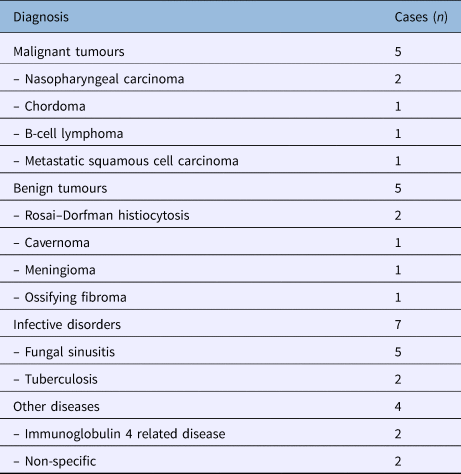
This latter patient, along with another in whom a non-diagnostic sample was obtained, were treated successfully as Tolosa–Hunt syndrome cases. This syndrome is a painful ophthalmoplegia with non-specific inflammation of the superior orbital fissure and cavernous sinus.Reference Hunt7,Reference Hunt, Meagher, Lefever and Zeman8 Five malignant tumours (24 per cent) were also identified, along with five benign tumours (24 per cent), seven infective cases (33 per cent) and two cases with immunoglobulin G4 related disease (10 per cent) (Table 1).
Diagnosis of patients*

* Those who underwent endoscopic biopsy of the orbital apex, optic canal and/or anterior cavernous sinus
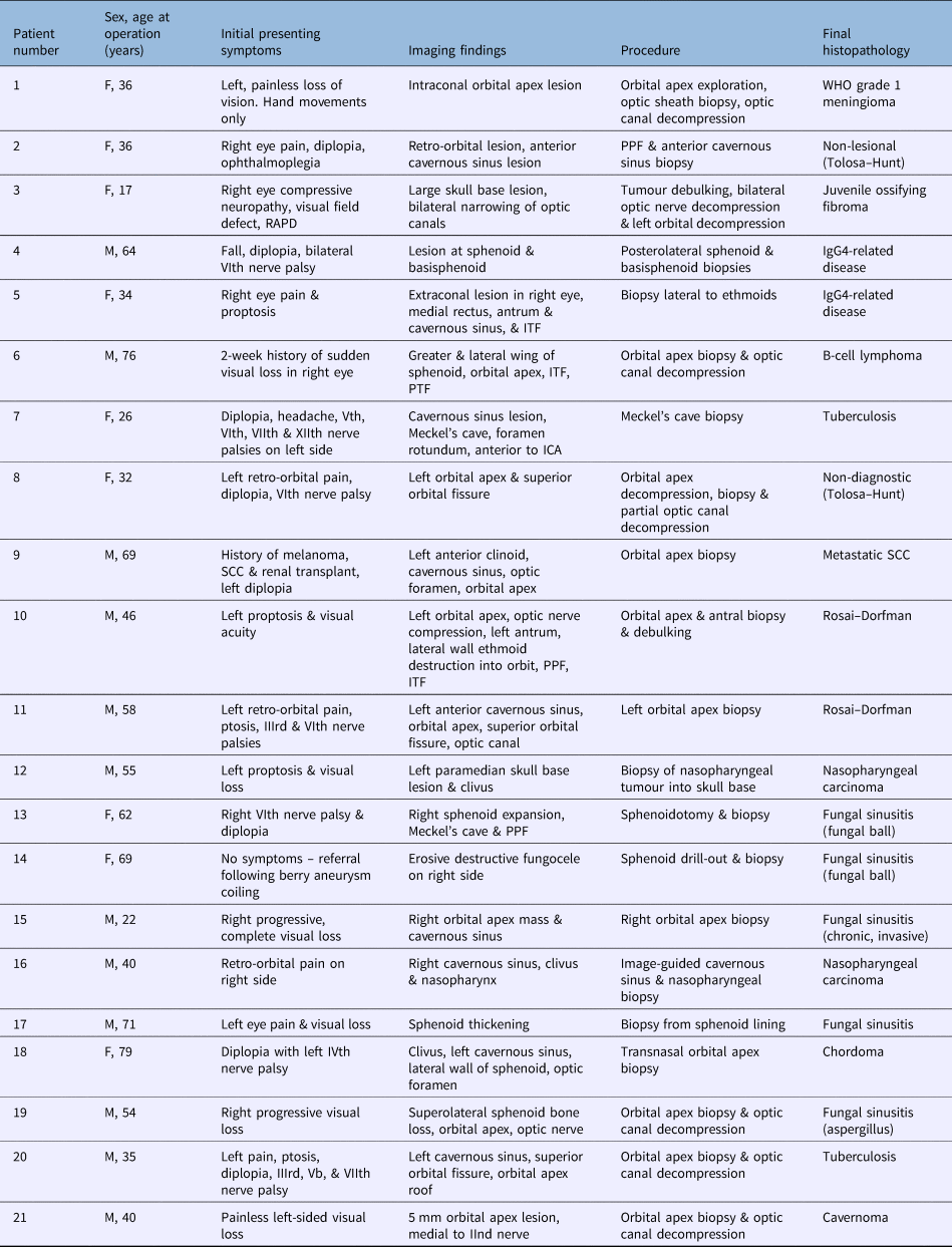
Prior to surgery, 11 patients (52 per cent) had left eye symptoms, 8 (38 per cent) had right eye symptoms and 1 (5 per cent) had bilateral symptoms. One patient (5 per cent) was asymptomatic at the time of presentation, but had an incidental finding of an erosive process in the right sphenoid, later found to be secondary to a fungal ball. The remaining patients all presented with ocular symptoms, namely visual loss, pain and concomitant cranial nerve palsies (Table 2). All patients underwent pre- and post-operative ophthalmological review.
Summary of patient findings

F = female; WHO = World Health Organization; PPF = pterygopalatine fossa; RAPD = relative afferent pupillary defect; M = male; IgG4 = immunoglobulin 4; ITF = infra-temporal fossa; PTF = pterygopalatine fossa; ICA = internal carotid artery; SCC = squamous cell carcinoma
The use of steroids pre- and post-operatively was highly subjective depending on the initial source of referral.
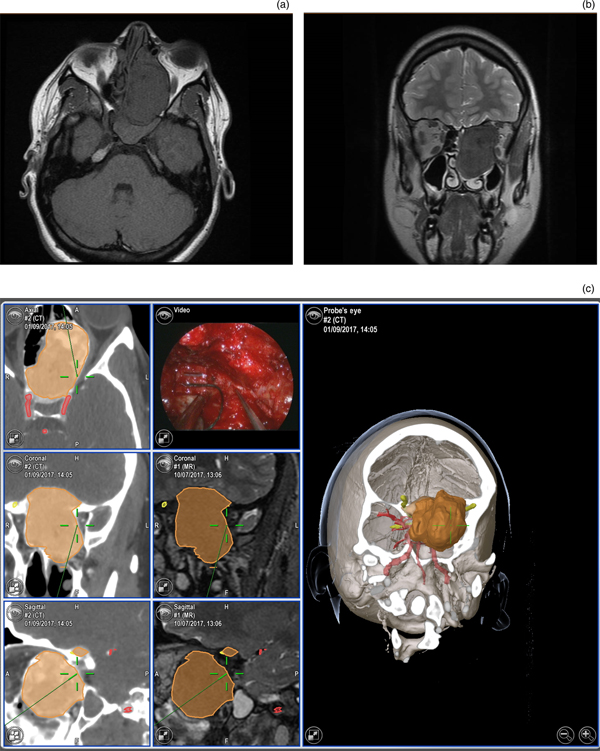
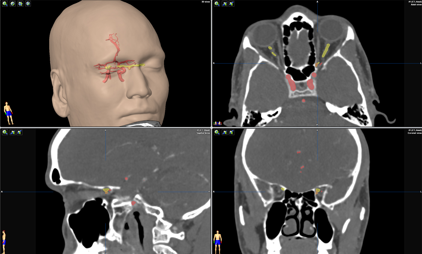
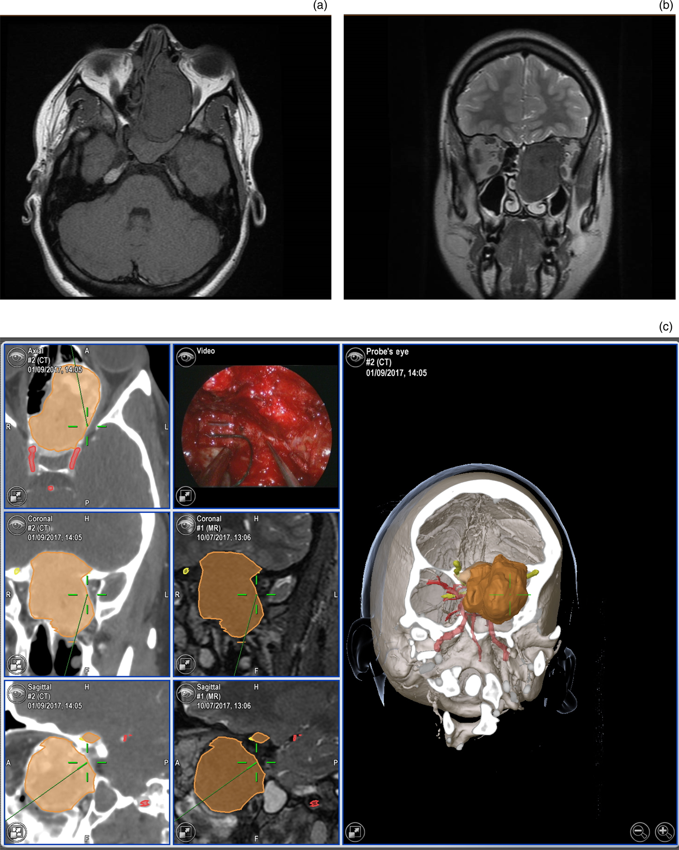
Post-operatively, there was neither improvement nor deterioration in visual acuity in any patient. Visual field loss improved in the patient (patient three) who underwent debulking of a large skull base juvenile ossifying fibroma, bilateral optic nerve decompressions and a left medial orbital decompression (Figure 2a–c). Her presentation was somewhat atypical in that she had a large benign fibro-osseous lesion, presenting with a visual field defect and a right compressive neuropathy. Intra-operatively, there was tumour replacing the sphenoid sinus. Following removal of disease indistinguishable from the bony tuberculum sella, there was a subsequent breach of the dura and arachnoid. Although the intra-operative leak was repaired with a nasoseptal flap, Duragen matrix (Integra Life-Sciences, Plainsboro, New Jersey, USA) and Duraseal sealant (Integra Life-Sciences), it persisted post-operatively. Hence, the patient was taken back to the operating theatre at day 3 post-procedure and underwent successful repair of the persistent leak. Six additional patients (including patient 21 (diagnosed with a cavernoma), whose imaging findings are shown in Figure 3), also had a contemporaneous optic nerve decompression and tissue biopsy without complications.
Images of patient three (juvenile ossifying fibroma), showing: (a) axial and (b) coronal magnetic resonance (MR) imaging sections of the lesion; and (c) image guidance pictures and an intra-operative endoscopic photograph. A = anterior; CT = computed tomography; R = right; L = left; P = posterior; H = head; F = foot
Images of patient 21 (cavernoma), showing: fused image guidance pictures demonstrating the lesion (orange), and its relation to the optic nerve (yellow) and vasculature (red). A = anterior; CT = computed tomography; R = right; L = left; P = posterior; H = head; F = foot

With regard to the source of referral, 10 patients were referred from neurology, 6 from other ENT units, 3 from neurosurgery and 2 from ophthalmology.
Discussion
Surgical approaches to the orbital apex have evolved over the last century with increased expertise in endoscopic techniques.Reference Sethi and Lau3 Traditional craniotomy approaches are associated with a much higher rate of debilitating complications, namely seizures, strokes, blindness and cerebrospinal fluid (CSF) leaks.Reference Murchison, Rosen, Evans and Bilyk9 Although the transconjunctival approach also affords a scar-free alternative,Reference Abuzayed, Tanriover, Gazioglu, Eraslan and Akar1 the exposure of the orbital apex remains limited, especially for very small, posteriorly based lesions.Reference Stokken, Gumber, Antisdel and Sindwani4
Acquiring skills in the endoscopic approach to the paranasal sinuses is a mandatory requirement of every UK otolaryngologist in training.10 The use of adjunctive image guidance has facilitated extended applications to the skull base and beyond. One such example includes the successful clipping of cerebral aneurysms.Reference Szentirmai, Hong, Mascarenhas, Salek, Stieg and Anand11
The present study demonstrates the wide-ranging histological picture of lesions of the orbital apex, optic canal and anterior cavernous sinus, with each one biopsied via an endoscopic transnasal approach. At the time of writing, 19 patients were alive; 1 (patient 9) had died of their metastatic squamous cell carcinoma and 1 (patient 12) was lost to follow up. Clinically, all but one of the patients presented with visual acuity loss and/or ophthalmoplegia.
The radiological features did not always point to a clear diagnosis, with several lesions looking similar on cross-sectional imaging. Eisen et al. (2000) aimed to assess the predictive value of pre-operative imaging on orbital tumours.Reference Eisen, Yousem, Loevner, Thaler, Bilker and Goldberg12 They identified that the most sensitive predictor of orbital invasion (i.e. implying a malignancy) was those lesions located adjacent to the periorbita along with extraocular muscle involvement. Nevertheless, they concluded that imaging should be used for surgical planning and cannot replace intra-operative assessment.Reference Eisen, Yousem, Loevner, Thaler, Bilker and Goldberg12 Ben Simon et al. (2005) reviewed the orbital imaging of 131 biopsy-proven orbital tumours.Reference Ben Simon, Annunziata, Fink, Villablanca, McCann and Goldberg13 Although they found certain characteristic findings in benign, malignant and inflammatory lesions (e.g. benign tumours were smaller, round or oval in shape), tissue diagnosis was frequently required and still deemed to be the ‘gold standard’ in assessment.
Five of the patients were found to have fungal disease, of which one case was chronically invasive. In this particular patient, there was no other evidence of chronic sinus disease on imaging, and radiological features could not differentiate it from a benign fibro-osseous lesion. There was also one patient found to have B-cell lymphoma. In each case, steroids could adversely affect the pathological accuracy, and cause a dysregulation of the immune system and thereby possibly promote progression. Moreover, in the malignant tumour cohort (5 of 21 cases), expectant management alone could have led to adverse outcomes.
One notable observation was the lack of significant improvement in visual status despite 7 of the 21 patients undergoing simultaneous optic nerve decompressions. A significant factor here is likely to be the time delay from the onset of symptoms to the point of referral. For example, patients 1 and 21 were both ultimately seen in the tertiary referral unit some six months after the onset of symptoms. Although there is a suggestion that decompressive surgery may have arrested the progression of visual loss in these patients, it is difficult to quantify.
Tissue diagnosis was successfully obtained in all but two patients. In only one of these patients (patient two) was there non-lesional material; in the other (patient eight), the sample yield was deemed to be lesional but non-diagnostic. Both patients were successfully treated as Tolosa–Hunt syndrome cases, receiving steroids. Alternative treatment strategies for this condition include azathioprine,Reference Hannerz14 methotrexateReference Smith and Rosenbaum15 and radiotherapy.Reference Mormont, Laloux, Vauthier and Ossemann16 In patient two, the lesion was located at the anterior cavernous sinus, and significant intra-operative bleeding may have led to an inadequate sample for diagnosis.
• In 10 out of 21 patients, endoscopic transnasal surgery was both diagnostic and part of definitive treatment (e.g. optic nerve decompression, fungal sinus surgery)
• Diagnosis was not established prior to tissue sampling in any of the cases
• No patient suffered a worsening of presenting visual symptoms
Although the majority of patients underwent biopsy without significant morbidity, there was one intra-operative CSF leak in patient three. This patient was atypical in that she had a large skull base lesion, which required a joint procedure with the neurosurgical team as the potential for CSF leak was anticipated. Despite intra-operative skull base reconstruction with a nasoseptal flap, she required a secondary minor procedure, which successfully closed the defect. All patients were counselled regarding the potential for CSF leak, but for the remaining patients, the lesions were small. The use of fused image guidance improved accuracy when pinpointing lesions, and in particular demonstrated its relation to the optic nerve and ophthalmic artery, which can be highly variable.Reference Ganiusmen, Citak, Samancioglu, Korkmaz and Binatli17
Conclusion
Orbital apex lesions are rare, and with endoscopic approaches still gaining popularity, there remains scant data in the literature. In experienced hands, anxiety regarding the morbidity associated with transnasal approaches for tissue biopsy may be reasonably allayed, particularly for lesions presenting medially to the optic nerve. The adjunctive use of image guidance techniques can prevent injury to vital structures including the optic nerve and ophthalmic artery. Ultimately, the present study demonstrates the broad spectrum of lesions encountered at this site, including malignancies; it also emphasises the need for specific treatment approaches, rather than a generic ‘one size fits all’ anti-inflammatory trial of steroid treatment.
Competing interests
None declared