Background
The role of radiotherapy for rectal cancer has been well established in terms of reducing the risk of local disease recurrence.Reference Kapiteijn, Marijnen and Nagtegaal1–Reference Glimelius3 Patients having a course of radiotherapy must be appropriately immobilised for stability and accuracy. Prone setups for radiotherapy to anatomical sites such as the rectum typically command large planning margins. This is to account for gross tumour movement compounded by intra-fractional patient movement in order to nullify geometric miss and reduce the risk of under-dosing the peripheral clinical target volume.Reference Dobbs, Barratt, Morris and Roques4 Although the likelihood of ensuring geometric accuracy increases with a larger planning target volume,Reference Koelbl, Vordermark and Flentje5 doses to normal tissue must be considered. Hence, the scope to assess an improvement in clinical outcomes by escalating radical doses is limited by these surrounding normal tissues. Only by curbing normal tissue dose by shrinking planning margins this may occur.Reference Dobbs, Barratt, Morris and Roques4, Reference van Herk6–Reference Brecevic, Leighton and Fisher9
A prone belly board device (BBD) is the standard immobilisation offered to patients with rectal cancer at our institution to minimise the aforementioned patient setup variables. Contoured specifically to displace the small bowel from the irradiated volume for a rectal tumour and to maintain a reliable patient position, the Medtec Contoura™ (Kalona, Iowa, USA) carbon fibre BBD provides an aid to radiotherapy planning and typical treatment techniques. Current radical approaches to treating T3–T4 rectal tumours with N0–N2 nodal disease make use of complex conformal radiotherapy with population-defined margins and doses ranging from 25·0 Gy in five daily fractions (pre-operative) up to 50·4 Gy in 28 daily fractions prescribed to the 100% isodose.Reference Dobbs, Barratt, Morris and Roques4
Further studies have examined the use of a local protocol with a prone BBD in terms of inter-fraction systematic (Σ) and random (σ) error.Reference Roels, Verstraete and Haustermans10–Reference Kasabasic, Ivkovic, Jurkovic and Belaj13 The systematic and random error at these institutions ranged from 1·6 to 3·7 mm and 1·7 to 3·0 mm, respectively. The radical treatment of lower gastrointestinal (GI) cancers at our institution were repatriated in 2010, as the new cancer service, which opened in June 2009, progressively adopted treatments from other regional centres. As such, an evaluation of a new technique and immobilisation training was necessary.
Task-specific radiographer training was the desired model because of the specificity to the desired nature of our repetitive practice and a need to standardise. Other institutions have utilised this model in their clinical setting and have found gains in both quality and efficiency. Those clinical settings include tomotherapy, radiographer-led volume contouring, cardiac pulmonary resusitation (CPR) and surgery.Reference Stewart, MacLure and George14–Reference Mashaud, Castellvi, Hollett, Hogg, Tesfay and Scott21
The aim of this study was to investigate whether task-specific training could impact upon setup reproducibility in belly board immobilised patients.
Materials and methods
This study was conducted at The Beacon Centre, Musgrove Park Hospital, Taunton, UK.
Patients
Sample
Owing to limited patient numbers, ten sequentially referred patients and their geometric displacements were evaluated in 2011. Then a ‘belly board training package’ was introduced and a further ten sequential patients geometric displacements were evaluated in 2012.
Inclusion criteria
• diagnosed advanced rectal cancer (T3–T4, N0–N3, M0);
• treated to 45–50·4 Gy in 25–28 fractions (long-course fractionation);
• patients were immobilised for radiotherapy in the prone position on the belly board.
Exclusion criteria
• neo-adjuvant radiotherapy schedules (short-course fractionation). Short courses of radiotherapy were excluded as insufficient geometric displacement data would be available for analysis.
Ethical consideration
This study was considered to be a service evaluation by the local research and development department at Musgrove Park Hospital, Taunton. In accordance with good clinical practice conduct, patient data were anonymised for the purpose of this study.
Radiotherapy technique
Patients were scanned on a Philips wide bore computed tomography (CT) scanner (5680 DA Best, The Netherlands). A conventional three–four beam technique was planned on the Pinnacle planning system, and treated on a 10 MV LinAc equipped with iViewGT™ (ELEKTA, Stockholm, Sweden) electronic portal imaging (EPI). The Medtec Contoura™ carbon fibre BBD was used for immobilisation.
Patient setup after training
The following radiographer training package was introduced at our institution. This was led by a designated radiographer who carried out all training where staff were coached and were able to practice setups in a closed environment:
• Explain to the patient the position they need to be in and ask them to climb onto the couch.
• Ask the patient to crawl on to the belly board to the level of the superior end of the leg separator and then lie down; they should then be asked to push themselves back down the board until told to stop (iliac crests are ∼1 cm inferior to the contoured edge of the insert and on the stable flat surface of the belly board). This draws the abdomen superiorly and allows for any scar/colostomy bag to be positioned within the contoured hole of the insert.
• For male patients, the radiographers should use an appropriate method of ensuring that external genitalia is appropriately positioned.
• The patient should be face down in the head support, with their arms raised and hands clasped above the support, elbows should not be resting on the side of the couch. Ensure the patient's face is comfortably positioned and their neck is straight. Twisting to the side will reduce daily reproducibility of the positioning.
• Ask the patient to raise their legs and place the knee-fix under their ankles in a reversed position (the high end is at the patient's toes). When they have lowered their legs, ensure the toes are together and the heels apart to aid the reproducibility of using lateral external skin marks to set up. The dorsal aspect of the feet should lay flat against the knee-fix.
• Ensure the patient is comfortable and alter the position of the three separate pieces of immobilisation equipment if and where necessary.
• Use surface anatomical markings to check for patient straightness and adjust as necessary. The bow-tie cushion may be used under the chest or knees for comfort. It must not be used under the pelvis or in any other position because this could introduce the possibility of rotation.
• The patient's positioning can be assessed using a three-finger technique as follows:
• stand to the side of the patient and place each hand on the patient's hips;
• hold each hand with the thumb, index and middle fingers extended and at right angles to each other;
• slide the middle finger under the patient and position at the patient's iliac crest. This will help to eliminate any roll because both hips should be in contact with the board;
• the index finger will check that the correct insert has been used by assessing the freedom of movement of the finger. It will also enable a check that the colostomy bag is encompassed within the insert;
• the thumb lies along the patient's side and checks the patient's yaw (Figure 1).
Figure 1Prone setup on indexed BBD.
Abbreviation: belly board device

Verification protocol
The standard lower GI imaging protocol at our institution was used. Orthogonal posterior and lateral EPI were captured at day 0, 1, 2 and weekly during treatment for comparison with the Pinnacle digitally reconstructed radiograph (DRR).
Surrogate bony structures of the pelvis were used for template matching. For the lateral view these included the pubic symphysis, inferior pubic ramus and acetablum to determine longitudinal and vertical displacement. For the posterior view these included the pubic symphysis, obturator foramen and pelvic brim to determine the lateral displacement.
Geometric displacement
The evaluated geometric displacements were those captured before correction to observe the effect of set-up reproducibility. Corrections were made online at a tolerance level of 5 mm as endorsed by The Royal College of Radiologists.7
Two therapy radiographers measured the geometric translational displacements independently. Their inter-observer measurements were authenticated for the purpose of this study using Spearman's ρ and Pearson correlation in SPSS 14. A correlation was considered significant at the p < 0·05 level. This process was performed against the Patient Group A cohort (before training) only and was not considered necessary to repeat against the Patient Group B (after training).
Systematic and random errors
Systematic and random errors were analysed with respect to the average geometric displacement with standard deviation per patientReference van Herk6, 7 between the Pinnacle DRR and the EPI before and after radiographer training.
Individual error
Σind = the mean average of each patients’ geometric displacements;
σind = the standard deviation of each patients geometric displacements.
Population error
Σpop = the standard deviation of patient groups systematic errors;
σpop = the root mean square of patient group random errors.
Results
Twenty retrospectively sought patients met the inclusion criteria. Fifteen male and five female patients with diagnosed rectal cancers (T3–T4, N0–N2, M0) and an age range of 41–77 years were selected. A total of 371 EPI were template matched.
Correlation analysis between therapy radiographers
Inter-observer measurements from Patient Group A were used to authenticate the EPI match data for the purpose of this study using Spearman's ρ and Pearson correlation. Before calculating statistical significance, the patient data (for both patient groups) was assessed as having a normal distribution via the one-sample Kolmogorov–Smirnov test (p = 0·253–0·999).
In the lateral correlation scatter graph, a tight correlation of geometric displacements is observed along the reference line. The correlation between matches by therapy radiographers was significant (p = 0·01) for the lateral, vertical and longitudinal directions (Figures 2–4). As two sets of data existed (therapy radiographer A and B matches) and due to the statistically significant correlation demonstrated, the displacement measurements of Radiographer A were chosen for analysis after being randomly selected in Excel.
Lateral correlation scatter graph (Pearson correlation =0·922, Spearman's ρ = 0·920).

Longitudinal correlation scatter graph (Pearson correlation = 0·899, Spearman's ρ = 0·862).
Vertical correlation scatter graph (Pearson correlation =0·818, Spearman ρ = 0·730).
Systematic and random error analysis
Lateral
Patient 09A had the largest systematic error (Σind = 4·1 mm). Patient 02B had the largest random error (σind = 5·1 mm; Figure 5).
Lateral displacement error bars before (Group A) and after training (Group B).
Longitudinal
Patients 03A and 09A had the largest individual systematic errors (Σind = 5·3 and 4·2 mm superior, respectively). The largest individual random errors were observed in patients 04A and 05A (σind = 6·0 and 4·9 mm, respectively; Figure 6).
Longitudinal displacement error bars before (Group A) and after training (Group B).
Vertical
Patient 06B had the largest individual systematic error (Σind = 3·7 mm anterior). The largest individual random errors were observed in patients 05A and 01B (σind = 5·4 and 5·3 mm, respectively; Figure 7).
Vertical displacement error bars before (Group A) and after training (Group B).
Discussion
The study's aim was to investigate the efficacy of task-specific training in setup reproducibility using verified geometric displacements as the main outcome measure. Limited patient numbers requiring lower GI radiotherapy led to very small sample sizes and so a power calculation was not deemed necessary for a service evaluation process. Before analysis of the geometric displacements, measurements made by two therapy radiographers were correlated for the validity of this study. The greatest level of correlation was observed in the lateral scatter graph (Figure 2; Pearson correlation = 0·922, Spearman's ρ = 0·920) based on a posterior–anterior (PA) EPI. A PA beams eye view (BEV) EPI could be easier to observe than a lateral BEV EPI due to the clarity of the pelvic bones across a typically smaller patient separation. Nonetheless, all three scatter graphs show a significant (p = 0·01) correlation between therapy radiographers (Figures 2–4). This warranted the appropriateness of using the data set for systematic and random error analysis.
In terms of pre- and post-training comparative displacements, the lateral error bar demonstrates a shift in translational direction from right to left (Σpop = 1·7–2·0 mm). In addition to showing no evidence for lateral setup improvement after training, the population had a tendency to remain within the local 5 mm verification tolerance (σpop = 2·2 and 2·8 mm, respectively). The consistency between patient groups could highlight that there are few quantifiable gains to lateral setup accuracy with radiographer setup training. The belly board equipment is noted to be good at limiting patient ‘roll’ and maintaining stability for a lateral position.
The observed longitudinal systematic and random displacements visibly improved after training (Σpop = 3·1–2·0 mm and σpop = 3·6 and 2·3 mm, respectively). Although positioning a patient laterally presents few obstacles, longitudinal positioning is the remaining influencing factor on accurate patient setup. It is noted that patient comfort is paramount to a reliable and reproducible setup. Specific consideration of male genital, bony landmark and belly positioning to achieve comfort was factored in to the training package, which may have given rise to the favourable results.
The observed trend for vertical systematic error remains inside the set tolerance (±5·0 mm). Variation in random error between patients was seen along the vertical axis for Patient Group A, where Patient Group B demonstrated more uniform magnitudes of random error.
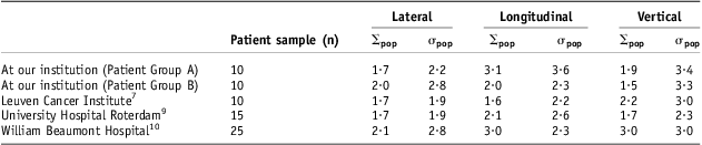
In terms of setup accuracy, previous studies that have looked into prone belly board immobilisation have demonstrated mixed results (Table 1). Lateral and vertical setup is observed to be universally reproducible and consistent between institutions. With training, longitudinal population systematic and random errors at our institution are shown to be more comparable with that seen elsewhere.
Observed systematic and random population errors at different institutions
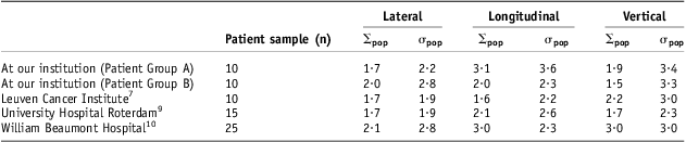
The concept of the BBD was the same at all four institutions. The actual devices used were different brands or made in-house at the host institution. This could imply that there are differences between the BBDs used. Locally, there are three different belly board inserts: flat, wide and narrow. The observed effect could be that one size is not suitable for all patients and so could have been scanned in an unstable position. These could have directly led to a position that was not reproducible in the longitudinal direction, although there is no evidence to authenticate this. It is not clear whether these comparative studies employed a task-specific training package.
It could be suggested that the radiographers were not familiar with this new piece of immobilisation. However, all radiographers were individually trained to use the BBD with an ‘approved setup protocol’. Task-specific training has demonstrated tangible improvements in standardising clinical techniques.Reference Dean and Routsis16–Reference Woollard, Whitfield, Newcombe, Colquhoun, Vetter and Chamberlain18 Gains in the number of tomotherapy fractions treated per day have been reported with the introduction of a comprehensive radiographer training package.Reference Kasabasic, Ivkovic, Jurkovic and Belaj13 This equated to 500 extra treatable fractions per year. Other training packages in radiographer-led volume contouring have noted reductions in outlining variation.Reference Stewart, MacLure and George14 Clinical skills retention is recognised as a key performance indicator in many healthcare professions. Notable studies have looked at the efficacy of annual CPR training with specific reference to skills deterioration.Reference Stefanidis, Acker and Todd Henniford19 Surgical laparoscopy training is another area that has demonstrated significant advances in skills retention with the introduction of systematic training programmes.Reference Kahol, Ashby, Smith and Ferarra20, Reference Mashaud, Castellvi, Hollett, Hogg, Tesfay and Scott21
In the absence of comparable studies in the literature, this study has shown that task-specific training can improve setup reproducibility.
Conclusion
There is evidence that the use of the prone belly board is reproducible, however, clinically significant variability was noted in the longitudinal direction in Patient Group A. It is recommended that task-specific refresher training should be carried out on a locally agreed basis.
Acknowledgement
The authors would like to thank all staff at the Beacon Centre, Taunton for their support and contributions towards completing this work. The authors specifically thank go Julie Walther, Vasu Ganesan, Kelly Mundy and Mandy Pitt.