Introduction
Zoonotic larval cestode infections were recently classified among the neglected tropical diseases (Budke et al., Reference Budke, White and Garcia2009; WHO, 2012). While human echinococcosis caused by E. granulosus is a serious life-threatening disease worldwide, polycystic echinococcosis (PE) caused by E. vogeli is an emergent zoonosis of great public health significance in the humid and tropical rainforests of South and Central America (D'Alessandro & Rausch, Reference D'Alessandro and Rausch2008; Siqueira et al., Reference Siqueira, Almeida, Suzuki, Lima, Machado-Silva and Rodrigues-Silva2010). Furthermore, the number of diagnosed cases of human PE may represent only the tip of the iceberg (D'Alessandro & Rausch, Reference D'Alessandro and Rausch2008).
Echincoccus vogeli is considered to be the most pathogenic species, responsible for a total of 179 reported human cases in the past four decades (Knapp et al., Reference Knapp, Chirica, Simonnet, Grenouillet, Bart, Sako, Itoh, Nakao, Ito and Millon2009; Siqueira et al., Reference Siqueira, Almeida, Suzuki, Lima, Machado-Silva and Rodrigues-Silva2010; Zegarra et al., Reference Zegarra, Cáceres and Piscoya2010). Echinococcus vogeli is transmitted between canines, such as bush dogs (Speothos venaticus) and domestic dogs (Canis familiaris), that harbour the adult tapeworm, and rodents, such as the Agouti paca, that harbour E. vogeli during the larval cestode stage (D'Alessandro & Rausch, Reference D'Alessandro and Rausch2008). Wild canines become infected with E. vogeli through the consumption of infected pacas, which they hunt (Jenkins et al., Reference Jenkins, Romig and Thompson2005), while humans become infected accidentally after ingesting eggs present in canine faeces, which causes polycystic lesions that are found most frequently in the liver and abdominal cavity (D'Alessandro & Rausch, Reference D'Alessandro and Rausch2008).
A number of studies have demonstrated that larval hook morphology is a valid criterion, being faster and more cost effective than traditional techniques for the identification of E. granulosus (Gholami et al., Reference Gholami, Irshadullah and Mobedi2011; Mowlavi et al., Reference Mowlavi, Salehi, Eshraghian, Rokni, Fasihi-Harandi, Mohajeran and Salahi-Moghaddam2012). Our previous study demonstrating the use of combining conventional and new morphological tools, such as light microscopy, confocal laser scanning microscopy, differential interference contrast and variable-pressure scanning electron microscopy, helps to resolve matters in regard to protoscoleces from the E. granulosus metacestode (Almeida et al., Reference Almeida, Rodrigues-Silva, Neves, Lessa, Sanchez and Machado-Silva2009). However, many of the phenotypic aspects of protoscoleces are unknown. Parasitological diagnosis of human PE is based on morphometric features of rostellar hooks (D'Alessandro & Rausch, Reference D'Alessandro and Rausch2008). The mean lengths of large and small hooks of E. vogeli are 41 and 33 μm respectively (D'Alessandro & Rausch, Reference D'Alessandro and Rausch2008).
The aim of the present study was to extend our knowledge and understanding of E. vogeli with reference to morphological and morphometric characteristics of the species, focusing on polymorphism of the rostellar hooks and the presence of calcareous corpuscles in the protoscolex.
Materials and methods
Collection and examination of cysts
The present investigation was carried out on hydatid cysts isolated from the livers and/or mesenteries of five female patients and three male patients, ranging in age from 4 to 49 years, from the cities of Feijó (08°09′51″S; 70°21′13″W), Sena Madureira (9°03′56″S; 68°39′25″W), Tarauacá (08°09′39″S; 70°45′57″W), Assis Brasil (10°56′29″S; 69°34′01″W) and Pauini (07°42′50″S; 66°58′33″W) in the Amazonas and Acre states, Brazil.
Cysts were not pooled as each cyst was considered a single sample. The rostellar hooks of protoscoleces were aspirated under sterile conditions together with the hydatid fluid from each cyst and then rinsed twice in a 0.85% NaCl solution. Each suspension of protoscoleces was passed through a sieve to remove larger debris. After this step, the material was transferred and stored at room temperature, in small tubes with 10% buffered formalin (Almeida et al., Reference Almeida, Rodrigues-Silva, Neves, Romani and Machado-Silva2007, Reference Almeida, Rodrigues-Silva, Neves, Lessa, Sanchez and Machado-Silva2009).
For light microscopy, protoscoleces were mounted on glass slides coated with polyvinyl lactophenol and crushed under a coverslip. Images were acquired using Image Pro Plus software (Media Cybernetics Inc., Silver Spring, Maryland, USA). For confocal laser scanning microscopy (CLSM), protoscoleces were stained with Langeron's carmine. Whole mounts were examined under a confocal microscope (LSM 510 – ZETA, Zeiss, Oberkochen, Germany) (Neves et al., Reference Neves, Costa-Silva, Martinez, Branquinho, Oliveira, Lenzi, Gomes and Machado-Silva2004). All slides were also examined under phase contrast by adding a prism (Zoom 3.8) to the laser scanning confocal microscope.
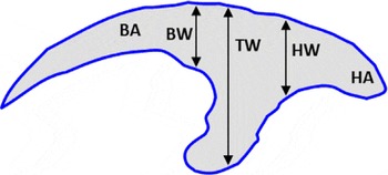
Several linear measurements and areas of both large and small hooks were analysed, including the total area, total length, total width, blade area, blade length, blade width, handle area, handle length and handle width (fig. 1). Images were captured with a digital camera (Nikon Eclipse E200 camera; Nikon, Chiyoda, Japan) connected to a microscope (Olympus BX41 microscope; Olympus, Tokyo, Japan). The camera output was processed and analysed with Image Pro Plus 3 image analysis software (Media Cybernetics). All measurements are in micrometres unless indicated otherwise.
Diagram of the rostellar hook of the protoscolex of Echinococcus vogeli with measurements (μm) of the total area (BA/HA), total width (TW), blade area (BA), blade width (BW), handle area (HA) and handle width (HW).

Data analysis
Data analysis was carried out with GraphPad InStat (GraphPad Instat Software Inc. version 3.01, California, USA). Statistical analyses included analysis of variance (ANOVA) followed by a post-hoc Tukey's test and Student's t-test. Statistical significance was assessed at P ≤ 0.05.
Results
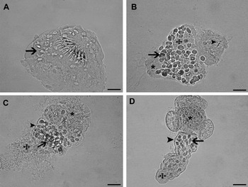
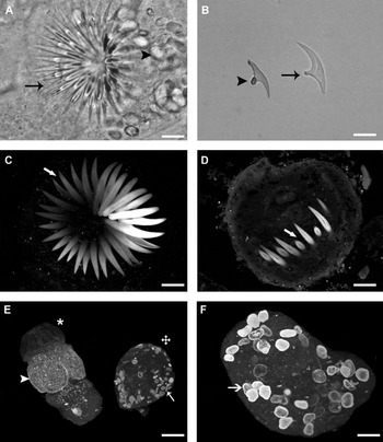
All microscopic techniques were necessary to examine the E. vogeli hooks and phenotypic aspects of protoscoleces. Using light microscopy, it was possible to visualize some protoscoleces during different developmental stages and also visualize the high density and differential distribution of calcareous corpuscles (fig. 2A–D), which are mainly located close to the rostellar hooks (fig. 3A). In protoscoleces, no differences are shown in organization of the hooks (figs 2A and 3A). There are two rows of hooks in the rostellar pad with a large hook intercalated by a small hook. Both large and small rostellar hooks vary morphologically, although all hooks possess a handle, blade and guard (fig. 3B). Large and small hooks possess a central amorphous pulp region. Large hooks frequently possess thin guards with an irregular surface between the guard and the handle, while small hooks are round and possess stout guards. In most cases, the surface between the guard and the handle is irregular. The blade does not show any relevant features.
Echinococcus vogeli metacestode to show stages of (A) the invaginated protoscolex with the rostellar pad and calcareous corpuscles irregularly localized; (B) evaginated protoscolex with suckers ( ), rostellum (arrowhead), neck region (*) and a high density of calcareous corpuscles (arrow); (C) evaginated protoscolex with scolex (*), neck region (arrowhead), developing proglottid (
 ) and cluster of calcareous corpuscles (arrow); (D) evaginated protoscolex showing the scolex with suckers (*), neck (arrowhead) with a lower density of calcareous corpuscles (arrow) than in (C) and the developing proglottid (
 ). All scale bars = 21 μm.

Light (A, B) and confocal laser scanning micrographs (C–F) of a whole mounts of the Echinococcus vogeli metacestodes, to show (A) large hooks in the upper row and small hooks in lower row (arrow) and calcareous corpuscles close to the rostellar hooks (arrowhead), scale bar = 15 μm; (B) polymorphism of large (arrow) and small (arrowhead) hooks, scale bar = 15 μm; (C) and (D) 1:1 arrangement of hooks in the rostellar pad (arrows), scale bars = 12 μm; (E) evaginated (*) and invaginated ( ) protoscoleces, calcareous corpuscles (arrow), and dorsal and ventral suckers (arrowhead), scale bar = 20 μm; (F) dense arrangement of calcareous corpuscles (arrow), scale bar = 12 μm.

CLSM images confirm the 1:1 arrangement of hooks in the rostellar pad (fig. 3C, D). There are protoscoleces in different stages of development with a high density of calcareous corpuscles (fig. 3E, F). In evaginated protoscoleces, we note the presence of both dorsal and ventral suckers, along with clusters of calcareous corpuscles in this region (fig. 3E).
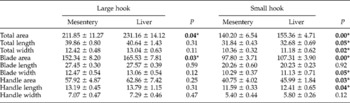
Morphometric approaches allowed the discrimination of differences between large and small hooks. All measurements are similar to those reported previously in the literature (table 1), although there are no previous data for mesentery samples. For the next step of this study, a comparison of morphometric characteristics of hooks from the liver and mesentery was undertaken (table 2). A phenotypic polymorphism was found in the linear and area biometrical parameters based on the analysis of 250 large and small hooks.
Rostellar hook morphometrics (μm) of Echinococcus vogeli metacestodes in the liver and mesentery (*) undertaken in the present study, compared with values reported by previous authors.

n, Number of hooks measured.
Comparative morphometrics (μm) with mean values ± standard deviation of large and small hooks of Echinococcus vogeli metacestodes, relative to location within the liver and mesentery.

Significant differences (in bold) given as P < 0.05.
When comparing hook measurements according to the infected organ, hooks from the liver are larger for all parameters analysed, with the exception of the blade length. When comparing small hooks with regard to the infected organ, all parameters analysed possess significant differences (P < 0.05), except for the blade length and the handle width. Large hooks possess only two significantly different characteristics, namely the blade area and total area (P < 0.05).
Discussion
The taxonomy of the Echinococcus genus has been resolved based on morphological characteristics viewed under light microscopy together with scanning and transmission electron microscopy (Rausch et al., Reference Rausch, Rausch and D'Alessandro1978, Reference Rausch, D'Alessandro and Ohbayashi1984; Kumaratilake et al., Reference Kumaratilake, Thompson and Eckert1986; Antoniou & Tselentis, Reference Antoniou and Tselentis1993). In addition, confocal microscopy provides new morphological features of E. granulosus (Almeida et al., Reference Almeida, Rodrigues-Silva, Neves, Lessa, Sanchez and Machado-Silva2009).
Because E. vogeli is related to all other Echinococcus taxa (Knapp et al., Reference Knapp, Nakao, Yanagida, Okamoto, Saarma, Lavikainen and Ito2011), this investigation demonstrated several phenotypic characteristics shared with E. granulosus. We showed that E. vogeli has two rows of hooks in the rostellar pad, arranged 1:1, with a large hook intercalated by a small hook (Rausch, Reference Rausch2006; Almeida et al., Reference Almeida, Rodrigues-Silva, Neves, Lessa, Sanchez and Machado-Silva2009). In addition, large and small hooks presented as a central, amorphous pulp region. Large hooks frequently had thin guards, in contrast to the slender guards observed in E. granulosus (Almeida et al., Reference Almeida, Rodrigues-Silva, Neves, Lessa, Sanchez and Machado-Silva2009). Small hooks were rounded and had stout guards, and the surface between the guard and the handle was irregular, confirming previous studies (Almeida et al., Reference Almeida, Rodrigues-Silva, Neves, Lessa, Sanchez and Machado-Silva2009). Differences in hook morphology are related to their function in the digestive systems of predatory carnivores (Antoniou & Tselentis, Reference Antoniou and Tselentis1993).
The present study confirmed that morphometry is a powerful tool for the analysis of rostellar hooks of cestodes belonging to the genus Echinococcus (Ponce-Gordo & Cuesta-Bandera, Reference Ponce-Gordo and Cuesta-Bandera1997; Almeida et al., Reference Almeida, Rodrigues-Silva, Neves, Lessa, Sanchez and Machado-Silva2009; Mowlavi et al., Reference Mowlavi, Salehi, Eshraghian, Rokni, Fasihi-Harandi, Mohajeran and Salahi-Moghaddam2012). Results from liver metacestodes show that the total lengths of small and large hooks agree with previous observations (D'Alessandro et al., Reference D'Alessandro, Rausch, Cuello and Aristizabal1979; Meneghelli et al., Reference Meneghelli, Martinelli, Velludo, Belluci, Magro and Barbó1992; Somocurcio et al., Reference Somocurcio, Sánchez, Náquira, Schilder, Rojas, Chacón and Yabar2004; Abdul-Hadi et al., Reference Abdul-Hadi, Chacón, Bruces, Gutierrez, Safar, Egui, Falco and Cantele2007; Knapp et al., Reference Knapp, Chirica, Simonnet, Grenouillet, Bart, Sako, Itoh, Nakao, Ito and Millon2009). From a phenotypic point of view, parameters relating to large and small hooks, such as total area, total width, blade area, blade length, blade width, handle area, handle length and handle width, can assist in differentiating E. granulosus metacestodes from various sites of infection (Almeida et al., Reference Almeida, Rodrigues-Silva, Neves, Romani and Machado-Silva2007). This investigation aimed to verify whether these parameters were reliable tools for distinguishing between hepatic and mesenteric metacestodes. Most importantly, we observed that small hooks provided a larger number of discriminatory criteria than large hooks and that blade length and handle width were not useful parameters, as had been reported previously for E. granulosus (Almeida et al., Reference Almeida, Rodrigues-Silva, Neves, Romani and Machado-Silva2007).
To the best of our knowledge, this is the first study describing measurements of mesenteric metacestodes. Surprisingly, all measurements of mesenteric metacestodes were less than those of hepatic metacestodes. A possible explanation for this is that E. vogeli metacestodes display phenotypic plasticity, in which morphological diversification may be organ-dependent or organ-induced. In this case, the liver and mesentery appear to represent heterogenic niches. Previously, we demonstrated that E. granulosus hooks recovered from lungs were larger than those collected from the liver (Almeida et al., Reference Almeida, Rodrigues-Silva, Neves, Romani and Machado-Silva2007). It is important to note that physiological and biochemical conditions are determinants of infection sites (Almeida et al., Reference Almeida, Rodrigues-Silva, Neves, Romani and Machado-Silva2007). Type I PE is the most common presentation of polycystic echinococcosis and includes the liver as a site of infection (D'Alessandro & Rausch, Reference D'Alessandro and Rausch2008), which suggests that the liver provides better physiological conditions for metacestode development than the mesentery.
Tissue from cestode species, such as Taenia solium (Xifeng & Baodong, Reference Xifeng and Baodong1998; Vargas-Parada et al., Reference Vargas-Parada, Merchant, Willms and Laclette1999; Yang, Reference Yang2004), E. granulosus metacestodes (Smith & Richards, Reference Smith and Richards1993; D'Alessandro, Reference D'Alessandro1997; Vargas-Parada & Laclette, Reference Vargas-Parada and Laclette1999; Martínez et al., Reference Martínez, Paredes, Stock, Saralegui, Andreu, Cabezón, Ehrlich and Galanti2005), T. taeniaeformis and Dipylidium caninum proglottids (Khalifa et al., Reference Khalifa, Mazen, Marawan and Thabit2011), contains mineral concretions termed calcareous corpuscles. An established body of literature states that calcareous corpuscles are of cellular origin (Pawlowski et al., Reference Pawlowski, Yap and Thompson1988; Xifeng & Baodong, Reference Xifeng and Baodong1998; Vargas-Parada et al., Reference Vargas-Parada, Merchant, Willms and Laclette1999), irregularly spherical or ovoid in shape (Smith & Richards, Reference Smith and Richards1993) and are of different sizes (Vargas-Parada & Laclette, Reference Vargas-Parada and Laclette1999).
In the present investigation, it appears that calcareous corpuscles from the protoscolex of E. vogeli follow a common, basic pattern, similar to that of E. multilocularis (Ohnishi & Kutsumi, Reference Ohnishi and Kutsumi1991) and E. granulosus (Smith & Richards, Reference Smith and Richards1993). The present results are consistent with earlier studies on the description of the cellular organization of developing stages of E. granulosus (Martínez et al., Reference Martínez, Paredes, Stock, Saralegui, Andreu, Cabezón, Ehrlich and Galanti2005). The latter indicated that cell buds generated during the formation of protoscoleces are concentrated mainly in suckers and the rostellar pad (Martínez et al., Reference Martínez, Paredes, Stock, Saralegui, Andreu, Cabezón, Ehrlich and Galanti2005).
Despite the observation that both invaginated and evaginated protoscoleces contain calcareous corpuscles, there are variations in their density and location, as shown by light and confocal microscopy. In the invaginated stage, protoscoleces containing the rostellar pad and calcareous corpuscles were irregularly localized, whereas in the evaginated stage, calcareous corpuscles were concentrated in the rostellum, suckers, neck region and developing proglottids. Furthermore, during morphogenesis, younger and evaginated protoscoleces contained a high density of calcareous corpuscles, but a reduced density of clusters was observed in protoscoleces with some evidence of strobilization.
Despite a number of exhaustive studies, the physiology of calcareous corpuscles in various cestode taxa remains poorly understood (Vargas-Parada & Laclette, Reference Vargas-Parada and Laclette1999). Calcareous corpuscles are the principal sources of calcium carbonate, magnesium and phosphate (Smith & Richards, Reference Smith and Richards1993), with a possible role in excretory activity (Vargas-Parada & Laclette, Reference Vargas-Parada and Laclette1999) or in the secretion of glycoproteins (Yang, Reference Yang2004). As their sites, such as suckers, rostellar pads, hooks and proglottids, present intense biological activity, it may be hypothesized that calcareous corpuscles are involved in parasite differentiation and growth (Rodrigues et al., Reference Rodrigues, Ferreira, Farias and Zaha1997). Although mechanisms by which calcareous corpuscles contribute to metacestode differentiation are not fully understood, these structures may act as a source of ions (Rodrigues et al., Reference Rodrigues, Ferreira, Farias and Zaha1997) or thioredoxin peroxidase, which suggests that they protect against oxidative damage, and may have other biological roles, such as energy transformation and differentiation (Rodrigues et al., Reference Rodrigues, Ferreira, Farias and Zaha1997; Li et al., Reference Li, Zhang, Loukas, Lin, Ito, Zhang, Jones and McManus2004).
To date, morphological characteristics of calcareous corpuscles from E. vogeli metacestodes have not been sufficiently studied. The present results, unlike previous studies, demonstrate that calcareous corpuscles are virtually absent from these parasite taxa (D' Alessandro, Reference D'Alessandro1997). A possible explanation is that the metacestodes studied were in different stages of maturation. In conclusion, it is suggested that morphological features of both large and small rostellar hooks of E. vogeli represent morphological adaptation to a varied environment within the vertebrate host. In these taxa, the present results have shown for the first time that morphological changes in calcareous corpuscles of metacestodes do occur at differing stages of maturation.
Acknowledgement
We would like to thanks Rodrigo Méxas from the Laboratório de Produção e Processamento de Imagem Científica, IOC – Fiocruz for preparing the images.
Financial support
The authors gratefully acknowledge the financial support of the Fundação Carlos Chagas Filho de Amparo a Pesquisa do Estado do Rio de Janeiro (FAPERJ) and the Universidade do Estado do Rio de Janeiro (UERJ). We would also like to thank the Conselho Nacional de Desenvolvimento Científico e Tecnológico (CNPq) for a scholarship (to J.R.M.S.).
Conflict of interest
None.
Ethical standards
The authors assert that all procedures contributing to this work comply with the ethical standards of the Instituto de Pesquisa Evandro Chagas, Fundação Oswaldo Cruz, Rio de Janeiro, RJ, Brazil and with the Helsinki Declaration of 1975, as revised in 2008, and have been approved by the institutional committee (no. 002.1.000.009-04).