I. INTRODUCTION
Chitin is a natural polysaccharide found in the shells of crustaceans, and in many other phyla. Chitin has been described as a “cellulose-like biopolymer” (Austin et al., Reference Austin, Brine, Castle and Zikakis1981). Chitin has many desirable properties, including toughness and biodegradability. Like cellulose, it is insoluble in ordinary solvents (Austin et al., Reference Austin, Brine, Castle and Zikakis1981).
The commercialization of chitin in shaped forms is dependent on both the discovery of a solvent and the identification of an appropriate end used. A non-degrading solvent for chitin, 5% lithium chloride in N,N-dimethyl acetamide, was discovered (Rutherford, Reference Rutherford1975; Austin, Reference Austin1977; Austin and Rutherford, Reference Rutherford and Austin1978), opening the way to the production of chitin fibers and films. One area in which chitin has shown considerable potential is in biomedical products. Chitin has been reported to accelerate wound-healing (Balassa and Prudden, Reference Balassa, Prudden, Muzzarelli and Pariser1978; Austin, Reference Austin1984), and has shown potential as a carrier for poorly soluble drugs (Sawayanagi et al., Reference Sawayanagi, Nambu, Nagai, Hirano and Tokura1982).
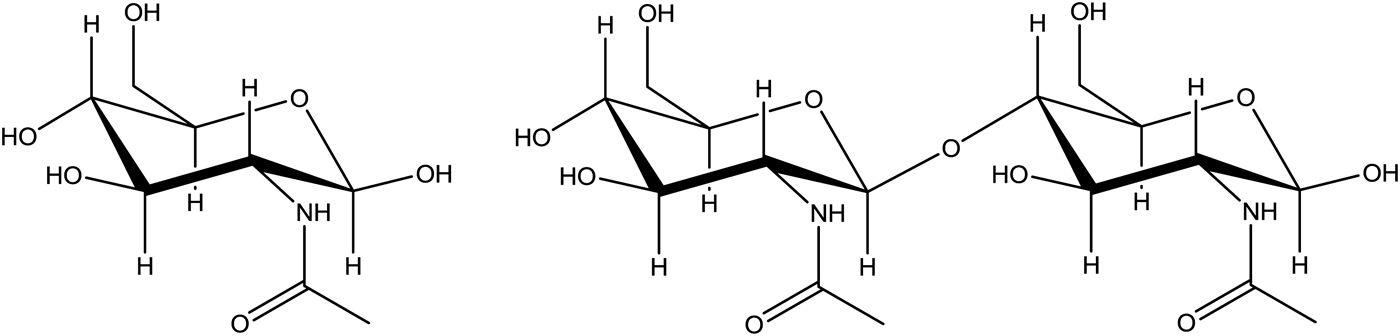
Although chitin is generally regarded as a polymer composed of linear chains of beta-(1,4)-linked N-acetylglucosamine (Figure 1) (N-acetyl-2-amino-2-deoxy-d-glucopyranose) this is an over-simplification, as chitin occurs as part of more complex systems (Brimacombe and Webber, Reference Brimacombe and Webber1964). Commercialization of chitin has been complicated by the fact that chitin almost always occurs in association with protein and many chitins are calcified. The original evidence for the association between chitin and protein was obtained by Fraenkel and Rudall (Reference Fraenkel and Rudall1947). They found a ratio of three amino acids to one chitobiose (the basic structural unit of chitin) in the cuticles of blowfly larvae. The occurrence of protein with chitin has been well documented (Carlstrom, Reference Carlstrom1957; Attwood and Zola, Reference Attwood and Zola1967; Brine, Reference Brine1979; Hunt and Nixon, Reference Hunt and Nixon1981; Brine and Austin, Reference Brine and Austin1981a). In the methods used to purify chitin, there are steps in which the material is treated with alkali to remove most, but not all, of the associated proteins (Muzzarelli, Reference Muzzarelli1977). Depending on the source of the chitin, de-calcification may also be required. Some commercial chitins add a de-colorization step to produce a more appealing product. Deacetylation of chitin produces chitosan (i.e., poly-glucosamine) a commercial product that finds uses in agriculture, winemaking, and medicine (Linden et al., Reference Linden, Stoner, Knutson and Gardner-Hughes2000).
N-acetylglucosamine (N-acetyl-2-amino-2-deoxy-D-glucopyranose, left) and chitobiose (right) the building block of chitin.

In this study, the chitin polymer is characterized by X-ray diffraction as it progresses through the purification procedure and the effect of various treatments is examined. Chitins from three sources (goose barnacles, horseshoe crabs, and a commercial chitin prepared from shrimp shells) are included in the study.
II. EXPERIMENTAL
A. Preparation of horseshoe crab chitin
Horseshoe crabs (Limulus polyphemus) were collected from the shores of the Delaware bay. The shells were air dried at room temperature. The dried material was ground in a blender. The material was examined during grinding and some film-like fragments were removed from some shell fragments for further study. These fragments are referred to as “native Limulus chitin”. A total of 200 g of the coarsely ground shells were suspended in 3 L of 0.5% sodium hydroxide for 1 h at room temperature. The solids were separated, washed with tap water, and resuspended in 3 L of 5.0% sodium hydroxide for 5 h. The solids were collected and suspended a second time in 3 L of 5.0% sodium hydroxide for 18 h. The product was collected on a Buchner funnel, washed with distilled water until neutral, air dried, and ground to 20 mesh in a Willy mill. This material is referred to as “Limulus chitin”.
B. Preparation of a chitin film
A chitin film was prepared by dissolving commercial chitin in the non-degrading solvent system (Rutherford, Reference Rutherford1975; Austin, Reference Austin1977; Austin and Rutherford, Reference Rutherford and Austin1978). A total of 2.0 g of KY-10 chitin (Hercules Inc.) was dissolved by slow stirring in 90 ml of dimethyl acetamide containing 5% lithium chloride. A film was prepared from this solution by casting onto a glass plate, allowing the film to setup for 20 min, and sliding the film into a t-butanol bath. The film was allowed to remain in the t-butanol bath overnight after which it was removed and allowed to air dry. The air-dried film was pale amber in color and flexible.
C. Preparation of a goose barnacle chitin
Goose barnacles (family Lepadidae) were collected in the area of Baltimore canyon approximately 150 km off the coast of Delaware and stored frozen for later study. After thawing, the chitinous shell was removed, washed several times with water, once with acetone, and then air dried. The dried material was ground to 20 mesh in a Willy mill. The ground sample was treated sequentially with EDTA, 0.01-M sodium hydroxide, 1-M sodium hydroxide, and 10% hydrochloric acid to remove weakly bound calcium, weakly bound protein, strongly bound protein, and strongly bound calcium, respectively. Samples were saved for later analysis after each step and designated B-1, B-2, B-3, and B-4, respectively. Material removed in the final decalcification step (i.e., 10% hydrochloric acid) was precipitated with acetone and designated B-3ppt.
D. Preparation of chitin products
Chitosan was obtained from Aldrich and Fluka and used as is. These materials were designated LMW (low molecular weight chitosan from Aldrich) and HMW (high molecular weight chitosan from Fluka).
Re-acylated chitosan was prepared by combining 2 g of Chitosan and 100-ml of concentrated acetic acid. This mixture was sheered in a blender for 2–3 min. A 25 ml of acetic anhydride was added to the mixture and allowed to react at room temperature for 1.5 h with continuous stirring. The solids were washed with two volumes of acetone to rapidly removed associated water followed by two volumes of water. The solids were collected on a Bűchner funnel, washed with absolute ethanol and dried at 60 °C.
E. X-ray diffraction data collection
X-ray diffraction data for powders were collected using a Bruker D2 Phaser. Data were collected from 10 to 90°2θ (unless noted otherwise) using a 0.02° step size. Data collection time was adjusted as necessary for the various samples analyzed.
X-ray diffraction data for films (i.e., native Limulus chitin, chitin thin film, and intact goose barnacles) were collected using a Bruker Proteum with a sample to detector distance of 5.0 cm. Images at different values of 2θ setting were collected and merged using the XRD2 plug-in for APEX software to cover the range of 10°–85° or greater. The merged images were integrated and saved as raw files for use in other applications.
III. RESULTS AND DISCUSSION
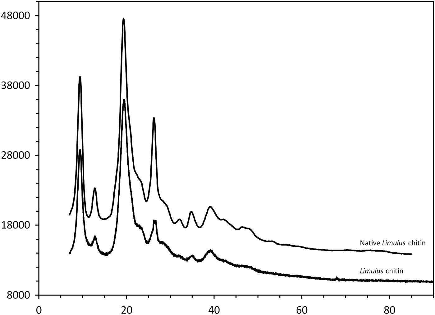
Chitin from the horseshoe crab (Limulus polyphemus) is simple to prepare as the shell of the horseshoe crab is not calcified. The method used in this study to prepare chitin from horseshoe crab shells is a modification of the method described by Rutherford (Reference Rutherford1975) and results in samples with a final protein content of 1.8–2.2% (Brine, Reference Brine1979; Austin et al., Reference Austin, Brine, Castle and Zikakis1981). The resulting chitin–protein complex is relatively undergraded as found by the high molecular weight and low de-acetylation (Brine and Austin, Reference Brine and Austin1981b) and thus should be a good reference material for chitin. The X-ray diffraction pattern of Limulus chitin is shown in Figure 2 with the pattern obtained from the unprocessed film (native Limulus chitin) isolated from the ground shells. Fine features in the diffraction pattern are preserved in the purified chitin – this is evidence that the alkali treatment used in purification is relatively non-degrading and is consistent with prior reports (Brine and Austin, Reference Brine and Austin1981b). A similar result is seen (Figure 3) in the chitin film prepared using the non-degrading solvent system (Rutherford, Reference Rutherford1975; Austin, Reference Austin1977; Austin and Rutherford, Reference Rutherford and Austin1978). The chitin used to prepare the film is a commercial chitin obtained from shrimp shells. The chitin in the film has a different origin and has gone through additional processing and dissolution but retains features present in native chitin. These features can be thought of as characteristic of chitin.
X-ray diffraction pattern of “native Limulus chitin” (top) and purified “Limulus chitin” (bottom). Note how well fine features in the diffraction pattern are preserved in purified chitin. This is evidence that the alkali treatment used in purification is relatively non-degrading.
X-ray diffraction pattern of “native Limulus chitin” (top) and a chitin film (bottom). Note how the fine features in the diffraction pattern are preserved in the chitin film as compared with native chitin.
Chitosan is produced by deacylation of chitin, generally by treatment with strong alkali at elevated temperatures (c.f. Tajik et al., Reference Tajik, Moradi, Rohani, Erfani and Jalali2008). Thus, preparation of chitosan generally requires deproteinization, demineralization, decolorization, and deacetylation of the source material to produce the desired product. Samples of two commercially available chitosans were selected for analysis based on reported differences in molecular weight (Figure 4). It might be expected that a low molecular weight (LMW) chitosan would be more degraded than a high molecular weight (HMW) chitosan and that this would manifest itself in the diffraction pattern as broader diffraction peaks, lower intensities for the diffraction peaks, or both. This does not seem to be the case as the LWM chitosan (Figure 4) shows multiple sharp peaks plus high-angle scattering not observed in the diffraction pattern for HMW chitosan (Figure 4). Since the two chitosans were produced by different suppliers and the source of the parent chitin is not identified the observed differences may be related to either the source material or the method of manufacture.
X-ray diffraction patterns of high molecular weight (HMW) and low molecular weight (LMW) chitosans, and re-acylated chitosan. The second phase present in the LMW sample (i.e., the sharp peaks) has not been identified.
Chitosan can be re-acylated by treatment with acetic anhydride in acetic acid. In theory this should result in the formation of chitin. The product of re-acylation was not soluble in dilute acetic acid, which is consistent with the product being more chitin-like. The diffraction pattern (Figure 4) is only slightly changed on re-acylation with a peak at about 39°2θ (also present in both chitosans, and two weak broad peaks at approximately 27 and 39°2θ. Diffraction peaks at these locations (27 and 39°) are present in chitin (see Figure 2), however, in chitin these peaks are both more intense and sharper. Thus, it would appear that chitin is processed to chitosan and the re-acylated order within the material is lost even though some of its physical properties (i.e., solubility) are regained.
Unlike chitin present in insects or horseshoe crabs the chitin present in crustaceans is generally highly calcified. Goose barnacles have a tough somewhat flexible shell. Extra stiffness in the goose barnacle shell is presumably due to calcification. This is obvious in the diffraction pattern of an isolated fragment of a goose barnacle plate that has not been subjected to any processing (Figure 5). As the material from the goose barnacles is processed through EDTA, dilute sodium hydroxide, concentrated sodium hydroxide, and hydrochloric acid (samples B-1, B-2, B-3, and B-4, respectively in Figure 6) the influence of calcium carbonate diminishes and the final material (fraction B-4) has a diffraction pattern nearly indistinguishable from that of native chitin (see Figure 2).
The X-ray diffraction pattern of an isolated fragment of a goose barnacle plate (solid line). Although not a perfect match it is clear this pattern is dominated by the diffraction pattern of calcium carbonate (red bars).
Goose barnacle chitin was prepared by sequential extraction of the shell with EDTA, dilute sodium hydroxide, concentrated sodium hydroxide, and hydrochloric acid resulting in materials B-1, B-2, B-3, and B-4 respectively.
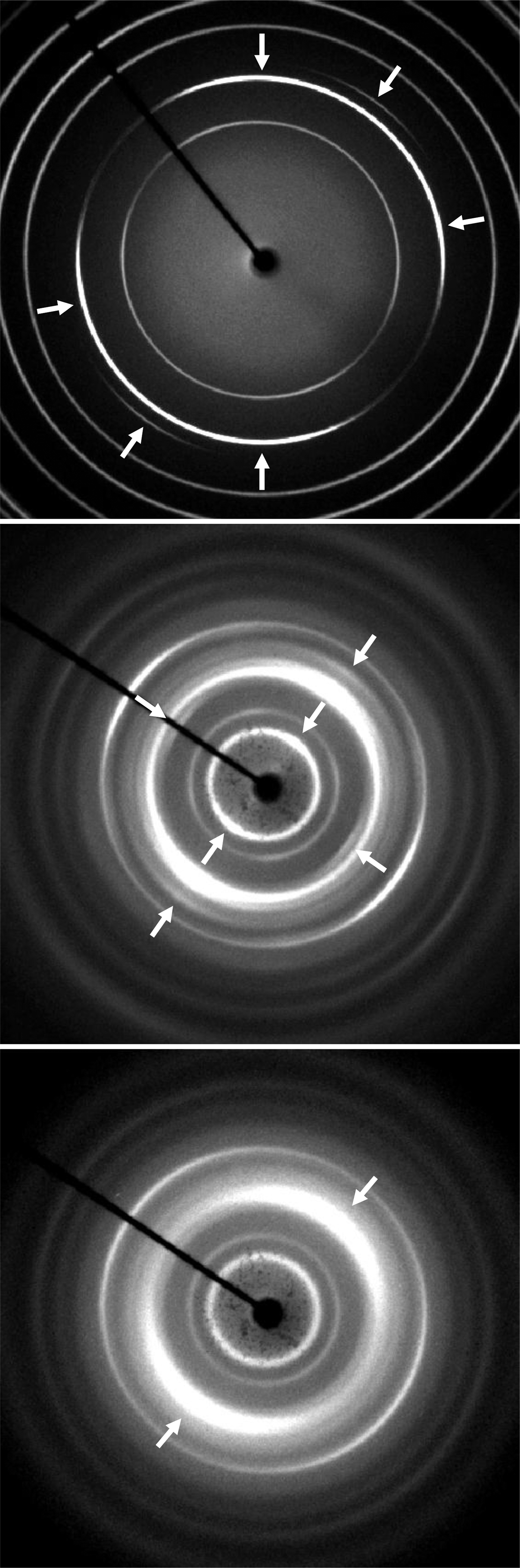
The native chitin containing materials also exhibit some preferred orientation. This is evident in the highly calcified material from goose barnacles (Figure 7, top), and the shell of L. polythemus (Figure 7, middle). Even a chitin film prepared in a non-degrading solvent system shows some evidence of preferred orientation (Figure 7, bottom). Other investigators have noted the additional order present in native materials (Brine & Austin, Reference Brine, Austin and Church1975). This additional order has been exploited to determine the structure of α-chitin in lobster and crab tendons (Minke and Blackwell, Reference Minke and Blackwell1978; Sikorski et al., Reference Sikorski, Hori and Wada2009). It is interesting that in this study the two “native” materials show the highest degree of order even though they represent very different materials (i.e., a diffraction pattern dominated by calcium carbonate vs. one which is almost a pure bio-polymer).
X-ray diffraction images of an isolated fragment of a goose barnacle (top), native Limulus chitin (middle), and a chitin film (bottom). Positions marked with arrows indicate regions that show evidence of preferred orientation.
IV. SUMMARY
Native chitin has a characteristic diffraction pattern that is not altered by mild treatments used to isolate relatively pure chitin, in addition, non-degrading solvents can be used to prepare chitin films which retain most of the characteristic patterns.
Although other investigators have shown differences in chitin preparation from different sources these differences are not present in the diffraction pattern of pure non-degraded chitins.
Processing chitin to chitosan results in large changes to the diffraction pattern. Some of the features present in the diffraction pattern of native chitin can be recovered (to a very limited extent) by re-acylation of chitosan.
Chitin present in crustaceans is generally highly calcified and the diffraction pattern of the unprocessed shell (or exoskeleton) is dominated by calcium carbonate. As the material is processed this pattern becomes less pronounced and the final product closely resembles that of chitin obtained from a non-calcified feed stock.
ACKNOWLEDGMENTS
This work was supported in part by the Naval Research Laboratory and the Office of Naval Research. The author thanks Dr W. Wong-Ng and Dr James Kaduk for their advice in preparation and revision of the manuscript.
Supplementary Materials and Methods
Supplementary material accompanies this paper on the Journal's website (http://journals.cambridge.org/PDJ).