Introduction
Seeds of some eastern Australian Grevillea species do not germinate readily under otherwise favourable conditions (Morris, Reference Morris2000) but germinate after fire in the field (Auld and Tozer, Reference Auld and Tozer1995). Seeds of these species demonstrate positive germination responses to smoke, or heat shock, with the greatest germination occurring if both are applied (Kenny, Reference Kenny2000; Morris, Reference Morris2000; Briggs and Morris, Reference Briggs and Morris2008). This pattern results because freshly shed seeds show dormancy, which is broken by exposure to one or both of the fire cues (Morris, Reference Morris2000; Morris et al., Reference Morris, Tieu and Dixon2000; Briggs and Morris, Reference Briggs and Morris2008). The seeds of Grevillea spp. that show this pattern lack an endosperm. The embryo is well developed and linear, surrounded by a water-permeable seed coat (Briggs et al., Reference Briggs, Morris and Ashford2005). Dissection of the embryos out of the seed coat results in full germination of normal seedlings (Kenny, Reference Kenny2000; Morris et al., Reference Morris, Tieu and Dixon2000; Ma et al., Reference Ma, Guo, Han and Yan2015). In the classification of dormancy types of Baskin and Baskin (Reference Baskin and Baskin2004), this combination of features is characteristic of seeds with non-deep physiological dormancy (PD).
Identifying the precise cause of non-deep PD is difficult (Baskin and Baskin, Reference Baskin and Baskin2014). In broad terms, the mechanism may lie in the embryo itself, in the covering structures (seed coat), or in some combination of the two. For the covering structures or seed coat, five types of dormancy are recognized: the seed coat may reduce the rate of water imbibition, restrict oxygen diffusion to the embryo, contain inhibitors and/or prevent the exit of inhibitors from the embryo, be changed by a dormancy-breaking cue, or physically restrain the embryo (Baskin and Baskin, Reference Baskin and Baskin2014).
Two of these potential mechanisms can be discounted for Grevillea since scarification of the seed coat makes little difference to water uptake, suggesting that the intact seed coat does not restrict imbibition (seven Grevillea spp.; Morris, Reference Morris2000; Morris et al., Reference Morris, Tieu and Dixon2000). Morris et al. (Reference Morris, Tieu and Dixon2000) observed no restriction on germination of dissected embryos re-inserted back into seed coats, suggesting a lack of germination inhibitors in the covering structures. Ma et al. (Reference Ma, Guo, Han and Yan2015) demonstrated that macerated extracts of the seed coat of Grevillea species inhibited germination of several agricultural seeds, but whether these seed-coat inhibitors restrict germination in intact Grevillea seeds has not been established. Furthermore, while there are internal barriers in the seed coat to the diffusion of molecules of large molecular weight (Briggs et al., Reference Briggs, Morris and Ashford2005) these are not weakened or eliminated by heat shock, smoke or their combination (Briggs and Morris, Reference Briggs and Morris2008), as was observed after smoking seeds of a fire-responding chaparral species that possesses a germination inhibitor located in the embryo (Egerton-Warburton, Reference Egerton-Warburton1998).
The remaining three seed-coat mechanisms remain untested in Grevillea spp. Restriction of oxygen diffusion would be possible in the seeds of Grevillea linearifolia, as the exotestal layer contains phenolic compounds (Briggs et al., Reference Briggs, Morris and Ashford2005), which have been shown to play such a role in other species (Baskin and Baskin, Reference Baskin and Baskin2014). The fire cues could have structural effects on the covering structures, as heat is known to do so in species with physical dormancy (Burrows et al., Reference Burrows, Virgona and Heady2009) and so could potentially affect seed-coat properties in Grevillea. The seed coat of G. linearifolia has two layers (outer testa, inner tegmen), parts of which could act as a mechanical constraint on the embryo. While the exotesta is made up of a single layer of thin-walled cells, and the mesotesta of several layers of thin-walled cells, the endotestal layer is made up of a single layer of lignified and unlignified palisade cells. The inner tegmen consists of several layers of non-lignified sclerenchyma cells. These last two layers would considerably strengthen the seed coat (Briggs et al., Reference Briggs, Morris and Ashford2005). When dissected G. linearifolia embryos were re-inserted into seed coats, the fit was loose rather than tight, and germination ensued (Morris et al., Reference Morris, Tieu and Dixon2000). This suggested that maintenance of the structural integrity of the seed coat in close alignment with the embryo may be important in preventing germination, and would match with the mechanical constraint model of seed-coat dormancy. If the seed coat acts as a mechanical constraint, there are species where this constraint is localized to the micropylar end of the seed (Chen, Reference Chen1968; Mehana and Martin, Reference Mehana and Martin1985; Delanoy et al., Reference Delanoy, Van Damme, Sceldeman and Beltran2006), and preliminary observations on dissected and/or scarified Grevillea seeds suggested that such localized constraint might be operating.
Mechanisms of non-deep physiological dormancy residing in the embryo can include the presence of inhibitors (which the covering structures prevent from diffusing out), or lack of sufficient growth potential by the embryo to break through the covering structures if a dormancy-breaking signal is absent. The embryos of Grevillea have not been tested for the presence of germination inhibitors, but permeability of the seed coat to potential inhibitors is not altered by the fire cues, as outlined above. The effect of fire cues on the growth potential (the force the radicle can exert) has not been tested, however. In a number of species, dormancy-breaking treatments have been found to increase the growth potential of the embryo (Baskin and Baskin, Reference Baskin and Baskin1971; Nabors and Lang, Reference Nabors and Lang1971; Weston et al., Reference Weston, Geneve and Staub1992; Feurtado et al., Reference Feurtado, Ren, Ambrose, Cutler, Ross, Abrams and Kermode2008) and thus the embryo's ability to break through the peri-embryonic tissues. Interactions between the embryo and the covering structures are also possible, such as weakening of the seed coat by a signal released by the embryo after dormancy has been broken. Hormonal signals such as gibberellic acid have been postulated to play this role (Kucera et al., Reference Kucera, Cohn and Leubner-Metzger2005). Smoke can have physiological effects on embryos, via hormonal pathways (Nelson et al., Reference Nelson, Flematti, Dixon and Smith2012).
There are several methods of testing the mechanical strength of peri-embryonic tissues. One is the application of compressive force to the intact seed. Reports in the literature using this approach are few and have been concerned with damage to the seed during mechanical processing. They have shown that the direction of applied force is important and that different values have been obtained when applied to the width and thickness (Akaami and Raji, Reference Akaami and Raji2006), or at different points along the vertical axis (Ogunjimi et al., Reference Ogunjimi, Aviara and Aregbesola2002). Furthermore, less force was required for hydrated than for dry seeds (Singh and Goswami, Reference Singh and Goswami1998).
A second approach has been to insert a probe into dissected seed coats to simulate the radicle's protrusive force. Only two studies of this nature have been carried out (Esashi and Leopold, Reference Esashi and Leopold1968; Nabors and Lang, Reference Nabors and Lang1971). A preliminary study was undertaken to trial this method for Grevillea seeds, but the results were unsatisfactory. However, the study indicated that there was a very tough layer present in the seed coat which would be a substantial barrier to any protrusive force acting on the micropylar region. The fractured seed coats from this study allowed an anatomical investigation of the rupturing of the seed coat in the micropylar region.
A third approach has been to determine the maximum thrust that can be developed by the hypocotyl/radicle (or embryonic) axis during the final stages of germination. The method to determine growth potential involves measuring the ability of partially or fully excised embryos to extract water over a range of decreasing water potentials generated by osmotica. The lowest water potential at which germination can still occur is taken as a measure of the maximum (positive) thrust that the embryo can generate (Nabors and Lang, Reference Nabors and Lang1971). This approach was adopted, both to estimate the maximum force that the embryo can generate, and to test for the effects of the fire cues on the force generated by the embryo (growth potential).
Seeds of Grevillea mature in thin-walled follicles over several weeks; at maturity the fruit darkens, ripens and splits open, releasing the seeds over a period of 24–48 h. As only a small percentage of the fruits on a plant are at this final stage at any one time, seed collection involves multiple repeat visits to field populations, and total seed numbers at the end of a collecting season can be in the low hundreds. This limited the number of treatments and replicates available for experiments. In previous work investigating dormancy in these species, seeds of G. linearifolia had been used (Briggs et al., Reference Briggs, Morris and Ashford2005; Briggs and Morris, Reference Briggs and Morris2008). Grevillea juniperina seeds were used for the current study, being more convenient to collect. Seeds of G. juniperina show the same limited germination in the absence of fire cues as G. linearifolia, but a strong response to application of the fire cues (Morris, Reference Morris2000).
To resolve further the mechanism of dormancy in Grevillea, questions were addressed as to whether the inhibitory effect of the covering structures is localized to particular parts of the seed coat; whether the fire cues, applied either singly or in combination, affect the growth potential of the embryo; what the mechanical strength of the seed coat is when compressive force is applied to seeds that are dry or hydrated, and either untreated or treated with fire cues; and the anatomical basis of fracture of the seed coat subjected to compressive force.
Materials and methods
Seed collection
Seeds of G. juniperina R. Br. were collected from roadside plants in field populations along Richmond Road, Marsden Park, and a private garden at Yarramundi, NSW, Australia. Seeds from both sources were mixed and kept in paper bags in air-conditioned, dry storage (20°C) until used (within 12 months of collection).
Seed coat and embryonic morphology of G. juniperina
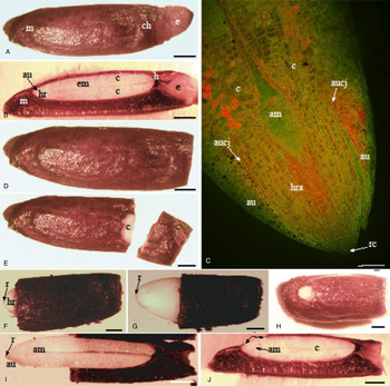
A preliminary investigation of the morphology and anatomy of G. juniperina seeds was conducted to compare with those of G. linearifolia, and to establish more precisely the anatomical dimensions and relationships of the radicle and the seed coat. The seeds of G. juniperina measured between 9 and 14 mm in length, with a distinct micropylar region and an eliosome at the chalazal end (Fig. 1A). Medial longitudinal sections (MLS) showed a hypostase at the chalazal end and ear-like extensions (called auricles) of the cotyledons which covered the embryonic axis (i.e. hypocotyl–radicle axis, root cap and apical meristems) at the micropylar end (Fig. 1B). The auricles extended over the hypocotyl–radicle (HR) axis, leaving the root cap and a short portion of the HR axis exposed (approximately 100 μm or greater) (Fig. 1C). This is similar to the anatomy of the embryo and seed coat already described for G. linearifolia (Briggs et al., Reference Briggs, Morris and Ashford2005).
(A) Whole seed of Grevillea juniperina (dorsal view); (m) micropylar region, (ch) chalazal region, (e) eliosome. Scale bar = 1 mm. (B) Median longitudinal section (MLS) of whole seed; (m) micropylar region, (em) embryo, (c) cotyledons, (hr) hypocotyl–radical axis, (au) auricles, (e) eliosome, (h) hypostase. Scale bar = 1 mm. (C) Confocal laser scanning image of an MLS through the micropylar end of a hand-cut embryo stained with acridine orange; seed coat removed. The embryo was treated with heat shock and smoke; (am) apical meristem, (au) auricles, (aucj) auricle/cotyledon junction, (c) cotyledon, (hra) hypocotyl–radicle axis, (rc) root cap. Scale bar = 145 μm. (D) Seed with eliosome portion removed. Scale bar = 1 mm. (E) Seed with chalazal end removed, exposing the ends of the cotyledons (c). Scale bar = 1 mm. (F) Seed with micropylar seed coat removed, exposing 1 mm of the embryo's hypocotyl–radicle axis (hr) and radicle (r). Scale bar = 1 mm. (G) Seed with micropylar seed coat removed, exposing 2.8 mm of embryo; (r) radicle. Scale bar = 1 mm. (H) Dorsal view showing a patch (1 mm × 1 mm) of embryo where the seed coat has been removed. Scale bar = 1 mm. (I) Median longitudinal section through the seed shown in (G), showing the extent of the exposed embryonic tissues; (am) apical meristem, (au) auricules, (r) radicle. Scale bar = 1 mm. (J) Median longitudinal section through the seed shown in (H), showing the relationship between the ‘window’ (arrows) and the embryo; (c) cotyledons, (am) apical meristem. Scale bar = 1 mm.

Effect of selective removal of seed coat on germination
A series of treatments was imposed which either removed the seed coat to expose the root cap and HR axis, or maintained the structural integrity of the seed coat over the root cap and HR axis. The overall aim was to test whether maintenance of the structural integrity of the seed coat over the micropylar region was important in allowing or preventing germination. Treatments consisted of: (1) the removal of the eliosome tissue only (control) (Fig. 1D); (2) the removal of the chalazal end of the seed to expose the cotyledons (CCEX) (Fig. 1E); (3) removal of c. 1 mm of the micropylar end of the seed coat to expose the HR axis (M1) (Fig. 1F); (4) removal of c. 2–3 mm of the micropylar end of the seed coat to further expose the HR axis and release the constriction of the seed coat on the basal portion of the cotyledons and auricles (M2) (Fig. 1G); (5) retention of the seed coat over the root cap and base of the auricles but removing a small area (approx. 1–2 mm2) of seed coat from the dorsal surface of the seedling above the HR axis (MD) (Fig. 1H). Treatments M1 and M2 removed the seed coat over the root cap and the HR axis (Fig. 1I); the remaining treatments maintained the structural integrity of the seed coat over the root cap and HR axis, but allowed movement of gases and any potential germination inhibitors between the embryo and the exterior (shown for MD treatment in Fig. 1J).
The seeds that were used had been kept in dry storage for 2–2.5 months. Surface sterilization and subsequent preparation of the seeds was carried out in a laminar flow cabinet using sterile conditions and sterile equipment. The number of replicates per treatment per experiment ranged from 4 to 5; the number of seeds per replicate was 5. For each replicate, the seeds were placed into a glass vial, covered with either sterile distilled water or 10 ml of sterile Triton X-100 solution for 1 h, rinsed with three changes of distilled water, covered with 10 ml of 75% ethanol and agitated for 8 s, decanted, covered with 10 ml of diluted bleach (equivalent to 0.525% NaOCl), left for 10 min with occasional agitation, decanted and rinsed with four changes of distilled water before the seeds were transferred to paper for modification to the seed coat. In all treatments and the controls, the eliosome tissue was removed and a new scalpel blade, filter paper and cutting dish were used for each replicate.
After treatment, all seeds were transferred to sterile Petri dishes and covered with 12 ml sterile distilled water, sealed with Parafilm® and inserted into Ziploc® bags before being placed into a Phytoton Climate Simulator, 12/12 dark/light at constant 20°C, 180 μE. Germination was determined when the radicle extended by 2 mm and germinated seeds were not removed, to reduce the incidence of fungal contamination. The experiment ran for 98 d.
Effects of fire cues on growth potential of the embryo
To investigate the effects of the fire cues on the growth potential of the embryo, seeds with the micropylar seed coat removed (referred to hereafter as ‘dissected seeds’) were used. Preliminary experiments investigating the effects of the combined fire cues on the growth potential of the embryo detected little difference between untreated and fire-cue treated dissected seeds over the range −0.5 to 0.0 MPa. Due to limited seed numbers, a fully replicated experiment was conducted over a range of water potentials <−0.5 MPa, using the fire cues applied singly, and in combination. The aim was to determine whether one, or both, or the combined fire cues affected the embryo's growth potential, and if so, to what extent. A factorial experiment combining germination treatments (controls, heat shock, smoke, heat shock + smoke) with water potential (0, −0.6, −0.75, −0.9 MPa) was established using dissected seeds of G. juniperina.
Fire-cue and water potential treatments
For the heat-shock treatment, intact seeds were dry heated at 80°C for 10 min. (Morris, Reference Morris2000). Seeds to be smoked were placed in a smoking cabinet and exposed to 10 min of cooled aerosol smoke produced by a bee-keeper's smoker. Seeds receiving both treatments were heated first, then smoked (Briggs and Morris, Reference Briggs and Morris2008). Replicates were treated individually to ensure independence (Morrison and Morris, Reference Morrison and Morris2000). Seeds were first surface sterilized as detailed above and all work was carried out under sterile conditions in a laminar flow cabinet. Seeds in the smoke and heat + smoke treatments were immersed for 24 h in distilled water to allow the smoke components to diffuse into the seed and embryo. All seeds then had the micropylar end of the seed coat removed. In all treatments and the controls, the eliosome tissue was also removed to reduce fungal infection.
After treatment and dissection, the seeds were transferred to a new sterile filter paper in 90-mm Petri dishes, covered with 12 ml of sterile distilled water (0.0 MPa) or polyethylene glycol (PEG) 8000 at water potentials of −0.6, −0.75 and −0.9 MPa. Solutions of decreasing water potential were prepared using the formula of Michel (Reference Michel1983). There were four replicate Petri dishes per treatment for −0.75 to 0.0 MPa water potentials, and five at −0.9 MPa. There were 4–5 seeds per dish (depending on available seed numbers). Petri dishes were sealed with Parafilm® and then sealed in individual Ziploc® bags before being placed into a growth cabinet under the same temperature and light conditions used for the seed coat removal experiment.
External force required to break the seed coat
To compare the resistance of the seed coat to compressive force, a circular metal probe (diameter 2 mm) with a flat end surface, was attached to a TA-XT2 food texture analyser (Food Technology Corporation, Sterling, Virginia, USA). Untreated intact seeds which were either dry (n = 5) or imbibed for 24 h (n = 10) were compressed at the micropylar, mid- and chalazal regions. The probe was automatically lowered on to the seed at a rate of 0.5 mm s−1 and the weight required to push the probe 0.3 mm into the seed (i.e. the depth from the dorsal surface to the tegmel layer of the coat) was measured.
To compare the effect of the combined fire cues on the resistance of the hydrated seed coat to compressive force, intact seeds that were either untreated or given heat shock and smoke (as described earlier) were imbibed in distilled water for 24 h, blotted and placed beneath the probe, such that the probe was centred 2 mm from the micropylar end of each seed. The probe was automatically lowered on to the seed at a rate of 0.5 mm s−1 and the weight required to push the probe 0.4 mm into the seed was recorded.
Anatomy of seed coat fracture
Selected compressed untreated seeds (hydrated or dry) were sectioned (median longitudinal section), stained with 0.1% Congo red (aq.) for 8 min, rinsed and mounted in glycerol. Untreated seeds and seeds treated with heat shock and with smoke were immersed whole into 0.1% Congo red (aq.) for 1 h, rinsed twice with distilled water and cross-sectioned. All sections were examined with a Leica Laser Scanning Confocal microscope (Leica SP5, Mannheim, Germany), with settings of: excitation 488 nm; emission 540–569, 588–638 nm; line average 8 or 16; 1024 × 1024 format. Dimensional measurements were obtained using Vernier calipers.
Analysis
Treatment means in the selective seed coat removal experiment were compared by one-way analysis of variance (ANOVA) using arcsin transformed germination data. Post-hoc comparison of means was undertaken by the Student Newman–Keul test.
For the water potential experiment, logistic regression was used to model the relationship between the probability of radicle extension (p) and water potential (WP), across the four treatments. The model is equivalent to:
 $$\log \displaystyle{\,p \over {1 - p}} = {\beta _0} + {\beta _1} \times {\rm WP}$$
where β 0 and β 1 are coefficients that may or may not depend on the pre-treatment. A quasi-binomial adjustment to logistic regression was made due to over-dispersion of data. This method estimates a dispersion parameter from the data, which is allowed for in the determination of the p values.
The predictions from the logistic model were plotted against water potential for each treatment, and used to estimate the proportion of the population that would show radicle extension at a given water potential. The only data on germination of intact seeds of G. juniperina are from Morris (Reference Morris2000); these data were combined with the predictive models to calculate the water potential giving the proportion of embryos showing radicle extension that equalled the reported germination of intact seeds. The absolute value of this water potential was taken as an estimate of the minimum force that embryos would need to generate, to give the observed germination of intact seeds.
The difference in force needed to penetrate into control and treated hydrated seeds was compared by one-way ANOVA (for means), and the Mann–Whitney test (for medians).
Results
Selective removal of the seed coat
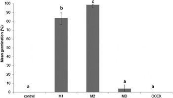
Germination of G. juniperina seeds was enabled if the micropylar part of the seed coat was removed, but prevented if it remained in situ. Differences between the seed coat removal treatments were highly significant (F 4,23 = 53.82, P < 0.001). Removal of a shorter length of the seed coat over the HR axis (treatment M1) resulted in 84% final germination (Fig. 2). Removal of a longer length of seed coat over the HR axis (treatment M2) led to a further significant increase in germination (Fig. 2). The remaining treatments that maintained the overall structural integrity of the micropylar end of the seed coat prevented germination. Making a small hole through the seed coat in the micropylar region (treatment MD) resulted in negligible germination (Fig. 2). Removing the chalazal end of the seed coat (treatment CCEX) or simply removing the elaisome resulted in no germination (Fig. 2).
Mean germination (back transformed %) of G. juniperina seeds subjected to one of the following treatments: control (eliosome removed); 1 mm of micropylar end of seed coat removed (M1); 2–3 mm of micropylar end of seed coat removed (M2); 1–2 mm2 removed from the dorsal side of the seed coat (MD); chalazal end of seed coat removed to expose the distal half of the cotyledons (CCEX). Means with different letters are significantly different at P < 0.05.

Growth potential of embryos at decreasing water potentials
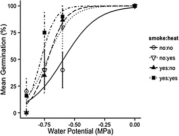
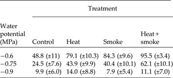
At a water potential of 0.0 MPa, the embryos in all dissected seeds showed radicle extension, irrespective of treatment (Fig. 3). The restrictive effect of decreasing water potential on radicle extension appeared as water potential declined. This effect was strongest in the controls, with just under half of embryos predicted to show radicle extension at −0.6 MPa, and one-quarter at −0.75 MPa in this treatment (Table 1, Fig. 3).
Mean germination (%) of G. juniperina embryos in dissected seeds over a range of decreasing water potential. Curves show predictions from the additive treatment model.

Proportion of embryos predicted to show radicle extension >2 mm over the range −0.6 to −0.9 MPa for control, heat, smoke and heat + smoke treatments. SE of estimate shown in brackets

Treatment with the fire cues significantly increased the growth potential of embryos (proportion extending their radicle), the analysis supporting an additive combination of effects rather than an interactive combination (Table 2). This effect was apparent between −0.6 and −0.75 MPa (Fig. 3). The effect of the fire cues in increasing the growth potential of embryos was most evident at −0.6 MPa, with 79–84% of embryos predicted to show radicle extension if either heat or smoke was applied singly, rising to 95% if both heat and smoke were applied (Table 1, Fig. 3). The increase in growth potential of embryos treated with fire cues was still apparent at −0.75 MPa, with 40–44% of heated or smoked embryos predicted to show radicle extension, rising to 62% of embryos if they received both cues (Table 1, Fig. 3). Differences in growth potential of embryos amongst the treatments disappeared by −0.9 MPa; the model predictions for the proportion of embryos extending their radicle were low, ranging from 8 to 14% across all treatments (Table 1, Fig. 3). Some embryos in the control, and heat and smoke treatment showed radicle extension at −0.9 MPa (Fig. 3).
Comparisons of models (no treatment differences, additive treatment effects, interactive treatment effects) for proportion of embryos showing radicle extension over a range of water potentials. F tests were used to account for quasi-binomial model

Estimation of the minimum force that embryos need to exert to attain the germination levels observed for intact seeds showed that embryos in control seeds would need to generate a force in excess of 0.9 MPa or more (estimated as 1.00 MPa) for 5% of intact seeds to germinate (Table 3). Treatment with the fire cues reduced the minimum pressure embryos would need to generate to give the germination observed for intact seeds, ranging from 0.84 MPa in the heat treatment down to 0.735 MPa for the heat + smoke treatment (Table 3).
Estimate of the water potential (WP) from the predictive models in Fig. 3 that corresponds to the proportion of intact G. juniperina seeds germinating in Morris (Reference Morris2000). SE shown in brackets

External compression of the seed coat
The compressive force required to penetrate 0.3 mm into untreated seeds was less for hydrated than for dry seeds, and varied with position along the longitudinal axis of the seed. The least mean compressive force (±SE) was required at the micropylar end; 517 (±23.5) g for hydrated seeds, compared to 1112 (±180) g for dry seeds. In the mid-region, the force rose to 942 (±42.1) g for hydrated seeds, compared to 1658 (±228) g for dry seeds. At the chalazal end, 592 (±42.6) g was required for hydrated seeds, and 1891 (±353) g for dry seeds.
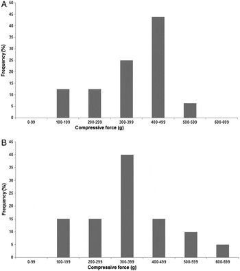
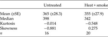
Application of the combined fire cues appeared to have no detectable effect on the force required to penetrate the seed coat, when tested after 24 h imbibition. The mean compressive force required to penetrate 0.4 mm into the micropylar region of seeds hydrated for 24 h was only 10 g lower for heated and smoked seeds compared to untreated seeds (Table 4; comparison of means, F 1,34 = 0.051, P = 0.81). The medians differed by more (lower by 56 g for heated and smoked seeds, Table 4, Fig. 4) although the difference was not significant (Mann–Whitney test).
Frequency distribution of the compressive force required to penetrate 0.4 mm into the micropylar region of imbibed G. juniperina seeds which were (A) untreated controls or (B) treated with heat and smoke.

Characteristics of the frequency distribution of peak force (g) needed to penetrate 0.4 mm into the micropylar region of 24-h hydrated seeds, which were untreated, or heated and smoked

Anatomy of seed coat fracture
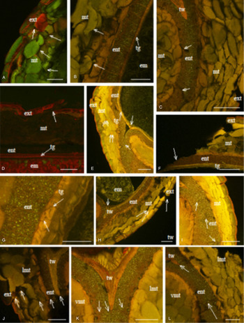
Medial longitudinal sections of seeds compressed when dry, revealed breaks in the dorsal exo- and mesotestal layers, with the fracture line following a path along the common walls and the dye clearly filling the intercellular spaces so created (Fig. 5A). Transverse sections through the micropylar region, showed the pathway of the dye through the testa and into the tegmen, but not passing the cuticle to enter into the embryo (Fig. 5B). Cell separation occurred between cells and cell layers in the exotesta and mesotesta; distinct fractures occurred across the cells forming the double palisade endotesta, but no fracture lines were found near, or in, the tegmel wedge (Fig. 5C). Examination of the dorsal chalazal region, i.e. near the hypostase, revealed similar fracture lines in the exo- and mesotestal layers (Fig. 5D). Structural integrity of the inner seed coat (tegmen) appeared to have been maintained (Fig. 5B, D).
Effects of compression upon seed coat structure of untreated seeds: (A–D) seeds compressed in the dry state, (E–L) seeds compressed when fully hydrated. Seeds in figures (A–E) were immersed in Congo red for 1 h to determine the fracture pathways. (A) Median longitudinal section of the dorsal tissue underlying the centre of the probe placed over the micropylar region. Fracture lines (arrows) follow the common wall rather than through the cells; (ext) exotesta, (mt) mesotesta. Scale bar = 75 μm. (B) Transverse section of the micropylar region, non-raphe side of seed. Upper lateral/dorsal testal cells flattened by the probe and mesotestal cells (mt) have separated (arrows), but no separation or fracture lines through the palisade endotesta (ent) or tegmel layers (tg). The dye penetrates through to the tegmel layers (tg) staining the walls orange. No staining of embryo (em) tissue. Scale bar = 100 μm. (C) Transverse section of the micropylar region, raphe side of seed. Separation between cells and cell layers of the exotesta (ext) and mesotesta (mt). Multiple, large fracture lines through the double-layered endotesta (ent) (arrows) but no fracture lines close to, or across the tegmel wedge (tw). Scale bar = 100 μm. (D) Median longitudinal section of the dorsal region in vicinity of the hypostase. Exotestal cells (ext) show major rupture with associated damage to the underlying mesotestal (mt) cells; (em) embryo, (tg) tegmen. Scale bar = 100 μm. (E) Transverse section through the mid-region of the seed. Separation of lateral exotestal cells (ext) and outer layers of mesotesta (mt) but no obvious fractures through the single-layered endotesta (ent) or adjacent tegmen (tg). Prominent fracture through the tegmel wedge (long arrows) and fine fractures through the double-layered endotesta (short arrow). All layers of the seed coat were well stained and the dye has accumulated in the spaces caused by the fractures (double arrows). Scale bar = 100 μm. (F) Transverse section through the mid-region of the seed, dorsal side. The fracture plane caused by the edge of the probe runs in a diagonal direction following the walls of the exotestal (ext) and mesotestal (mt) layers. However, it crossed through (arrow) rather than around, the endostestal (ent) cells, and diagonally through the tegmen (tg). Scale bar = 100 μm. (G) Transverse section through the mid-region of seed, upper lateral raphe side. Stress fracture (arrow) in the tegmel (tg) layers. Note the interlocking nature of these cells (between the arrows); (ent) endotesta. Scale bar = 25 μm. (H) Transverse section through the mid-region of seed, non-raphe side. Breaks occurred in the exotestal (ext) layer (long arrow) with disruption to the contents (double arrows) of the outer layers of lateral mesotesta (mt). No obvious damage to the endotestal (ent) or tegmel (tg) layers except for the tegmel wedge (tw) where short breaks were found (short arrow); (em) embryo. Scale bar = 100 μm. (I) Transverse section through the mid-region of seed, non-raphe side of the seed shown in Fig. E. Prominent multiple fracture lines (arrows) through the double-layered endotesta (ent). Separation of exotestal (ext) cells but mesotestal cells (mt) have not been displaced, although intercellular spaces have developed along the common wall junctions, indicating some cellular separation. Scale bar = 100 μm. (J) Transverse section through the micropylar region, non-raphe side. Note the continuous line of fracture (arrows) extending from the exotesta (ext), between the cells of the lateral mesotesta (lmt), across the palisade endotesta (ent) and across the tip of the tegmel wedge (tw). Scale bar = 100 μm. (K) Transverse section through the micropylar region, non-raphe side. Enlargement of tegmel wedge (tw) and endotesta; (lmt) lateral mesotesta, (vmt) ventral mesotesta. Fracture line through the tip of the wedge and abutting endotestal cells. Scale bar = 75 μm. (L) Transverse section through the micropylar region, non-raphe side. Enlargement of double-layered endotesta (ent) showing large fracture across the cells (long arrow) and a finer fracture across the tip of the tegmel wedge (tw) (short arrow). Note the nearby exotestal cells are missing; (lmt) lateral mesotesta, (vmt) ventral mesotesta. Scale bar = 100 μm.

For seeds that were hydrated when compressed, similar fracture lines were found in the mid-region (Fig. 5E–I) and micropylar regions (Fig. 5J–L). However, additional fracture lines lying very close to and across the tegmel wedge also occurred (Fig. 5E, H, K, L). Transverse sections revealed no difference between the raphe and non-raphe sides of the seed, but the degree of cell separation of the exo- and endotesta was greater in hydrated seeds. When tissue at the edge of the probe was examined (Fig. 5F), the fracture pathway followed the cell wall junctions rather than breaking across the cells. Only the more rigid, lignified palisade endotestal cells (Fig. 5I–L) and the interlocking cells of the tegmen (Fig. 5G) showed across-cell fractures.
Discussion
Selective removal of the seed coat
The role of the covering structures in contributing to non-deep physiological dormancy in G. juniperina seeds was apparent when the seed coat was selectively removed. Restriction of germination by the seed coat was localized to the micropylar region, as there was little or no germination if the micropylar part of the seed coat remained undisturbed and in place, whereas if it was removed, germination occurred. A positive germination response to selective removal of part of the seed coat or endosperm over the micropylar region has been found in Nemophila insigni (Chen, Reference Chen1968) and Prunus persica (Mehana and Martin, Reference Mehana and Martin1985).
The role of the structural integrity of the micropylar seed coat in maintaining dormancy is further shown by the lack of germination response if only a small dorsal area of this part of the seed coat is removed, leaving the rest intact. The lack of germination after removal of a small dorsal area of the micropylar seed coat or the removal of the chalazal end of the seed coat also shows that the seed coat was not imposing dormancy by restricting gas exchange between the embryo and the exterior, nor was it retaining germination inhibitors inside the seed. A positive germination response would have been expected to these two treatments if either mechanism was operating, but no such response occurred. Furthermore, there was an increase in germination as a greater length of the auricles was exposed, in the M2 compared to the M1 treatment. A possible mechanism underlying these observations may be linked to the mechanical constraint maintained by the auricles that cover the hypocotyl–radicle axis in the intact seed. As living tissue, these cells would swell during hydration. Given the structural rigidity of the seed coat, imposed by the palisade layer and the non-lignified sclerenchyma of the tegmen (Briggs et al., Reference Briggs, Morris and Ashford2005), swelling of the auricles would exert inwards constraining pressure upon the hypocotyl–radicle axis if the micropylar part of the seed coat remained in place. Removal of the seed coat from the auricles would release the compressive force exerted upon the hypocotyl–radicle axis, permitting their expansion and extension; removing more of the seed coat over the auricles would release it further.
Fire-cue effects on growth potential of the embryo
Incubating dissected embryos over a range of water potentials showed that both smoke and heat shock increased their growth potential, with such effects being physiological. The embryo can play a part in non-deep physiological dormancy (Baskin and Baskin, Reference Baskin and Baskin2014), and a physiological response to the fire cues by embryos of G. juniperina demonstrates this principle. Bioactive components in smoke have been linked to physiological pathways in plants (Light et al., Reference Light, Daws and Van Staden2009; Nelson et al., Reference Nelson, Flematti, Dixon and Smith2012). Other dormancy-breaking factors that have been demonstrated to increase the growth potential of embryos include red light (Carpita et al., Reference Carpita, Nabors, Ross and Petretic1979; De Miguel and Sanchez, Reference De Miguel and Sanchez1992) and low temperatures (Baskin and Baskin, Reference Baskin and Baskin1971; Feurtado et al., Reference Feurtado, Ren, Ambrose, Cutler, Ross, Abrams and Kermode2008). This is the first report of smoke and heat shock increasing embryonic growth potential in any species. Physiological effects of heat-shock temperatures up to 100°C were postulated to underlie curvilinear germination responses of other species tested for heat and smoke effects (Thomas et al., Reference Thomas, Morris and Auld2007).
The range of negative water potential over which the fire cues were effective in increasing the embryonic growth potential was −0.6 to −0.75 MPa. Preliminary trials had found little evidence of any treatment effect from 0 to −0.5 MPa (unpublished data), and the effect of fire cues was not apparent at −0.9 MPa. The predictive models suggest that over the range −0.6 to −0.75 MPa, heat shock and smoke had a more or less equal effect on the growth potential of the embryo, and that the combined cues had a greater effect. Thus, at any given water potential within this range, treatment with the fire cues meant that a higher proportion of the embryos could generate sufficient thrust to extend their radicle. The maximum thrust achievable by embryos appeared to lie beyond 0.9 MPa, irrespective of treatment with fire cues or not. This is a major contrast with the effect of the dormancy-breaking stimulus (red light) in Grand Rapids lettuce seed, where treatment with red light increased both the proportion of seeds that could achieve a thrust of given magnitude, and the maximum thrust possible (Nabors and Lang, Reference Nabors and Lang1971). These authors estimated that treatment with red light increased the maximum thrust generated by the embryos by 0.75 MPa, and that to break through the intact seed coat, a force equivalent to 0.4–0.95 MPa was required (Nabors and Lang, Reference Nabors and Lang1971).
Despite the apparent lack of a difference in the maximum growth potential achieved by embryos after treatment with fire cues, there is another important implication that follows from the growth potential vs. water potential response curves in our study: the fire cues appear to have led to weakening of the seed coat in G. juniperina, in addition to their effects on the embryonic growth potential. This conclusion is derived from the calculation of the minimum force needed by embryos to give the observed percentage germination of intact seed. The calculation suffers from the limitation of combining data across two separate experiments; however, the rank order of the response of intact Grevillea seeds to fire cues from a wider range of experiments and species is that germination is lowest in controls, intermediate if seeds are treated with heat or smoke singly (usually heat ≤ smoke), and highest if treated with heat and smoke (supplementary Table S1; Kenny, Reference Kenny2000; Morris, Reference Morris2000; Morris et al., Reference Morris, Tieu and Dixon2000; Briggs and Morris, Reference Briggs and Morris2008). The two fire cues usually combine additively to increase germination of intact seeds in these experiments, and sometimes synergistically. So, irrespective of the precise values used, the qualitative conclusion from combining the pattern of germination percentages of intact seeds with the growth potential response curves of embryos from our study is that embryos in intact seeds can germinate using lower values of thrust than control seeds. This can only occur if there has been weakening of the seed coat after exposure to the fire cues. The estimate of the greatest extent of the reduction compared to controls was 0.27 MPa for heated and smoked seeds. Estimated reductions were less for the cues applied singly (0.16 MPa for heat, 0.23 MPa for smoke). The rank order of the estimated reduction in thrust required matches the rank order of increasing germination of intact seeds. Weakening of the seed coat could be caused either by the fire cues directly, or by the fire cues inducing physiological changes in the embryos that then allow them to weaken the seed coat, as has been demonstrated in other cases of non-deep physiological dormancy (Kucera et al. Reference Kucera, Cohn and Leubner-Metzger2005; Arana et al., Reference Arana, De Miguel and Sánchez2006).
Compressive force and seed coat strength
The structural properties of the seed coat clearly changed with hydration, as observed for other species, with a reduction in compressive weight required to achieve a given penetration for wet compared to dry seeds (Baskin and Baskin, Reference Baskin and Baskin2014). Variation in this compressive force along the length of the seed was also evident.
Treatment with fire-related cues did not appear to directly affect the structural properties of the seed coat, as the compressive weight required to penetrate it did not change after treatment with heat and smoke. Our study was limited by low numbers of seeds, tested after 24 h of imbibition only, was restricted to compression, and the findings should be regarded as preliminary. It gives quantitative data that can be used to guide future studies to detect changes in coat strength of known magnitude with sample sizes large enough for a more powerful test.
Anatomy of seed coat fracture
Very few studies have been carried out testing the compressive force needed to break the seed coat and none of these has examined the resulting breaks microscopically.
Plant tisues are composite materials and therefore behave in a non-linear, viscoelastic manner (Niklas et al., Reference Niklas, Spatz and Vincent2006). They are usually highly deformable and anisotrophic (Niklas et al., Reference Niklas, Spatz and Vincent2006), a condition that is conferred by the overall shape of the tissue/organ, different cell types within a tissue, the composition of the cell walls at different locations and the degree of adhesion between cells, as well as the amount of tissue occupied by intercellular spaces – all of these factors will determine the response of the tissue to a compressive force (Vincent, Reference Vincent1990). When apples and potatoes were subjected to compressive force using a wedge, the fracture line ran between the cells of the apple but through the cells of the potato (Vincent, Reference Vincent1990). In apple, there were numerous air-filled intercellular spaces but no similar spaces occurred in the potato tissue, where the cells were also isotrophic. The pathway of the fracture is suggested to be determined by the air spaces, which blunt the fracture and deflect it into another direction (Vincent, Reference Vincent1990).
In G. juniperina seeds, intercellular spaces lie at the junctions of cells forming the mesotestal layers and exotesta/mesotesta boundary. Microscopic examination of the compressed control seeds (whether compressed dry or fully hydrated) revealed that the fracture lines ran along the middle lamella of the exo- and mesotestal cells, with the change in direction being guided by the intercellular spaces. However, once the fracture reached the lignified, internally reinforced palisade endotesta, the stronger adhesive forces between these cells, together with a lack of air spaces, caused the fracture to cross through into the cell and often diagonally across the tegmel layers. These tegmel layers are formed from thick-walled, non-lignified cells with a small lumen; the outer edges of the cell walls are shaped like flanges that interlock when forming a single layer. Interlocking of edges is also present when the tegmen is double layered in the lateral fold that forms the tegmel wedge. The interlocking of edges, lack of intercellular spaces and small lumen would confer greater strength to these cells and the layer as a whole. More energy is required to break across cells rather than go around via cell walls (Vincent, Reference Vincent1990). The cells showing cross-breakages in our study were the single-layered tegmen, the double-layered tegmel wedge (both sclerenchyma) and the reinforced lignified palisade layer. These would be the areas of maximum resistance for the radicle to overcome at emergence.
Conclusions
The current study showed clearly that both the seed coat and the response of the embryo to smoke and heat shock play roles in controlling germination in G. juniperina. Both observations fit with the known mechanisms of non-deep physiological dormancy, in which the covering structures, and/or the embryo, control germination (Baskin and Baskin, Reference Baskin and Baskin2014). Removing the micropylar part of the seed coat allows germination, whereas if the micropylar seed coat remains in place, the fire cues can break the dormancy imposed by the seed coat. The restrictive effect of the seed coat fits best with the mechanical constraint model for covering structures, and the response of the embryonic growth potential to the fire cues is most likely physiological. An implication of the growth potential response curves of embryos and the observed germination percentages of intact seeds is that the fire cues weaken the seed coat. Our limited investigation of seed coat strength suggested that neither of the fire cues directly weakened the mechanical strength of the seed coat, independently of any effects on the embryo, although this finding needs confirmation. If the fire cues cause the embryos to weaken the seed coat, the pathway by which this occurs remains to be elucidated. If identified, it would be an example of an interaction between the embryo and the covering structure playing a role in non-deep physiological dormancy (Baskin and Baskin, Reference Baskin and Baskin2014).
Supplementary material
To view supplementary material for this article, please visit http://dx.doi.org/10.1017/S0960258516000088
Acknowledgements
Seeds were collected under the NSW Office of Environment and Heritage, Scientific Licence SL101040.
Financial Support
This work was supported by a grant from the Australian Flora Foundation (2006/03), and by ex gratia contributions from Penrith Lakes Development Corporation.
Conflicts of Interest
None.