




No CrossRef data available.
Hormonal control of tick development and reproduction
Published online by Cambridge University Press: 19 April 2005
Abstract
Ecdysteroids (moulting hormones), juvenoids and neuropeptides in ticks are reviewed but, by far, the emphasis is on the former since this class of hormones has been the subject of most investigations. In immature stages of ticks, ecdysteroids have been shown to regulate moulting and to terminate larval diapause. Although there is a paucity of information on the molecular action of ecdysteroids in ticks, their action appears to be via a heterodimeric ecdysone/ultraspiracle receptor, as in insects. The role of ecdysteroids in sperm maturation in adult males is considered. In females, ecdysteroids function in the regulation of salivary glands, of production of sex pheromones and of oogenesis and oviposition. There is evidence for ecdysteroid production in the integument and pathways of hormone inactivation are similar to those in insects. Ecdysteroids also function in embryogenesis. Although evidence for the occurrence and functioning of juvenile hormones in ticks has been contradictory, in recent thorough work it has not been possible to detect known juvenile hormones in ticks, nor to demonstrate effects of extracts on insects. Factors (neuropeptides) from the synganglion affect physiological processes and limited immunocytochemical studies are reviewed. Sigificantly, a G-protein-coupled receptor has been cloned, expressed, and specifically responds to myokinins.
Information
- Type
- Research Article
- Information
- Copyright
- © 2004 Cambridge University Press
INTRODUCTION
The developmental hormone systems of insects and crustaceans are probably best understood of all the arthropods (for reviews, see Gilbert, Iatrou & Gill, 2004; Wainwright & Rees, 2001). In other arthropod classes, information concerning the identification and functional significance of hormones is fragmentary or non-existent. The endocrine regulation of development and reproduction in ticks (acarines) has been reviewed (Oliver & Dotson, 1993; Lomas & Rees, 1998; Chang & Kaufman, 2004) and the reader is referred to these for further detail. However, there is a relative lack of new work in this field.
As alluded to in other articles in this Supplement, blood meals are critical in ticks for triggering various events, including the endocrine system. In adult female ixodid ticks, the transition between the slow feeding phase and the rapid engorgement phase (that has been defined as the critical weight; Harris & Kaufman, 1984; Lindsay & Kaufman, 1988; Weiss & Kaufman, 2001) seems to be a critical control point for regulation of many endocrine events, including salivary gland degeneration, vitellogenesis and egg production (see article by Kaufman, in this Supplement). Thus, females prematurely removed from the host below the critical weight retain a host-seeking strategy and can reattach to a host if given the opportunity, do not undergo salivary gland degeneration and will not lay eggs. However, females prematurely removed above the critical weight are unable to reattach to a host, undergo salivary gland degeneration and will lay as many eggs as the acquired blood meal will support.
In this article, aspects of the endocrine regulation of development and reproduction will be considered. Various factors involved in regulating reproduction are also considered in the article by Kaufman in this Supplement.
ECDYSTEROIDS
From evidence primarily based on a combination of the observed effects of exogenously administered moulting hormones (ecdysteroids) and the correlation of changes in ecdysteroid titres with physiological events, it appears that the functions of ecdysteroids in ticks are largely similar to those in insects and crustaceans. However, in ticks there is still a lack of information on direct effects of the hormones on individual gene transcription. The physiological roles of ecdysteroids in ticks have been reviewed (Diehl, Connat & Dotson, 1986; Sonenshine, 1991; Oliver & Dotson, 1993; Lomas & Rees, 1998).
Immature stages
Moulting
There is a good body of evidence that ecdysteroids, particularly 20-hydroxyecdysone, regulate moulting in ticks as they do in insects and crustaceans (Fig. 1). It has been shown that feeding in vitro of nymphs of the argasid, Ornithodoros porcinus, with porcine blood containing 20-hydroxyecdysone, accelerated moulting (cited in Solomon, Mango & Obenchain, 1982). Surprisingly, similar treatment of nymphs of O. parkeri with 20-hydroxyecdysone (Campbell & Oliver, 1984) or O. moubata with the incompletely hydroxylated ecdysteroid, 22,25-dideoxyecdysone (Diehl et al. 1986) did not affect the moulting period, although the ticks underwent additional moults without taking a further blood meal. However, as with O. porcinus, in nymphs of the ixodid, Hyalomma dromedarii, topical application of 20-hydroxyecdysone speeded up moulting (Khalil et al. 1984). As might be expected, the sensitivity of ticks is highly dependent upon the dose of ecdysteroid administered, the precise developmental time, and the method of exposure (Sonenshine, 1991). By injection of 20-hydroxyecdysone into female O. parkeri, a dose-dependent induction of apolysis and cuticle formation has been demonstrated (Pound, Oliver & Andrews, 1984).
There is also substantial evidence for a close temporal correlation between haemolymph ecdysteroid titres and the moulting process. Haemolymph and whole-body ecdysteroid titres in fifth-stage nymphs of the argasid O. moubata have been carefully correlated with the moulting processes which are induced by the blood meal (Germond, Diehl & Morici, 1982). Between days 2 and 3 post-feeding, when the ecdysteroid titres were low, a few procuticle lamellae were deposited and mitosis initiated. Ecdysteroid titres then began to increase (days 3–4), with the mitotic period ending on day 4. The hormone titres then rose sharply simultaneously with apolysis and the formation of the exuvial space (days 4–5), with highest titres being observed during deposition of the new epicuticle (days 5–6). The titres began to decrease concomitantly with the beginning of procuticle deposition and digestion of the old cuticle (day 6), and started to decrease to low values shortly before ecdysis (days 9–10). Similar correlations have been demonstrated in nymphs of O. parkeri (Zhu et al. 1994) and in the ixodids, Amblyomma hebraeum (Diehl, Germond & Morici, 1982) and A. variegatum (Stauffer & Connat, 1990), with probably a similar situation in larvae of O. moubata (cited in Diehl et al. 1986; Dotson, Connat & Diehl, 1991).
Using well-synchronised A. variegatum, all the integumental events were realised along an anterior-posterior gradient. Of particular significance was the fact that a small peak of ecdysteroid preceded the major peak of hormone during this nymphal-adult moulting cycle. Many structural changes were observed in the integument, salivary glands and dermal glands during this moulting cycle, but additionally, the muscles of the mouthparts and the pharynx underwent a temporary de-differentiation at the end of the first small peak of ecdysteroids. At the end of the second peak of hormone, when the ecdysteroid titre dropped to basal levels, these muscles reappeared in an adult differentiated state (Stauffer & Connat, 1990). This occurrence of two ecdysteroid peaks is reminiscent of the larval-pupal moult in holometabolous insect species.
In all tick species investigated, at high ecdysteroid titres, the principal hormone is 20-hydroxyecdysone, the presumed major active hormone, accompanied by ecdysone (see Delbecque, Diehl & O'Connor, 1978; Dees, Sonenshine & Breidling, 1984a). As in insects, the capacity to transform ecdysone into 20-hydroxyecdysone is widely distributed in tissues, including the midgut (cited in Diehl et al. 1986) and fat body (Zhu, Oliver & Dotson, 1991a).
Certain argasids, but not ixodids, have been induced experimentally to supermoult by administration of ecdysteroids, although adult ticks do not normally moult (Diehl et al. 1986). Apparently, sensitivity to induction of supermoulting depends on the species of tick, the hormone and the type and timing of application. Supermoulting in O. moubata and O. parkeri frequently resulted in failure of ecdysis, which rendered subsequent feeding impossible (Connat et al. 1983a; Campbell & Oliver, 1984). However, in O. porcinus, supermoulting resulted in healthy specimens which could go on to feed, supermoult and oviposit (Mango, Odhiambo & Galun, 1976). Interestingly, the speculation was made in this case that supermoulting could occur in nature when the host feeds on ecdysteroid-rich plants.
Termination of larval diapause
It has been demonstrated that administration of ecdysteroids leads to termination of larval diapause in the ixodids, Dermacentor albipictus and Rhipicephalus sanguineus, although the significance of this to the in vivo situation is unclear (Wright, 1969; Sannasi & Subramoniam, 1972).
Ecdysteroid receptors
In insects, where the molecular action of ecdysteroids is best understood of the Arthropods, this action is mediated by a heterodimeric receptor composed of two members of the nuclear superfamily of receptors, the ecdysone receptor (EcR) and ultraspiracle (USP) (Henrich, Rybczynski & Gilbert, 1999; Koslova & Thummel, 2000; Riddiford, Cherbas & Truman, 2001; Henrich, 2004). USP is the insect homologue of the vertebrate Retinoid X Receptor (RXR) genes, which have a critical role in many nuclear signalling pathways (Mangelsdorf & Evans, 1995). In the case of numerous nuclear receptor ligands, such as ecdysteroids, they exert diverse effects on development, growth and physiological processes (Thummel, 2001, 2002). Such a diversity of effects is frequently due to the existence of multiple forms of the receptor protein that differentially transduce the ligand signals. Multiple forms of receptors can arise from expression of related gene families, to yield receptor sub-types, from alternative RNA processing of a single gene to yield receptor isoforms, or from a combination of both mechanisms (Leid, Kastner & Chambon, 1992).
All nuclear receptors possess a characteristic modular structure (Fig. 2) that includes a variable N-terminal domain (A/B), a highly conserved DNA-binding domain, C, a variable hinge region, D, a conserved ligand-binding domain, E, together with a variable C-terminal region (F) of unknown significance and may be present in some, but not all nuclear receptors (Fig. 2).

Fig. 1. Formulae of ecdysone and 20-hydroxyecdysone.

Fig. 2. The modular structure characteristic of all nuclear receptors. A/B, variable N-terminal domain; C, a highly conserved DNA-binding domain; D, a variable hinge region; E, a conserved ligand-binding domain, and; F, a variable C-terminal region that is present in some, but not all nuclear receptors.
cDNAs encoding three ecdysteroid receptor isoforms that have common DNA- and ligand-binding domains linked to distinct amino termini have been isolated from the ixodid tick, A. americanum (Guo et al. 1997). The DNA- and ligand-binding domains share an average of 86 and 64% identity, respectively, with such domains from insect EcR proteins. In these A. americanum EcRs, the amino termini are highly divergent and they also lack the ‘F’ C-terminal domain occurring in the insect EcRs. Analysis of the cDNAs showed that RNA processing is complex and in addition to producing transcripts with unique amino termini, produces EcR transcripts with different 5′ and 3′ untranslated regions, as well as splicing variants having incomplete open reading frames. Results of Northern blot analyses suggested both stage- and tissue-specific regulation of the EcR mRNA expression.
The vertebrate RXRs are encoded by a multigene family, whereas the insect RXR homologue, Ultraspiracle (USP), is encoded by a single gene. To determine the situation in acarines, cDNAs encoding two RXR homologues, AamRXR1 and AamRXR2 have been isolated from A. americanum (Guo et al. 1998). The DNA-binding domains share ~95% and 87% identity, respectively, with such domains from insect USP and vertebrate RXR proteins. Surprisingly, ligand-binding domains of the AamRXRs are more similar to such domains from vertebrate RXRs than to insect USP ligand-binding domains (~71 vs. ~52%, respectively). The biological significance of this is unclear. Furthermore, Northern blot and RT–PCR analysis revealed both unique and overlapping patterns of AamRXR1 and AamRXR2 expression during development. Both AamRXR1 and AamRXR2 proteins, when paired with any of the three AamEcR isoforms, or the Drosophila EcRB1 isoform, can activate transcription of ecdysone response element-containing reporters. Thus, despite differences in the ligand-binding domains of vertebrate RXRs, AamRXRs, and insect USP proteins, all can pair with either insect or tick EcRs, yielding a functional ecdysteroid receptor in vitro in response to the ecdysteroid, muristerone A (Guo et al. 1998). The foregoing studies on A. americanum EcRs and RXRs have been reviewed (Palmer, Harmon & Laudet, 1999).
As we have seen, the DNA-binding domains of arthropod USPs and their vertebrate homologues, the RXRs, are highly conserved. However, interestingly the ligand-binding domain sequences divide into two district groups: (1) sequences from members of the holometabolous higher insect orders, the Diptera and Lepidoptera, and (2) sequences from vertebrates, a fiddler crab (Uca pugilator), a tick (A. americanum) and a hemimetabolous orthopteran insect, the locust, Locusta migratoria (Hayward et al. 1999). The reason for this evolutionarily sharp divergence of the lepidopteran and dipteran sequences is unknown.
Determination of total body ecdysteroid titres during development in A. americanum revealed that one ecdysteroid peak was observed following larval apolysis. However, two distinct ecdysteroid peaks occurred in the nymphal moulting cycle, the first following apolysis and the second occurring at about the time of ecdysis. Determination of the whole body profiles of the EcR and RXR mRNAs by RT–PCR showed that they were both correlated with the hormone titre (Palmer et al. 2002). However, using an electrophoretic gel mobility shift assay, it has been demonstrated that AamEcR-AamRXR1 (but not AamEcR-AamRXR2) exhibits broad DNA-binding specificity, suggesting that functional differences exists between the RXR1 and RXR2 proteins.
Ecdysteroid inactivation
Before considering ecdysteroid inactivation in ticks, it is germane to summarise such mechanisms in insects, since these are better understood. Ecdysteroid inactivation in insects may occur via several different routes, depending on species, stage in development and tissue (Lafont et al. 2004). The major ecdysteroid inactivation pathways include: (1) Conjugate (ester) formation, primarily polar phosphates or apolar fatty acyl derivatives. Such conjugates were originally identified as inactive maternal, storage ecdysteroid 22-esters in adult females/newly laid eggs for utilization in early stages of embryogenesis before de novo synthesis of hormone in embryos. However, analogous phosphate conjugates (at the C-2, C-3, or C-22 positions of the ecdysteroid) and fatty acyl esters (see Fig. 3) are formed in immature stages of insects; (2) An apparently fairly universal pathway of ecdysteroid inactivation involves ecdysteroid 26-oic acid formation via 26-hydroxyecdsyteroid (see Fig. 5); (3) In many insect systems, irreversible 3-epiecdysteroid formation is emphasised. This occurs via ecdysone oxidase-catalyzed 3-dehydroecdysteroid formation that then undergoes irreversible 3-dehydroecdysteroid 3α-reductase-catalyzed reduction to 3-epiecdysteroid (see Fig. 4). Analogous reactions occur with both ecdysone and 20-hydroxyecdysone.

Fig. 3. Formula of ecdysteroid 22-long chain fatty acyl esters.

Fig. 4. Conversion of ecdysone into 3-epiecdysone.

Fig. 5. Formula of ecdysteroid 26-oic acid.
Most of the work on ecdysteroid inactivation/metabolism in nymphs has been undertaken in O. moubata. In last-instar nymphs, injected [3H]ecdysone was efficiently converted into 20-hydroxyecdysone, the transformation having wide tissue distribution but which is particularly active in the midgut (Bouvier, Diehl & Morici, 1982; cited in Diehl et al. 1986). 20-Hydroxyecdysone is further transformed into putative 20,26-dihydroxyecdysone and all three ecdysteroids are subsequently converted into polar compounds of unknown structures. Significantly, this polar pathway was particularly active when the titre of endogenous ecdysteroids was decreasing, with accumulation of polar products in the digestive tract, thus suggesting its importance for inactivation of endogenous nymphal ecdysteroids. However, it was not operative with ingested ecdysteroids nor in adult females (see below).
However, apolar ecdysteroid 22-long-chain fatty acyl esters are also quickly formed from injected ecdysteroids, but even more rapidly produced from ingested hormones (Diehl et al. 1985; Connat, Diehl & Thompson, 1986a). Such apolar conjugates were then gradually converted into slightly more polar conjugates that are uncharacterized. Interestingly, most ingested ecdysteroid was retained in the digestive tract, where the midgut cells transformed it into apolar conjugates that accumulated in the midgut lumen (Diehl et al. 1986). It had been suggested that the apolar metabolic pathway may serve to inactivate ecdysteroids ingested in blood from hosts feeding on phytoecdysteroid-containing plants (Diehl et al. 1985). Such apolar esters are important and predominant endogenous components in all nymphal stages of O. moubata, particularly in the fourth and fifth stages (Connat et al. 1997). Interestingly, although 20-hydroxyecdysone predominated over ecdysone in all nymphal stages, the proportion of the latter was enhanced in the later three nymphal stages. The ecdysteroid metabolic pathways in fed nymphs of the ixodid, A. hebraeum, were similar to the ones in O. moubata (Diehl et al. 1985).
Undoubtedly, formation of ecdysteroid 22-fatty acyl esters is a prominent metabolic route in ticks. Formation of such esters is widespread in arthropods (Connat & Diehl, 1986; Kubo et al. 1987; Robinson et al. 1987).
Adult males
Sperm maturation
In male ticks, sperm production and maturation has two phases (for review see Sonenshire, 1991), spermatogenesis (the development of haploid spermatids by meiotic and mitotic division) and spermiogenesis (the growth and maturation of spermatids). Spermatogenesis begins during the nymphal-adult moult and in a few exceptional species proceeds to completion during that moult so that unfed males have mature prospermia and can mate (see Lomas & Rees, 1998). However, in most species, spermatogenesis stops after the development of secondary spermatogonia during adult ecdysis and remains arrested until the adult male begins feeding.
Although the mechanism of reinitiation of sperm development is uncertain, ecdysteroids appear to be involved. Ecdysteroids occur in males (Oliver, 1986a; Oliver & Dotson, 1993) and injection of 20-hydroxyecdysone into unfed males strongly stimulated germ cell DNA synthesis; similarly, injection of a crude synganglial extract could mimic this effect (Oliver, l986a). It is significant that the accumulation of differentiated spermatocytes in D. variabilis nymphs is biphasic and parallels the ecdysteroid titres reported in A. hebraeum (Dumser & Oliver, 1981; Diehl et al. 1982). There is evidence for ecdysteroid synthesis in insect testis under the influence of a brain peptide (Loeb et al. 1987; Jarvis, Earley & Rees, 1994), but such steroid synthesis has not been demonstrated in testes of ticks.
The final growth and maturation of spermatids (spermiogenesis) can be divided further into a growth and elongation phase (resulting in prospermia) and a capacitation phase (sperm maturation; El-Said et al. 1981). Spermatogenesis continues with sperm growth and elongation, resulting in mature prospermia throughout the testis after five days of feeding (Khalil, 1970). The final phase of sperm development, capacitation, requires 24 hours and is triggered by a low molecular weight protein(s) from the male accessory gland (El-Said et al. 1981; Shepherd, Oliver & Hall, 1982).
For further discussion of various factors from male ticks involved in reproduction, see the article by Kaufman in this Supplement.
Adult females
Regulation of salivary glands
In female ixodid ticks, salivary glands perform several important functions relating to tick feeding and maintaining osmoregulation (for reviews see Kaufman, 1989; Sauer et al. 1995; Sauer, Essenberg & Bowman, 2000). A major function is to secrete excess fluid from the blood meal back to the host, resulting in concentration of the blood meal and regulation of the haemolymph volume. A detailed account of tick salivary gland function and regulation is given in the chapter by Bowman & Sauer in this Supplement.
After the female tick has engorged, the salivary glands degenerate, an event characterized by the appearance of autophagic vacuoles (Harris & Kaufman, 1981). This autolytic degeneration of the salivary glands is triggered by 20-hydroxyecdysone (Harris & Kaufman, 1985; Kaufman, 1991). This hormone also triggers vitellogenesis (Sankhon et al. 1999) and, more recently, a role for ecdysteroid in inhibiting re-attachment to the host has been demonstrated (Weiss & Kaufman, 2001).
The molecular action of ecdysteroid, particularly the role of the receptors, EcR and USP, in salivary gland degeneration has been studied in Amblyomma hebraeum, where the glands degenerate within four days of engorgement. Extensive evidence has been furnished for the existence of a salivary gland EcR in A. hebraeum: (1) [3H]ponasterone A binds to a salivary gland protein (Mao, McBlain & Kaufman, 1995); (2) the specific ponasterone A-binding protein binds to a Drosophila ecdysone response element, hsp27 EcRE, in a sequence-specific manner, the binding being enhanced by biologically active ecdysteroids; (3) monoclonal antibodies against Drosophila EcR and ultraspiracle protein (USP) cross-react with counterparts in tick salivary gland extracts by Western blotting; and (4) the monoclonal antibody against USP retards the hsp27 EcRE-tick EcR complex in a gel mobility shift assay (Mao & Kaufman, 1998).
In A. hebraeum, the profile of the functional ecdysteroid receptor (EcR/USP) determined by [3H] ponasterone A binding, gel mobility shift assays and Western blotting, has been correlated with the haemolymph ecdysteroid titre during the feeding period and six days post-engorgement (Mao & Kaufman, 1999). EcR was undetectable in unfed ticks, but following onset of feeding, specific ponasterone A binding and two major EcR bands detected by Western blots appeared. Both measurements were sustained throughout the feeding period, but declined after detachment when the salivary glands were degenerating. A discrete DNA-binding band, shown by gel mobility shift assay using Drosophila hsp27 EcRE as probe, intensified when haemolymph ecdysteroid titre reached its peak during the rapid phase of feeding, but declined along with decreasing EcR/USP levels and with specific ligand binding activity following engorgement. These results, taken together, substantiate a physiological role for ecdysteroid, acting via nuclear receptors, in salivary gland degeneration. Furthermore, the latter study suggested a role for the small haemolymph ecdysteroid peak during the rapid phase of feeding in initiating salivary gland degeneration, that may constitute a signal somewhat analogous to the ‘commitment peak’ of ecdysteroid in larval Lepidoptera and Drosophila that triggers the behavioural and molecular changes leading to metamorphosis (Mao & Kaufman, 1999).
Regulation of sex pheromone production
The various types of pheromone systems in ticks are considered in the article by Sonenshine in this Supplement, with previous reviews also being available (Sonenshine 1986, 1991; Hamilton, 1992). This short section will consider the involvement of ecdysteroids, particularly in regulation of sex pheromone production.
Evidence has been furnished that ecdysteroids may regulate aspects of sex pheromone production in some species. Production of the attractant sex pheromone, 2,6-dichlorophenol, by the foveal glands begins shortly after the nymphal-adult moult. An increase in 2,6-dichlorophenol production in unfed Hyalomma dromedarii females has been demonstrated when they were stimulated by exogenous 20-hydroxyecdysone prior to moulting (Dees, Sonenshine & Breidling, 1984b, 1985; also see Sonenshine, 1991). Furthermore, a similar increase was observed in unfed females and males when treated with 22,25-dideoxyecdysone (Jaffe et al. 1986).
In addition, ecdysteroids may also be components of pheromone systems per se. In Dermacentor, the genital sex pheromone consists of long-chain saturated fatty acids together with ecdysone and 20-hydroxyecdysone (Allan et al. 1988; Taylor, Sonenshine & Phillips, 1991c).
Regulation of oogenesis and oviposition
In most ticks, feeding and mating are required for oogensis and oviposition to occur. Ixodid females must copulate in order to complete the blood meal, whereas argasid females generally feed to repletion whether or not they have copulated. Whereas ixodid ticks undergo one gonotrophic cycle and die, argasids have several such cycles. It is clear that mating triggers factors regulating oogenesis and oviposition, with involvement of interactions between nervous, endocrine and reproductive systems (for earlier reviews, see Connat et al. 1986b; Oliver, 1986b; Oliver & Dotson, 1993; Lomas & Rees, 1998).
Egg maturation in all arthropods involves synthesis of the major egg yolk protein, vitellogenin (Vg) and its uptake in modified form, vitellin (Vn) by oocytes. In ticks, Vg is synthesized outside the ovary, released into the haemolymph and transported to the ovary, where it is selectively taken up as Vn (Chinzei & Yano, 1985; Rosell-Davis & Coons, 1989), which is a haemoglycolipoprotein (Chinzei, Chino & Takahashi, 1983; Rosell & Coons, 1991; James & Oliver, 1997).
In argasids, there is evidence that the fat body is a site of Vg production (e.g. Chinzei & Yano, 1985; for review see Chinzei, 1986). Clearly, vitellogenesis is under hormonal control in ticks, but there are appreciable conflicting data. In addition to neuropeptides, ecdysteroids and juvenile hormones are the main candidate hormones (see Oliver & Dotson, 1993; Lomas & Rees, 1998). However, the status of the occurrence of juvenile hormones in ticks is uncertain (see below).
In the argasid, O. moubata, Vg production is stimulated by a Vitellogenesis Inducting Factor (VIF) released from the synganglion following the time of coxal fluid secretion during feeding and continues to be released for approximately one hour after feeding (Chinzei et al. 1992). Although the mechanism of action of VIF is uncertain, it has been suggested to trigger the release of ‘Fat body Stimulating Factor’ (FSF), which in turn, stimulates vitellogenesis in the fat body (Chinzei & Taylor, 1990). Although the identity of the FSF is unknown (see Oliver & Dotson, 1993), some evidence exists that it may be a juvenile hormone (Pound & Oliver, 1979; Connat, Ducommun & Diehl, 1983b) with stronger evidence that it may be an ecdysteroid (Sankhon et al. 1999; Taylor, Nakajima & Chinzei, 2000; Ogihara, 2003). Furthermore, evidence has been obtained in three other argasid species that an Egg Development Stimulating Factor (EDSF) from the synganglion may be involved in incorporation of Vg into oocytes (Argas arboreus, Shanbaky & Khalil, 1975; Argas hermanni, Shanbaky et al. 1990; O. parkeri, Oliver et al. 1992).
Administration of ecdysteroids to females of the argasid, O. moubata, not only induced supermoulting, but inhibited oogenesis and caused resorption of previously formed eggs (Diehl et al. 1986). Such action of ecdysteroids has been incorporated into a working hypothesis for possible control of the gonotrophic cycle in O. moubata (Connat, Dotson & Diehl, 1987a). Analogous inhibition of oogenesis by ecdysteroids has also been reported in ixodids, although there are conflicting results as well (see Diehl et al. 1986). Although such inhibitory effects of ecdysteroids on oogenesis might at first sight appear to be difficult to reconcile with the notion of ecdysteroid being the vitellogenic hormone (FSF), the situation is not so different from that in Drosophila where 20-hydroxyecdysone may trigger oocyte resorption or vitellogenesis at certain different precise stages of oogenesis (Soller, Bownes & Kubli, 1999).
In ixodids, there is evidence that the prime sites of Vg synthesis are the fat body and midgut (Rosell & Coons, 1992; see Oliver & Dotson, 1993). Evidence has been furnished in Hyalomma dromedarii for the involvement in Vg production of a synganglion factor similar to VIF in argasids (Shriefer, 1991). Furthermore, in Ixodes scapularis and D. variabilis, a synganglion factor, analogous to EDSF in argasids, appears to stimulate Vg uptake by oocytes (Oliver & Dotson, 1993). Since high doses of 20-hydroxyecdysone also stimulate Vg uptake in I. scapularis, the possibility exists that the synganglion factor might be similar to the ecdysteroidotropic neurohormone (EtNH) identified in A. hebraeum synganglion (Lomas, Turner & Rees, 1997; see ‘Neuroendocrine’ section below).
For correlation of ecdysteroid titres with oocyte maturation, vitellogenesis and some other events in various species, see Lomas & Rees (1998). Recently, good evidence has been furnished for the regulation of vitellogenesis by ecdysteroid in two ixodid species. Using fat body organ cultures and backless explants of unfed female D. variabilis, it has been shown that 20-hydroxyecdysone stimulates vitellogenin production in the fat body trophocytes, whereas the juvenile hormone analogue, methoprene, was without significant effect (Sankhon et al. 1999). Similarly, it has been demonstrated in A. hebraeum that the natural rise in haemolymph Vg concentration lagged slightly behind the rise in haemolymph ecdysteroid and that Vg synthesis was stimulated by injections of 20-hydroxyecdysone into non-vitellogenic females (Friesen & Kaufman, 2002). The pyrethroid insecticide, cypermethrin, stimulates vitellogenesis in O. moubata by stimulating release of the normal VIF from the synganglion, that subsequently triggers the release of FSF, which stimulates yolk synthesis in fat body (Taylor et al. 1991a). However, in A. hebraeum, cypermethrin inhibits vitellogenesis and egg development, possibly in part, due to inhibiting release of 20-hydroxyecdysone (Friesen & Kaufman, 2003). The reason for this discrepancy between these argasid and ixodid species is unclear.
Ecdysteroid inactivation
As alluded to above, ecdysteroid 22-fatty acyl esters are prominent metabolites in immature stages of ticks. The situation is similar in some cases in adult females. For example, in R. appendiculatus, apolar esters occur throughout oviposition and are incorporated, with free ecdysteroids, into the eggs (Magee, Jones & Rees, 1996). Incubation in vitro of ovaries of O. moubata with [3H]ecdysone yielded 20-hydroxyecdysone and apolar 22-fatty acyl esters (Connat, Diehl & Morici, 1984). Furthermore, such esters accumulated in eggs of female O. moubata injected with [3H]ecdysone or 20-hydroxyecdysone during vitellogenesis (Connat, Dotson & Diehl, 1988). The demonstration that such esters remained unchanged during embryonic development and during the moulting cycle of larvae led to the conclusion that in O. moubata, apolar ecdysteroid conjugates are inactivation metabolites that are not re-utilized during development. However, in the ixodid species, Boophilus microplus, the situation is apparently different (see Embryogenesis section below). In this species, the tissue distribution of the production of fatty acyl esters from [3H]ecdysone in vitro was widespread, including Malpighian tubules, ovaries, gut, and fat body (Wigglesworth, Lewis & Rees, 1985). These esters were definitively identified as ecdysteroid 22-fatty acyl esters in eggs of B. microplus (Crosby et al. 1986a). The fact that these esters are utilised in early embryogenesis (see below), suggests that they are within the egg as opposed to be merely components of the egg wax. Similarly, in A. hebraeum, putative ecdysone 22-fatty acyl ester formation occurred rapidly in all tissues incubated in vitro with [3H]ecdysone (Connat, Lafont & Diehl, 1986c). Malpighian tubules and gut had the highest 20-hydroxylase activity, although only low amounts of 20-hydroxyecdysone were formed. In this work, a massive conversion of ecdysone into 3-epiecdysone was observed in ovaries, together with detection of 3-dehydroecdysone the expected intermediate (Fig. 4). In view of the large accumulation in the ovaries of only free endogenous hormones (ecdysone and 20-hydroxyecdysone; Connat et al. 1985), and of incorporation of only free ecdysteroids into the newly laid eggs in in vivo labelling experiments (Connat et al. 1987), the extensive formation of apolar esters in these ovary incubations is an enigma. As suggested for R. appendiculatus (Whitehead et al. 1986), it was surmised (Connat et al. 1987) that in vivo, the ecdysteroids may be bound to vitellogenins/vitellins and thus be protected from metabolism. Surprisingly, 3-epiecdysteroids were also not detected during [3H]ecdysone metabolism in vivo (Connat et al. 1987). It is conceivable that the epimerization enzymes occur in the oocytes for utilisation in vivo during embryogenesis, when 3-epiecdysteroid conjugates are formed as hormone inactivation products (cited in Connat et al. 1986c). The foregoing results demonstrating apolar ester and 3-epiecdysteroid formation in vitro but not in vivo, demonstrate that experiments in vitro may not always accurately reflect the situation in vivo.
The foregoing results, taken together with a comparative study of ecdysteroids in newly laid eggs of various tick species (Connat & Dotson, 1988) indicate that ecdysteroids in eggs either occur in the free form (A. hebraeum and A. variegatum), or as apolar fatty acyl esters (O. moubata and B. microplus), or a mixture of both forms (R. appendiculatus and H. dromedarii). In Amblyomma species, the significance of incorporation of the free ecdysteroids into the eggs is unclear.
After the identification of ecdysteroid 22-fatty acyl esters in newly laid eggs of ticks, such conjugates were also characterized in eggs of some insect species (for review, see Isaac & Slinger, 1989). Such esters may possibly serve as storage forms of maternal ecdysteroids, releasing free hormone during embryogenesis (Slinger & Isaac, 1988; Dinan, 1997), analogous to the situation for 22-phosphate conjugates in locusts (Rees & Isaac, 1984).
Embryogenesis
In the ixodid, B. microplus, electron microscopic evidence was obtained during embryogenesis of the successive formation of three embryonic membranes/cuticles before larval cuticle deposition (T. Crosby, D. Lewis & H. H. Rees, unpublished observations). It was difficult to correlate changes in free ecdysteroid titre during embryogenesis with specific cuticular events, probably due to the lack of synchronisation in the developing embryos. However, the drastic reduction in the amounts of the maternal ecdysteroid 22-fatty acyl esters incorporated into the eggs in early embryogenesis suggests that they can at least serve as a source of free ecdysteroids until the embryos have differentiated the synthetic machinery (T. Crosby, H. H. Rees & L. O. Lomas, unpublished observations). Obviously, this does not preclude the existence of other sources of hormone. During embryogenesis in B. microplus there is successive formation of ecdysteroid 26-oic acids together with a new class of apolar esters that are more polar on reversed-phase HPLC than the 22-fatty acyl esters, with both types of compound being presumed hormone inactivation products. The complete structure of the latter apolar esters is unclear, but evidence suggests that they may be fatty acyl derivatives of the 26-oic acids (Crosby et al. 1986b).
As alluded to earlier, in another ixodid, A. hebraeum, free maternal ecdysteroids are incorporated into the eggs. In this species, the embryonic cuticular events appear similar to B. microplus, with no significant peaks of hormone being detected (Dotson, Connat & Diehl, 1995). However, during embryogenesis, maternally incorporated [3H]ecdysteroid was converted into 22-fatty acyl esters and 3-epimers of the latter, both types of metabolites being regarded as hormone inactivation products. Significantly, the free ecdysteroids are apparently completely inactivated by the time of hatching. Various possible functions for the maternal free ecdysteroids have been suggested (Dotson et al. 1995): they may be involved in oocyte maturation, such as a role in eggshell production; an involvement in production of the first two embryonic membranes/cuticles, before the free hormones are inactivated and; the ecdysteroids could act as a feeding deterrent to predators. In this respect, it is interesting that pycnogonids have comparatively large concentrations of ecdysteroids in their integument, where they are believed to function as a feeding deterrent (Tomaschko, 1994). However, it is not clear how such ecdysteroids are prevented from causing developmental effects.
During a study of embryogenesis and larval development in the argasid species, O. moubata, formation of three embryonic ‘cuticles’/envelopes was observed, with the larval cuticulin commencing on day 8 of embryonic development and procuticle deposition continuing after hatching until apolysis of the larval cuticle (Dotson et al. 1991). Whereas no distinct peaks in immunoreactive ecdysteroids were observed during deposition of the three envelopes, a very small peak occurred coincident with shortening of the germ band, with a second distinct peak coinciding with deposition of the larval epicuticle. As stated previously, maternal ecdysteroid 22-fatty acyl esters are deposited in newly laid eggs of O. moubata. However, in contrast to the situation in B. microphus, the embryos apparently did not hydrolyse the maternal ecdysteroid apolar esters during development, but apparently synthesised ecdysteroids, at the times of the peaks and these then appeared to be esterified (Dotson et al. 1991). Furthermore, embryos could metabolise [3H]ecdysone in vitro into 22-fatty acyl esters, 20,26-dihydroxyecdysone (an intermediate in 26-oic acid formation) and 20-hydroxyecdysone. Significantly, 20-hydroxylation first became evident in 2-day-old embryos, with highest activity occurring during increasing endogenous ecdysteroid titres (Dotson, Connat & Diehl, 1993). Although fatty acylation occurred in all stages, it was more pronounced during periods of low endogenous ecdysteroid titres. It has been suggested that ecdysteroid fatty acyl ester may represent a detoxification mechanism in O. moubata eggs, although their true significance is uncertain (Dotson et al. 1993).
Sites of ecdysteroid production
Although various organs had been proposed to synthesise ecdysteroids in ticks (for review see Oliver & Dotson, 1993), definitive evidence has been furnished for ecdysone synthesis in nymphal integument of O. parkeri, D. variabilis and I. scapularis (Zhu, Oliver & Dotson, 1991b). Significantly, when the integument of O. parkeri was incubated alone, only ecdysone was produced, whereas when both integument and fat body were incubated together, large amounts of 20-hydroxyecdysone, together with a small amount of ecdysone were obtained, suggesting that the fat body is a site of 20-hydroxylation. Similarly, in adult female A. hebraeum, the integument is competent to synthesise ecdysteroids, but in this case synthesis is dependent on the presence of a synganglial peptide (Lomas, Turner & Rees, 1997; also see ‘Neuroendocrine’ section below). In immature stages of insects, the prothoracic glands are a major site of ecdysteroid synthesis, and it may be significant that both these glands and the epidermis are of ectodermal origin.
JUVENOIDS
Juvenile hormones (JHs; juvenoids) in insects function in immature stages in preventing metamorphosis (Riddiford, 1996) and in adults control aspects of reproduction (Goodman & Granger, 2004). In females, vitellogenin synthesis, its secretion into the haemolymph and uptake into the oocytes, are controlled by JH and by ecdysteroid in some dipterans (Bellés, 1998). Outside the Lepidoptera, the most commonly occurring JH is JH-III. In Crustacea, the juvenoid appears to be the unepoxidised derivative of JH-III, methyl farnesoate (for review, see Wainwright & Rees, 2001).

Fig. 6. Formula of juvenile hormone III.
In ticks, there is appreciable conflicting evidence regarding the occurrence and functioning of a juvenoid or JH-like compound. Although there is much indirect evidence, sometimes based on application of pharmacological doses of juvenoids, suggesting the functioning of such a compound, there are also contradictory reports. Such evidence has been thoroughly reviewed (Solomon et al. 1982; Sonenshine, 1991; Oliver & Dotson, 1993; Lomas & Rees, 1998).
It is difficult to assess the significance of conflicting reports of effects of juvenoids on moulting in ticks (Solomon et al. 1982). For example, JH-I delayed moulting in nymphs of O. porcinus (cited in Solomon et al. 1982) and H. dromedarii (Khalil et al. 1984). Furthermore, JH-III led to a dose-dependent reversal of 20-hydroxyecdysone-induced supermoulting in mated females of O. porcinus with promotion of normal oviposition (Obenchain & Mango, 1980).
There is appreciable evidence indicating that ticks possess a gonadotrophin having a somewhat similar function to insect JH. Administration of synganglion homogenate from fed, mated females induced oviposition in O. moubata (Aeschlimann, 1968), which is in accordance with ligation experiments implicating the synganglion as the source of gonadotropin (Shanbaky & Khalil, 1975). Similarly, it has been suggested that the synganglion-lateral organ complex is the putative site of JH/gonadotropin production (Binnington, 1981; Marzouk, Mohamed & Khalil, 1985). However, in some of these cases, neurosecretory products, in addition to hormones, may be involved in the regulatory processes being examined.
Experiments examining the effects of exogenous JHs have furnished indirect evidence suggesting a role for JH-like compounds in reproduction in ticks. Interestingly, topical application of a JH-mimic led to termination of reproductive diapause in female A. arboreus (Bassal & Roshdy, 1974), with similar application of JH-I and JH-III inducing maturation and oviposotion in fed, virgin female O. porcinus (Obenchain & Mango, 1980). However, a role of JH in egg development in the ixodid A. hebraeum could not be demonstrated (Lunke & Kaufman, 1993).
JH analogues, JH-I or JH-III induced vitellogenesis and oviposition in fed, virgin female O. moubata (Connat et al. 1983b). However, in contrast, in other experiments using unfed O. moubata or O. parkeri, vitellogenesis was not stimulated by JH and analogues (Chinzei, Taylor & Ando, 1991; Taylor et al. 1991b).
Interestingly, application of precocene-2, a compound which suppresses JH production in insect corpora allata, sterilised the agasid species, Argas persicus, O. coriaceus and the ixodid R. sanguineus (Leahy & Booth, 1980). In O. parkeri, such precocene-2-induced sterility was partially reversed by JH-III application (Pound & Oliver, 1979). In contrast, clear-cut effects of lower doses of precocene analogues on fecundity in O. moubata were not observed (Connat & Nepa, 1990).
Many of the foregoing studies used fairly high doses of JHs and analogues and, thus, the effects observed may not necessarily be direct. However, collectively, they are suggestive of the occurrence of a JH-related hormone in ticks.
Various pieces of indirect biochemical evidence also support the occurrence of JH-like compounds in ixodid ticks. For example, HPLC fractionation of haemolymph from adult female D. variabilis and H. dromedarii with collection of fractions for JH-radioimmunoassay yielded peaks of immunoreactivity corresponding to JH-III (Sonenshine et al. 1989). However, as indicated by the authors, owing to likely cross-reactivity of the antiserum with other compounds related to JH-III, conclusions could not be drawn regarding the identity of the immunoreactive material.
It is known that specific JH-binding proteins (JHPs) are involved in JH transport in insect haemolymph. When [3H]EFDA, a photoreactive analogue of JH-III was used, [3H]EFDA-labeled proteins were demonstrated in haemolymph of mated, vitellogenic females, but not of virgin females of D. variabilis. The fact that JH-III displaced the [3H]EFDA binding, suggested that the binding proteins recognised JH-like substances in vitellogenic female ticks (Kulcsar, Prestwich & Sonenshine, 1989).
A major route of JH inactivation in insects involves hydrolysis by a haemolymph JH-specific esterase, with tissue epoxide hydrolase-catalysed conversion to JH-diol also occurring (Hammock, 1985; Chang & Kaufman, 2004). It has been shown in adult female D. variabilis that incubation of JHs with haemolymph yielded primarily JH-acid, demonstrating the occurrence of esterase activity, whereas with whole body homogenates, appreciable epoxide hydrolase activity was observed as well (Venkatesh et al. 1990). The fact that these enzymic activities were influenced by feeding and mating, with different profiles being observed for the JH ester hydrolysis and general esterase activity, is consistent with a conceivable role of these JH-metabolising enzymes in the control of JH titre during development and reproduction.
An insect-type JH has never been identified in ticks. Recently, a thorough investigation by a variety of approaches did not provide direct evidence for the presence of such a JH or methyl farnesoate in either an ixodid (D. variabilis) or argasid (O. parkeri) tick species (Neese et al. 2000). These authors provide strong evidence that indicates that ticks do not synthesize or have measurable amounts of farnesol, methyl farnesoate, JH-I, JH-II, JH-III, or JH-III bisepoxide and do not appear to use these hormones to regulate nymphal-adult metamorphosis or vitellogenesis. Although based on negative evidence, this thorough study does strongly indicate that known juvenoids do not occur and function in ticks. Furthermore, tick egg and larval extracts had no apparent juvenilizing activity in insects. In contrast, in other work, tick extracts were reported to have juvenilizing activity on insects, although a known juvenoid could not be detected in haemolymph of B. microplus by gas chromatography-mass spectrometry (Connat, 1987). There are also further recent reports on the effects of a JH analogue, pyriproxyfen, on the lone star tick, A. americanum, that include disruption of moulting (Donahue et al. 1997; Strey, Teel & Langnecker, 2001).
NEUROENDOCRINE SYSTEMS
Nervous system
In ticks, there is a highly condensed, fused nerve mass where the cerebral ganglia and ventral nerve cord, with its associated segmental ganglia, have coalesced into a perioesophageal synganglion (Obenchain & Oliver, 1975; Binnington, 1987; Sonenshine, 1991). The whole synganglion is ensheathed by an acellular neurilemma and by perineural glial cells (Binnington, 1987). Peripheral nerves branch laterally from the synganglion and innervate all organs throughout the body. Paired discrete neurohaemal/endocrine organs of unknown function, the lateral segmental organs and the retrocerebral organ complex, are associated with the synganglion and peripheral nerves. Unlike the situation in ticks, in other arthropods the body is segmented and discrete neuronal segments have been preserved in the form of segmental ganglia.
Neuroendocrine system
There is a paucity of information on the products of the neuroendocrine system in ticks, with no neuropeptides fully identified or characterized (for reviews see Oliver & Dotson, 1993; Lomas & Rees, 1998). Neurosecretory centres within the synganglion have been identified primarily by paraldehyde fuchsin or vital dye (e.g. methylene blue) staining of histological sections. In the synganglion of D. variabilis and O. parkeri, eighteen such centres have been identified (Pound & Oliver, 1982). It has been shown that a system of neurosecretory tracts extends between the foregoing identified neurosecretory cells, between the supraoesophageal and suboesophageal regions and between the retrocerebral organ complex (Obenchain & Oliver, 1975; see Sonenshine, 1991 for review). Furthermore, the neurilemma is innervated by axons from the neurosecretory cells that terminate in a diffuse network of neurosecretory terminals, suggesting a general release of neurohormones from the neurilemma. Interestingly, during periods of desiccation (unfed stages) and after engorgement, secretory products accumulate along the axonal pathways within the suboesophageal and supraoesophageal regions of the synganglion and extend to the perineural layers, including the retrocerebral organ complex (Obenchain & Oliver, 1975; Binnington & Oliver, 1982). However, the significance of the accumulated secretory products has not been elucidated. The involvement of neuropeptides in regulation of vitellogenesis and oogenesis has already been considered in the section on regulation of oogenesis and oviposition (above).
Limited immunocytochemical studies have been undertaken to address the occurrence of possible neuropeptides in tick synganglia using antisera raised against peptides from non-tick species. It has been shown that three of the eighteen paraldehyde fuchsin–positive synganglion regions of female O. parkeri (Pound & Oliver, 1982) reacted to an anti-bovine insulin antibody (Zhu et al. 1991a). The occurrence of immunoreactivity as well in the extracellular surface of the neurilemma of the synganglion suggested a possible neurohaemal site. Similar, insulin-like, immunoreactivity has been demonstrated in the synganglion of nymphal and adult D. variabilis (Davis, Dotson & Oliver, 1994). The occurrence of such insulin-like immunoreactivity may be particularly significant, since bovine insulin and the neuropeptide bombyxin, a prothoracicotropic hormone that stimulates ecdysteroid synthesis in prothoracic glands, from the silkworm, Bombyx mori, have a high degree of amino acid sequence identity (Iwami et al. 1989).
In another study, using a monoclonal antibody against the neuropeptide, allatostatin I from the cockroach, Diploptera punctata, immunoreactivity has been demonstrated in the synganglion of D. variabilis females (Zhu & Oliver, 2001). Immunoreactive cells were widely distributed within the syngnanglion. The observation that weak immunoreactivity and fewer immunoreactive cells were apparent in newly moulted females compared to one month old, unfed females, suggests that the immunoreactive products may be depleted during moulting and synthesized in females before feeding. Although the cockroach-type of allatostatins apparently only inhibit juvenile hormone (JH) biosynthesis in cockroaches and crickets, they have additional physiological roles in certain insect species, such as antimyotropic effects and the inhibition of vitellogenein release by fat body (for reviews see Gäde, Hoffmann & Spring, 1997; Hoffmann, Meyering-Vos & Lorenz, 1999). It may be that the wide distribution of allatostatin-like immunoreactive cells in the synganglion of D. variabilis females suggests that the secretory products of those cells may likewise have multiple functions.
In immature stages of insects, ecdysteroid synthesis in prothoracic glands is stimulated by prothoracicotropic hormone (PTTH) produced by neurosecretory cells in the brain. Similarly, a synganglial peptide(s) (ecdysteroidotropic neurohormone, EtNH) has been identified in adult females of the ixodid tick, A. hebraeum, that regulates ecdysteroid synthesis (Lomas, Turner & Rees, 1997). As previously demonstrated in immature stages of ixodid and argasid ticks (Zhu et al. 1991b), the epidermal tissue of adult female A. hebraeum synthesises ecdysteroids, but in this case, the synthesis is dependent on the presence of a synganglial peptide(s) in the incubation mixture (Lomas et al. 1997). However, ligation experiments to examine the role of synganglial factors in stimulating ecdysteroid synthesis in nymphs of the argasid, O. parkeri were inconclusive (see Oliver & Dotson, 1993).
The ecdysteroidotropic neurohormone (EtNH) from A. hebraeum synganglion, referred to above, stimulates ecdysteroid synthesis in both a time- and dose-dependent manner. Furthermore, the effect of this peptide on epidermal ecdysteroidogenesis can be mimicked either by experimental elevation of endogenous cAMP concentration or by cAMP analogues, suggesting that cAMP may be involved in the action of the peptide (Lomas et al. 1997), in an analogous manner to the action of prothoracicotropic hormone (PTTH) on prothoracic glands in insects (Smith et al. 1996).
Particularly significant is the cloning and functional analysis of the first neuropeptide receptor from the Acari, the myokinin or leucokinin-like peptide G-protein-coupled receptor (Holmes et al. 2000, 2003). The leucokinins are a family of neuropeptides that have been demonstrated in several arthropod and invertebrate groups that have myotropic and diuretic activity in insects, and may also serve as neuromodulators of the central nervous system (Nässel, 1996). The cDNA encoding a leucokinin-like peptide receptor was cloned from larvae of B. microplus (Holmes et al. 2000) and subsequently expressed in CHO–KI cells (Holmes et al. 2003). Several myokinin peptides at nanomolar concentrations specifically induced intracellular calcium release from intracellular stores in such transformed cells. The detection of receptor mRNA in all life stages of B. microplus indicates that myokinin peptides may have a critical role in the physiology of the tick (Holmes et al. 2000). However, the receptor remains an orphan, since no endogenous ligand has been isolated as yet.
It is also significant that a factor, possibly a neuropeptide, from the synganglion of the lone star tick (A. americanum) induces a phosphoinositide signalling pathway in salivary glands in vitro (McSwain et al. 1989).
CONCLUDING REMARKS
It is evident that much of the literature cited is relatively old and there are major open questions in every aspect of the review; some of these are highlighted below. In the case of ecdysteroids, direct evidence for the functional significance of the peaks in haemolymph ecdysteroid titres during the life cycle is generally lacking. Understanding of the hormone biosynthetic and inactivation pathways (including their regulation) that are responsible for production of these mandatory changes in ecdysteroid profile is poor. There is evidence for the production of ecdysteroids in the ‘integument’, but it is not known whether this capacity is widespread in ‘epidermal’ cells or is more restricted to a discrete sub-population of cells. Although ecdysteroid action in ticks is mediated by a heterodimeric receptor composed of the ecdysone receptor (EcR) and ultraspiracle (USP) homologues, information on downstream events is practically devoid.
Current evidence strongly suggests that known insect-type juvenoids do not occur and function in ticks, but it is always difficult to draw firm conclusions from negative evidence. Presumably, it is still just conceivable that a novel (unique) type of juvenoid could exist and function in ticks.
As indicated in this review, uncharacterized neuropeoptides/peptides appear to be involved in many different types of regulatory pathways. The continued improvement in techniques with enhanced sensitivity should make isolation and characterization of such peptides feasible.
There is no doubt that the absence of complete genome sequences for at least two representative species of ticks is a major impediment to furthering our understanding of the foregoing processes in these important vectors of disease.
ACKNOWLEDGEMENTS
I thank The Wellcome Trust and the BBSRC for financial support of work from our laboratory and Dr Hajime Takeuchi for the figures.
References
REFERENCES
AESCHLIMANN, A. A. (
1968).
La ponte chez Ornithodoros moubata Murray (Ixodoidea: Argasidae).
Revue Suisse Zoologie
75, 1033–1039.Google Scholar
ALLAN, S. A., PHILLIPS, J. S., TAYLOR, D. &
SONENSHINE, D. E. (
1988).
Genital sex pheromones of ixodid ticks: evidence for the role of fatty acids from the anterior reproductive tract in mating of Dermacentor variabilis and Dermacentor andersoni.
Journal of Insect Physiology
34, 315–323.CrossRefGoogle Scholar
BASSAL, T. T. M. &
ROSHDY, M. A. (
1974).
Argas (Persicargas) arboreus: juvenile hormone analog termination of diapause and oviposition control.
Experimental Parasitology
36, 34–39.CrossRefGoogle Scholar
BELLÉS, X. (
1998).
Endocrine effectors in insect vitellogenesis. In
Recent Advances in Arthropod Endocrinology
(ed. Coast, G. M. & Webster, S. G.), pp.
71–90.
Cambridge, Cambridge University Press.
BINNINGTON, K. C. (
1981).
Ultrastructural evidence for the endocrine nature of the lateral organs of the cattle tick Boophilus microplus.
Tissue and Cell
13, 475–490.CrossRefGoogle Scholar
BINNINGTON, K. C. (
1987).
Histology and ultrastructure of the acarine synganglion. In
Arthropod Brain: Its Evolution, Development, Structure, and Functions
(ed. Gupta, A. P.), pp.
95–109.
Oxford, Pergamon Press.
BINNINGTON, K. C. &
OLIVER, J. H. Jr. (
1982).
Structure and function of the circulatory, nervous and neuroendocrine systems of ticks. In
Physiology of Ticks
(ed. Obenchain, F. D. & Galun, R.), pp.
351–398.
Oxford, Pergamon Press.CrossRef
BOUVIER, J., DIEHL, P. A. &
MORICI, M. (
1982).
Ecdysone metabolism in the tick Ornithodoros moubata (Argasidae, Ixodoidea).
Revue Suisse de Zoologie
89, 967–976.CrossRefGoogle Scholar
CAMPBELL, J. D. &
OLIVER, J. H. Jr. (
1984).
Membrane feeding and developmental effects of ingested β-ecdysone on Ornithodoros parkeri (Acari: Argasidae). In
Acarology VI, Vol. 1
(ed. Griffiths, D. A. & Bowman, C. E.), pp.
393–399.
Chichester, Ellis Horwood.
CHANG, E. S. &
KAUFMAN, W. R. (
2004).
Endocrinology of crustaceans and arachnids. In
Comprehensive Insect Science – Vol. 3 Endocrinology
(ed. Gilbert, L. I., Iatrou, K. & Gill, S.), pp.
805–842.
Amsterdam, Elsevier.
CHINZEI, Y. (
1986).
Vitellogenin biosynthesis and processing in a soft tick, Ornithodoros moubata. In
Host Regulated Development Mechanisms in Vector Arthropods
(ed. Borovsky, D. & Spielman, A.), pp.
18–24.
Vero Beach, Florida, University of Florida Press IFAS.
CHINZEI, Y., CHINO, H. &
TAKAHASHI, K. (
1983).
Purification and properties of vitellogenin and vitellin from a tick Ornithodoros moubata.
Journal of Comparative Physiology
152, 13–21.CrossRefGoogle Scholar
CHINZEI, Y. &
TAYLOR, D. (
1990).
Regulation of vitellogenesis induction by engorgement in the soft tick (Ornithodoros moubata).
Advances in Invertebrate Reproduction
5, 565–570.Google Scholar
CHINZEI, Y., TAYLOR, D. &
ANDO, K. (
1991).
Effects of juvenile hormone and its analogs on vitellogenin synthesis and ovarian development in Ornithodoros moubata (Acari: argasidae).
Journal of Medical Entomology
28, 506–513.CrossRefGoogle Scholar
CHINZEI, Y., TAYLOR, D., MIURA, K. &
ANDO, K. (
1992).
Vitellogenesis induction by synganglion factor in adult female tick, Ornithodoros moubata (Acari: Argasidae).
Journal of the Acarological Society of Japan
1, 15–26.CrossRefGoogle Scholar
CHINZEI, Y. &
YANO, I. (
1985).
Fat body is the site of vitellogenin synthesis in the soft tick Ornithodoros moubata.
Journal of Comparative Physiology, B
155, 671–678.CrossRefGoogle Scholar
CONNAT, J.-L. (
1987).
Aspects endocrinologiques de la physiologie du dévelopment et de la reproduction chez le tiques. PhD thesis, University of Burgogne.
CONNAT, J.-L., DELBECQUE, J.-P., ALABOUVETTE, J. &
PITOIZET, N. (
1997).
Evolution of ecdysteroids and of their apolar conjugates during the post-embryonic development of the tick Ornithodoros moubata.
Archives of Insect Biochemistry and Physiology
35, 159–168.3.0.CO;2-A>CrossRefGoogle Scholar
CONNAT, J.-L. &
DIEHL, P. A. (
1986).
Probable occurrence of ecdysteroid fatty acid esters in different classes of arthropods.
Insect Biochemistry
16, 91–97.CrossRefGoogle Scholar
CONNAT, J.-L., DIEHL, P. A., DUMONT, N., CARMINATI, S. &
THOMPSON, M. J. (
1983 a).
Effects of exogenous ecdysteroids on the female tick, Ornithodoros moubata: Induction of supermolting and influence on oogenesis.
Zeitschrift für Angewandte Entomologie
96, 520–530.Google Scholar
CONNAT, J.-L., DIEHL, P. A., GFELLER, H. &
MORICI, M. (
1985).
Ecdysteroids in females and eggs of the ixodid tick Amblyomma hebraeum.
International Journal of Invertebrate Reproduction and Development
8, 103–116.CrossRefGoogle Scholar
CONNAT, J-L., DIEHL, P. A. &
MORICI, M. (
1984).
Metabolism of ecdysteroids during the vitellogenesis of the tick Ornithodoros moubata (Ixodoidea: Argasidae): accumulation of apolar metabolites in the eggs.
General and Comparative Endocrinology
56, 100–110.CrossRefGoogle Scholar
CONNAT, J.-L., DIEHL, P. A. &
THOMPSON, M. J. (
1986 a).
Possible inactivation of ingested ecdysteroids by conjugation with long-chain fatty acids in the female tick Ornithodoros moubata (Acarina: Argasidae).
Archives of Insect Biochemistry and Physiology
3, 235–252.Google Scholar
CONNAT, J.-L. &
DOTSON, E. M. (
1988).
Comparative investigation of the egg ecdysteroids of ticks using radioimmunoassay and metabolic studies.
Journal of Insect Physiology
34, 639–645.CrossRefGoogle Scholar
CONNAT, J.-L., DOTSON, E. M. &
DIEHL, P. A. (
1987).
Metabolism of ecdysteroids in the female tick Amblyomma hebraeum (Ixodoidea: Ixodidae): accumulation of free ecdysone and 20-hydroxyecdysone in the eggs.
Journal of Comparative Physiology B
157, 689–699.CrossRefGoogle Scholar
CONNAT, J.-L., DOTSON, E. M. &
DIEHL, P. A. (
1988).
Apolar conjugates of ecdysteroids are not used as a storage form of molting hormone in the argasid tick Ornithodoros moubata.
Archives of Insect Biochemistry and Physiology
9, 221–235.CrossRefGoogle Scholar
CONNAT, J.-L., DUCOMMUN, J. &
DIEHL, P. A. (
1983 b).
Juvenile hormone-like substances can induce vitellogenesis in the tick Ornithodoros moubata (Acarina: Argasidae).
International Journal of Invertebrate Reproduction
6, 285–294.Google Scholar
CONNAT, J.-L., DUCOMMUN, J., DIEHL, P. A. &
AESCHLIMANN, A. (
1986 b).
Some aspects of the control of the gonotrophic cycle in the tick Ornithodoros moubata (Ixodoidea, Argasidae). In
Morphology, Physiology, and Behavioral Biology of Ticks
(ed. Sauer, J. R. & Hair, J. A.), pp.
194–216.
Chichester, Ellis Horwood.
CONNAT, J.-L., LAFONT, R. &
DIEHL, P. A. (
1986 c).
Metabolism of [3H]ecdysone by isolated tissues of the female ixodid tick Amblyomma hebraeum (Ixodoidea: Ixodidae).
Molecular and Cellular Endocrinology
47, 257–267.Google Scholar
CONNAT, J.-L. &
NEPA, M.-C. (
1990).
Effects of different anti-juvenile hormone agents on the fecundity of the female tick Ornithodoros moubata.
Pesticide Biochemistry and Physiology
37, 266–274.CrossRefGoogle Scholar
CROSBY, T., EVERSHED, R. P., LEWIS, D., WIGGLESWORTH, K. P. &
REES, H. H. (
1986 a).
Identification of ecdysone 22-long chain fatty acyl esters in newly laid eggs of the cattle tick Boophilus microplus.
Biochemical Journal
240, 131–138.Google Scholar
CROSBY, T., WIGGLESWORTH, K. P., LEWIS, D. &
REES, H. H. (
1986 b).
Moulting hormones in the development of the cattle tick, Boophilus microplus. In
Host Regulated Developmental Mechanisms in Vector Arthropods
(ed. Borovsky, D. & Spielman, A.), pp.
37–45.
Vero Beach, University of Florida-IFAS.
DAVIS, H. H., DOTSON, E. M. &
OLIVER, J. H. Jr. (
1994).
Localization of insulin-like immunoreactivity in the synganglion of nymphal and adult Dermacentor variabilis (Acari: Ixodidae).
Experimental and Applied Acarology
18, 111–122.CrossRefGoogle Scholar
DEES, W. H., SONENSHINE, D. E. &
BREIDLING, E. (
1984 a).
Ecdysteroids in the American dog tick, Dermacentor variabilis (Acari: Ixodidae), during different periods of tick development.
Journal of Medical Entomology
21, 514–523.Google Scholar
DEES, W. H., SONENSHINE, D. E. &
BREIDLING, E. (
1984 b).
Ecdysteroids in Hyalomma dromedarii and Dermacentor variabilis and their effects on sex pheromone activity. In
Acarology VI
(ed. Griffiths, D. A. & Bowman, C. E.), pp.
405–413.
Chichester, Ellis Horwood.
DEES, W. H., SONENSHINE, D. E. &
BRIEDLING, E. (
1985).
Ecdysteroids in the camel tick, Hyalomma dromedarii (Acari: Ixodidae) and comparison with sex pheromone activity.
Journal of Medical Entomology
22, 22–27.CrossRefGoogle Scholar
DELBECQUE, J. P., DIEHL, P. A. &
O'CONNOR, J. D. (
1978).
Presence of ecdysone and ecdysterone in the tick Amblyomma hebraeum Koch.
Experientia
34, 1379–1381.CrossRefGoogle Scholar
DIEHL, P. A., CONNAT, J.-L. &
DOTSON, E. M. (
1986).
Chemistry, function, and metabolism of tick ecdysteroids. In
Morphology, Physiology and Behavioral Biology of Ticks
(ed. Sauer, J. R. & Hair, J. H.), pp.
165–193.
Chichester, Ellis Horwood.
DIEHL, P. A., CONNAT, J.-L., GIRAULT, J. P. &
LAFONT, R. (
1985).
A new class of apolar ecdysteroid conjugates: esters of 20-hydroxy-ecdysone with long-chain fatty acids in ticks.
International Journal of Invertebrate Reproduction and Development
8, 1–13.CrossRefGoogle Scholar
DIEHL, P. A., GERMOND, J. E. &
MORICI, M. (
1982).
Correlations between ecdysteroid titres and integument structure in nymphs of the tick, Amblyomma hebraeum Koch (Acarina: Ixodidae).
Revue Suisse de Zoologie
89, 859–868.CrossRefGoogle Scholar
DINAN, L. (
1997).
Ecdysteroids in adults and eggs of the house cricket, Acheta domesticus (Orthoptera: Gryllidae).
Comparative Biochemistry and Physiology
116B, 129–135.CrossRefGoogle Scholar
DONAHUE, W. A., TEEL, P. D., STREY, O. F. &
MEOLA, R. W. (
1997).
Pyriproxyfen effects on newly engorged larvae and nymphs of the lone star tick (Acari: Ixodidae).
Journal of Medical Entomology
34, 206–211.CrossRefGoogle Scholar
DOTSON, E. M., CONNAT, J.-L. &
DIEHL, P. A. (
1991).
Cuticle deposition and ecdysteroid titres during embryonic and larval development of the argasid tick Ornithodoros moubata.
General and Comparative Endocrinology
82, 386–400.CrossRefGoogle Scholar
DOTSON, E. M., CONNAT, J.-L. &
DIEHL, P. A. (
1993).
Metabolism of [3H]ecdysone in embryos and larvae of the tick Ornithodoros moubata.
Archives of Insect Biochemisty and Physiology
23, 67–78.CrossRefGoogle Scholar
DOTSON, E. M., CONNAT, J.-L. &
DIEHL, P. A. (
1995).
Ecdysteroid titre and metabolism and cuticle deposition during embryogenesis of the ixodid tick Amblyomma hebraeum (Koch).
Comparative Biochemistry and Physiology
110B, 155–166.CrossRefGoogle Scholar
DUMSER, J. B. &
OLIVER, J. H. Jr. (
1981).
Kinetics of spermatogenesis, cell cycle analysis, and testis development in nymphs of the tick Dermacentor variabilis.
Journal of Insect Physiology
27, 743–753.CrossRefGoogle Scholar
EL-SAID, A., SWIDERSKI, Z., AESCHLIMANN, A. &
DIEHL, P. A. (
1981).
Fine structure of spermiogenesis in the tick Amblyomma hebraeum (Acari: Ixodidae): late stages of differentiation and structure of the mature spermatozoon.
Journal of Medical Entomology
18, 464–476.CrossRefGoogle Scholar
FRIESEN, K. J. &
KAUFMAN, W. R. (
2002).
Quantification of vitellogenesis and its control by 20-hydroxyecdysone in the ixodid tick, Amblyomma hebraeum.
Journal of Insect Physiology
48, 773–782.CrossRefGoogle Scholar
FRIESEN, K. J. &
KAUFMAN, W. R. (
2003).
Cypermethrin inhibits egg development in the ixodid tick, Amblyomma hebraeum.
Pesticide Biochemistry and Physiology
76, 25–35.CrossRefGoogle Scholar
GÄDE, G., HOFFMANN, K. H. &
SPRING, J. (
1997).
Hormonal regulation in insects: facts, gaps, and future directions.
Physiological Reviews
77, 963–1032.CrossRefGoogle Scholar
GERMOND, J.-E., DIEHL, P. A. &
MORICI, M. (
1982).
Correlations between integument structure and ecdysteroid titres in fifth-stage nymphs of the tick, Ornithodoros moubata.
General and Comparative Endocrinology
46, 255–266.CrossRefGoogle Scholar
GILBERT, L. I., IATROU, K. &
GILL, S. (Eds.) (
2004).
Comprehensive Insect Science – Vol. 3 Endocrinology.
Amsterdam, Elsevier.
GOODMAN, W. &
GRANGER, N. (
2004).
The juvenile hormone. In
Comprehensive Insect Science – Vol. 3 Endocrinology (
ed. Gilbert, L. I., Iatrou, K. & Gill, S.), pp.
319–408.
Amsterdam, Elsevier.
GUO, X., HARMON, M. A., LAUDET, V., MANGELSDORF, D. J. &
PALMER, M. J. (
1997).
Isolation of a functional ecdysteroid receptor homologue from the ixodid tick, Amblyomma americanum (L.).
Insect Biochemistry and Molecular Biology
27, 945–962.CrossRefGoogle Scholar
GUO, X., XU, Q., HARMON, M. A., JIN, X.
LAUDET, V., MANGELSDFORF, D. J. &
PALMER, M. J. (
1998).
Isolation of two functional retinoid X receptor subtypes from the Ixodid tick, Amblyomma americanum (L.).
Molecular and Cellular Endocrinology
139, 45–60.CrossRefGoogle Scholar
HAMILTON, J. G. C. (
1992).
The role of pheromones in tick biology.
Parasitology Today
8, 130–133.CrossRefGoogle Scholar
HAMMOCK, B. D. (
1985).
Regulation of juvenile hormone titer: degradation. In
Comprehensive Insect Physiology, Biochemistry and Pharmacology, Vol. 7 (
ed. Kerkut, G. A. & Gilbert, L. I.), pp.
431–472.
Oxford, Pergamon.
HARRIS, R. A. &
KAUFMAN, W. R. (
1981).
Hormonal control of salivary gland degeneration in the ixodid tick Amblyomma hebraeum.
Journal of Insect Physiology
27, 241–243.CrossRefGoogle Scholar
HARRIS, R. A. &
KAUFMAN, W. R. (
1984).
Neural involvement in the control of salivary gland degeneration in the ixodid tick.
Amblyomma hebraeum. Journal of Experimental Biology
109, 281–290.Google Scholar
HARRIS, R. A. &
KAUFMAN, W. R. (
1985).
Ecdysteroids: possible candidates for the hormone which triggers salivary gland degeneration in the ixodid tick Amblyomma hebraeum.
Experientia
41, 740–742.CrossRefGoogle Scholar
HAYWARD, D. C., BASTIANI, M. J., TRUEMAN, J. W. H., TRUMAN, J. W., RIDDIFORD, L. M. &
BALL, E. E. (
1999).
The sequence of Locusta RXR, homologous to Drosophila Ultraspiracle, and its evolutionary implications.
Development Genes and Evolution
209, 564–571.CrossRefGoogle Scholar
HENRICH, V. (
2004).
The ecdysteroid receptor (EcR). In
Comprehensive Insect Science – Vol. 3 Endocrinology (
ed. Gilbert, L. I., Iatrou, K. & Gill, S.), pp.
245–285.
Amsterdam, Elsevier.
HENRICH, V. C., RYBCZYNSKI, R. &
GILBERT, L. I. (
1999).
Peptide hormones, and puffs: mechanisms and models in insect development.
Vitamins and Hormones – Advances in Research and Applications
55, 73–125.Google Scholar
HOFFMANN, K. H., MEYERING-VOS, M. &
LORENZ, M. W. (
1999).
Allatostatins and allatotropins: is the regulation of corpora allata activity their primary function?
European Journal of Entomology
96, 255–266.Google Scholar
HOLMES, S. P., BARHOUMIT, R., NACHMAN, R. J. &
PIETRANTONIO, P. V. (
2003).
Functional analysis of a G protein-coupled receptor from the Southern cattle tick Boophilus microplus (Acari: Ixodidae) identifies it as the first arthropod myokinin receptor.
Insect Molecular Biology
12, 27–38.CrossRefGoogle Scholar
HOLMES, S. P., HE, H., CHEN, A. C., IVIE, G. W. &
PIETRANTONIO, P. V. (
2000).
Cloning and transcriptional expression of a leucokinin-like peptide receptor from the Southern cattle tick, Boophilus microplus (Acari: Ixodidae).
Insect Molecular Biology
9, 457–465.CrossRefGoogle Scholar
ISAAC, R. E. &
SLINGER, A. J. (
1989).
Storage and excretion of ecdysteroids. In
Ecdysone (
ed. Koolman, J.), pp.
250–253.
Stuttgart, G. Thieme.
IWAMI, M., KAWAKAMI, A., ISHIZAKI, H., TAKAHASI, S. Y., ADACHI, T., SUZUKI, Y., HAGAWASA, H. &
SUZUKI, A. (
1989).
Cloning of a gene encoding bombyxin, an insulin-like brain secretory peptide of the silkmoth Bombyx mori with prothoracicotropic activity.
Development Growth and Differentiation
31, 31–37.CrossRefGoogle Scholar
JAFFE, H., HAYES, K. K., SONENSHINE, D. E., DEES, W. H., BEVERIDGE, M. &
THOMPSON, M. J. (
1986).
Controlled release reservoirs system for the delivery of insect steroid analogues against ticks.
Journal of Medical Entomology
23, 685–691.CrossRefGoogle Scholar
JAMES, A. M. &
OLIVER, J. H. Jr. (
1997).
Purification and partial characterization of vitellin from the black-legged tick Ixodes scapularis.
Insect Biochemistry and Molecular Biology
27, 639–649.CrossRefGoogle Scholar
JARVIS, T. D., EARLEY, F. G. &
REES, H. H. (
1994).
Ecdysteroid biosynthesis in larval testes of Spodoptera littoralis.
Insect Biochemstry and Molecular Biology
24, 531–537.CrossRefGoogle Scholar
KAUFMAN, W. R. (
1989).
Tick–host interaction: a synthesis of current concepts.
Parasitology Today
5, 47–56.CrossRefGoogle Scholar
KAUFMAN, W. R. (
1991).
Correlation between haemolymph ecdysteroid titre, salivary gland degeneration and ovarian development in the ixodid tick, Amblyomma hebraeum Koch.
Journal of Insect Physiology
37, 95–99.CrossRefGoogle Scholar
KHALIL, G. M. (
1970).
Biochemistry and physiological studies on certain ticks (Ixodoidea). Gonad development and gametogenesis in Hyalomma (H.) anatolicum excavatum Koch (Ixodidae).
Journal of Parasitology
56, 596–610.Google Scholar
KHALIL, G. M., SONENSHINE, D. E., HANAFY, H. A. &
ABDELMONEM, A. E. (
1984).
Juvenile hormone I effects on the camel tick, Hyalomma dromedarii (Acari: Ixodidae).
Journal of Medical Entomology
21, 561–566.CrossRefGoogle Scholar
KOZLOVA, T. &
THUMMEL, C. S. (
2000).
Steroid regulation of postembryonic development and reproduction in Drosophila.
Trends in Endocrinology and Metabolism
11, 276–280.CrossRefGoogle Scholar
KUBO, I., KOMATSU, S., ASAKA, Y. &
DE BOER, G. (
1987).
Isolation and identification of apolar metabolites of ingested 20-hydroxyecdysone in frass of Heliothis virescens larvae.
Journal of Chemical Ecology
13, 785–794.CrossRefGoogle Scholar
KULCSAR, P., PRESTWICH, G. G. &
SONENSHINE, D. E. (
1989).
Detection binding proteins for juvenile hormone-like substances in ticks by photoaffinity labelling. In
Host Regulated Developmental Mechanisms in Vector Arthropods (
ed. Borovsky, D. & Spielman, A.), pp.
18–23.
Vero Beach, University of Florida Press, IFAS.
LAFONT, R., DAUPHIN-VILLEMANT, C., WARREN, J. &
REES, H. H. (
2004).
Ecdysteroid chemistry and biochemistry. In
Comprehensive Insect Science – Vol. 3 Endocrinology (
ed. Gilbert, L. I., Iatrou, K. & Gill, S.), pp.
125–195.
Amsterdam, Elsevier.
LEAHY, M. G. &
BOOTH, K. S. (
1980).
Precocene induction of tick sterility and ecdysis failure.
Journal of Medical Entomology
17, 18–21.CrossRefGoogle Scholar
LEID, M., KASTNER, P. &
CHAMBON, P. (
1992).
Multiplicity generates diversity in the retinoic acid signalling pathway.
Trends in Biochemical Sciences
17, 427–433.CrossRefGoogle Scholar
LINDSAY, P. J. &
KAUFMAN, W. R. (
1988).
Action of some steroids on salivary gland degeneration in the ixodid tick.
A. americanum. Journal of Insect Physiology
34, 351–359.CrossRefGoogle Scholar
LOEB, M. J., BRANDT, E. P., WOODS, C. W. &
BORKOVEC, A. B. (
1987).
An ecdysiotropic factor from brains of Heliothis virescens induces testes to produce immunodetectable ecdysteroid in vitro.
Journal of Experimental Zoology
243, 275–282.CrossRefGoogle Scholar
LOMAS, L. O. &
REES, H. H. (
1998).
Endocrine regulation of development and reproduction in acarines. In
Recent Advances in Arthropod Endocrinology (
ed. Coast, G. M. & Webster, S. G.), pp.
91–124.
Cambridge, Cambridge University Press.
LOMAS, L. O., TURNER, P. C. &
REES, H. H. (
1997).
A novel neuropeptide–endocrine interaction controlling ecdysteroid production in ixodid ticks.
Proceedings of the Royal Society of London B
264, 589–596.CrossRefGoogle Scholar
LUNKE, M. D. &
KAUFMAN, W. R. (
1993).
Hormonal control of ovarian development in the tick Amblyomma hebraeum Koch (Acari: Ixodidae).
Invertebrate Reproduction and Development
23, 25–38.CrossRefGoogle Scholar
MAGEE, R. M., JONES, L. D. &
REES, H. H. (
1996).
Ecdysteroids in relation to adult development and reproduction in female Rhipicephalus appendiculatus (Acari: Ixodidae).
Archives of Insect Biochemistry and Physiology
31, 197–206.3.0.CO;2-X>CrossRefGoogle Scholar
MANGELSDORF, D. J. &
EVANS, R. M. (
1995).
The RXR heterodimers and orphan receptors.
Cell
83, 841–850.CrossRefGoogle Scholar
MANGO, C., ODHIAMBO, T. R. &
GALUN, R. (
1976).
Ecdysone and the super tick.
Nature
260, 318–319.CrossRefGoogle Scholar
MAO, H. &
KAUFMAN, W. R. (
1998).
DNA binding properties of the ecdysteroid receptor in the salivary gland of the female ixodid tick, Amblyomma hebraeum.
Insect Biochemistry and Molecular Biology
28, 947–957.CrossRefGoogle Scholar
MAO, H. &
KAUFMAN, W. R. (
1999).
Profile of the ecdysteroid hormone and its receptor in the salivary gland of the adult female tick, Amblyomma hebraeum.
Insect Biochemistry and Molecular Biology
29, 33–42.CrossRefGoogle Scholar
MAO, H., McBLAIN, W. A. &
KAUFMAN, W. R. (
1995).
Some properties of the ecdysteroid receptor in the salivary gland of the ixodid tick, Amblyomma hebraeum.
General and Comparative Endocrinology
99, 340–348.CrossRefGoogle Scholar
MARZOUK, A. S., MOHAMED, F. S. A. &
KHALIL, G. M. (
1985).
Neurohemal-endocrine organs in the camel tick, Hyalomma dromedarii (Acari: Ixodoidea: Ixodidae).
Journal of Medical Entomology
22, 385–391.CrossRefGoogle Scholar
McSWAIN, J. L., TUCKER, J. S., ESSENBERG, R. C. &
SAUER, J. R. (
1989).
Brain factor induced formation of inositol phosphates in tick salivary glands.
Insect Biochemistry
19, 343–349.CrossRefGoogle Scholar
NÄSSEL, D. R. (
1996).
Neuropeptides, amines, and amino acids in an elementary insect ganglion: functional and chemical anatomy of the unfused abdominal ganglion.
Progress in Neurobiology
48, 325–420.CrossRefGoogle Scholar
NEESE, P. A., SONENSHINE, D. E., KALLAPUR, V. L., APPERSON, C. S. &
ROE, R. M. (
2000).
Absence of insect juvenile hormones in the American dog tick, Dermacentor variabilis (Say) (Acari: Ixodidae), and in Ornithodoros parkeri Cooley (Acari: Argasidae).
Journal of Insect Physiology
46, 477–490.CrossRefGoogle Scholar
OBENCHAIN, F. D. &
MANGO, C. K. A. (
1980).
Effects of exogenous ecdysteroids and juvenile hormones on female reproductive development in Ornithodoros p. porcinus.
American Zoologist
20, Abstract No. 1192.Google Scholar
OBENCHAIN, F. D. &
OLIVER, J. H. Jr. (
1975).
Neurosecretory system of the American dog tick, Dermacentor variabilis (Acari: Ixodidae). II. Distribution of secretory cell types, axonal pathways and putative nerohemal-neuroendocrine associations: comparative histological and anatomical implications.
Journal of Morphology
145, 269–294.Google Scholar
OGIHARA, K. (
2003).
Ecdysteroid hormone titer and expression of ecdysone receptor mRNA as related to vitellogenesis in the soft tick, Ornothodoros moubata (Acari: Argasidae). Master of Agricultural Science Thesis, University of Tsukuba, Japan.
OLIVER, J. H. Jr. (
1986 a).
Relationship among feeding, gametogenesis, mating and syngamy in ticks. In
Host Regulated Development Mechanisms in Vector Arthropods (
ed. Borovsky, D. & Spielman, A.), pp.
93–99.
Vero Beach, University of Florida Press, IFAS.
OLIVER, J. H. Jr. (
1986 b).
Induction of oogenesis and oviposition in ticks. In
Morphology, Physiology and Behavioral Biology of Ticks (
ed. Sauer, J. R. & Hair, J. A.), pp.
233–247.
Chichester, Ellis Horwood.
OLIVER, J. H. Jr. &
DOTSON, E. M. (
1993).
Hormonal control of molting and reproduction in ticks.
American Zoologist
33, 384–396.CrossRefGoogle Scholar
OLIVER, J. H. Jr., ZHU, X. X., VOGEL, G. N. &
DOTSON, E. M. (
1992).
Role of synganglion in oogenesis of the tick Ornithodoros parkeri (Acari: Argasidae).
Journal of Parasitology
78, 93–98.CrossRefGoogle Scholar
PALMER, M. J., HARMON, M. A. &
LAUDET, V. (
1999).
Characterization of EcR and RXR homologues in the Ixodid tick, Amblyomma americanum (L.).
American Zoologist
39, 747–757.CrossRefGoogle Scholar
PALMER, M. J., WARREN, J. T., JIN, X., GUO, X. &
GILBERT, L. I. (
2002).
Developmental profiles of ecdysteroids, ecdysteroid receptor mRNAs and DNA binding properties of ecdysteroid receptors in the Ixodid tick. Amblyomma americanum (L.).
Insect Biochemistry and Molecular Biology
32, 465–476.CrossRefGoogle Scholar
POUND, J. M. &
OLIVER, J. H. Jr. (
1979).
Juvenile hormone: evidence of its role in the reproduction of ticks.
Science
206, 355–357.CrossRefGoogle Scholar
POUND, J. M. &
OLIVER, J. H. Jr. (
1982).
Synganglial and neurosecretory morphology of female Ornithodoros parkeri (Cooley) (Acari: Argasidae).
Journal of Morphology
173, 159–177.CrossRefGoogle Scholar
POUND, J. M., OLIVER, J. H. Jr. &
ANDREWS, R. H. (
1984).
Induction of apolysis and cuticle formation in female Ornithodoros parkeri (Acari: Argasidae) by hemocoelic injections of β-ecdysone.
Journal of Medical Entomology
21, 612–614.CrossRefGoogle Scholar
REES, H. H. &
ISAAC, R. E. (
1984).
Biosynthesis of ovarian ecdysteroid phosphates and their metabolic fate during embryogenesis in Schistocerca gregaria. In
Biosynthesis, Metabolism and Mode of Action of Invertebrate Hormones. (
ed. Hoffmann, J. & Porchet, M.), pp.
181–195.
Berlin, Springer Verlag.CrossRef
RIDDIFORD, L. M. (
1996).
Juvenile hormone: the status of its “status quo” action.
Archives of Insect Biochemistry and Physiology
32, 271–286.3.0.CO;2-W>CrossRefGoogle Scholar
RIDDIFORD, L. M., CHERBAS, P. &
TRUMAN, J. W. (
2001).
Ecdysone receptors and their biological actions.
Vitamins and Hormones – Advances in Research and Applications
60, 1–73.Google Scholar
ROBINSON, P. D., MORGAN, E. D., WILSON, Y. D. &
LAFONT, R. (
1987).
The metabolism of ingested and injected [3H]ecdysone by final instar larvae of Heliothis armigera. Physiological Entomology
12, 321–330.CrossRefGoogle Scholar
ROSELL, R. &
COONS, L. B. (
1991).
Purification and partial characterization of vitellin from the eggs of the hard tick Dermacentor variabilis.
Insect Biochemistry
21, 871–885.CrossRefGoogle Scholar
ROSELL, R. &
COONS, L. B. (
1992).
The role of the fat body, midgut and ovary in vitellogenin production and vitellogenesis in the female tick Dermacentor variabilis.
International Journal for Parasitology
22, 341–349.CrossRefGoogle Scholar
ROSELL-DAVIS, R. &
COONS, L. B. (
1989).
Relationship between feeding, mating, vitellogenin production and vitellogenesis in tick Dermacentor variabilis.
Experimental and Applied Acarology
7, 95–105.CrossRefGoogle Scholar
SANKHON, N., LOCKEY, T., ROSELL, R. C., ROTHSCHILD, M. &
COONS, L. (
1999).
Effect of methoprene and 20-hydroxyecdysone on vitellogenin production in cultured fat bodies and backless explants from unfed female Dermacentor variabilis.
Journal of Insect Physiology
45, 755–761.CrossRefGoogle Scholar
SANNASI, A. &
SUBRAMONIAM, T. (
1972).
Hormonal rupture of larval diapause in the tick Rhipicephalus sanguineus (Lat.).
Experientia
28, 666–667.CrossRefGoogle Scholar
SAUER, J. R., ESSENBERG, R. C. &
BOWMAN, A. S. (
2000).
Salivary glands in ixodid ticks: control and mechanism of secretion.
Journal of Insect Physiology
46, 1069–1078.CrossRefGoogle Scholar
SAUER, J. R., McSWAIN, J. L., BOWMAN, A. S. &
ESSENBERG, R. C. (
1995).
Tick salivary gland physiology.
Annual Review of Entomology
40, 245–267.CrossRefGoogle Scholar
SCHRIEFER, M. E. (
1991).
Vitellogenesis in Hyalomma dromedarii (Acari: Ixodidae): a model for analysis of endocrine regulation in ixodid ticks. PhD Dissertation, Old Dominion University & East Virginia Medical School, Norfolk.
SHANBAKY, N. M. &
KHALIL, G. M. (
1975).
The sub-genus Persicargus (Ixodoidea: Argasidae: Argas). 22. The effect of feeding on hormonal control of egg development in Argas (Persicargas) arboreus.
Experimental Parasitology
37, 361–366.Google Scholar
SHANBAKY, N. M., MANSOUR, M. M., MAIN, A. J., EL-SAID, A. &
HELMY, N. (
1990).
Hormonal control of vitellogenesis in Argas (Argas) hermanni (Acari: Argasidae).
Journal of Medical Entomology
27, 968–974.CrossRefGoogle Scholar
SHEPHERD, J., OLIVER, J. H. Jr. &
HALL, J. D. (
1982).
A polypeptide from male accessory glands which triggers maturation of tick spermatozoa.
International Journal of Invertebrate Reproduction
5, 129–137.CrossRefGoogle Scholar
SLINGER, A. J. &
ISAAC, R. E. (
1988).
Ecdysteroid titers during embryogenesis of the cockroach, Periplaneta americana.
Journal of Insect Physiology
34, 1119–1125.CrossRefGoogle Scholar
SMITH, W. A., VARGHESE, A. H., HEALY, M. S. &
LOU, K. J. (
1996).
Cyclic AMP is a prerequisite messenger in the action of big PTTH in the prothoracic glands of pupal Manduca sexta.
Insect Biochemistry and Molecular Biology
26, 161–170.CrossRefGoogle Scholar
SOLLER, M., BOWNES, M. &
KUBLI, E. (
1999).
Control of oocyte maturation in sexually mature Drosophila females.
Developmental Biology
208, 337–351.CrossRefGoogle Scholar
SOLOMON, K. R., MANGO, C. K. A. &
OBENCHAIN, F. D. (
1982).
Endocrine mechanisms in ticks: effects of insect hormones and their mimics on development and reproduction. In
Physiology of Ticks (
ed. Obenchain, F. D. & Galun, R.), pp.
399–438.
Oxford, Pergamon.CrossRef
SONENSHINE, D. E. (
1986).
Tick pheromones: an overview. In
Morphology, Physiology, and Behavioural Biology of Ticks (
ed. Sauer, J. R. & Hair, J. A.), pp.
342–360.
Chichester, Ellis Horwood.
SONENSHINE, D. E. (
1991).
Biology of Ticks, Vol. 1.
New York, Oxford University Press.
SONENSHINE, D. E., ROE, R. M., VENKATESH, K., APPERSON, C., WINDER, B., SCHRIEFER, M. E. &
BAEHR, J. C. (
1989).
Biochemical evidence of the occurrence of a juvenoid in ixodid ticks. In
Host Regulated Developmental Mechanisms In Vector Arthropods (
ed. Borovsky, D. & Spielman, A.), pp.
9–17.
Vero Beach, University of Florida Press, IFAS.
STAUFFER, A. &
CONNAT, J.-L. (
1990).
Anterioposterior gradient during nymphal-adult moulting cycle of the tropical bont tick, Amblyomma variegatum (Acarina: Ixodidae). Correlations between ecdysteroid titers and integument structure.
Roux's Archives of Developmental Biology
198, 309–321.Google Scholar
STREY, O. F., TEEL, P. D. &
LONGNECKER, M. T. (
2001).
Effects of pyriproxyfen on off-host water-balance and survival of adult lone star ticks (Acari: Ixodidae).
Journal of Medical Entomology
38, 589–595.CrossRefGoogle Scholar
TAYLOR, D., CHINZEI, Y., ITO, K., HIGUCHI, N. &
ANDO, K. (
1991 a).
Stimulation of vitellogenesis by pyrethroids in mated and virgin female adults, and fourth instar females of Ornithodoros moubata.
Journal of Medical Entomology
28, 322–329.Google Scholar
TAYLOR, D., CHINZEI, Y., MIURA, K. &
ANDO, K. (
1991 b).
Vitellogenin synthesis, processing and hormonal regulation in the tick, Ornithodoros parkeri (Acari: Argasidae).
Insect Biochemistry
21, 723–733.Google Scholar
TAYLOR, D., NAKAJIMA, Y. &
CHINZEI, Y. (
2000).
Ecdysteroids and vitellogenesis in the soft tick, Ornithodoros moubata (Acari: Argasidae). In
Proceedings of the 3rd International Conference on “Ticks and Tick-borne Pathogens: Into the 21st Century” (
ed. Kazimírová, M., Labuda, M. & Nuttall, P. A.), pp.
223–227. Bratislava, Slovak Republic, Institute of Zoology, Slovak Academy of Sciences.
TAYLOR, D., SONENSHINE, D. E. &
PHILLIPS, J. S. (
1991 c).
Ecdysteroids as a component of the genital sex pheromone in two species of hard ticks, Dermacentor variabilis (Say) and Dermacentor andersoni Stiles (Acari: Ixodidae).
Experimental and Applied Acarology
12, 275–296.Google Scholar
THUMMEL, C. S. (
2001)
Molecular mechanisms of developmental timing in C. elegans and Drosophila.
Developmental Cell
1, 453–465.CrossRefGoogle Scholar
THUMMEL, C. S. (
2002).
Ecdysone-regulated puff genes 2000.
Insect Biochemistry and Molecular Biology
32, 113–120.CrossRefGoogle Scholar
TOMASCHKO, K.-H. (
1994).
Ecdysteroids from Pycnogonium litorale (Arthropoda, Pantopoda) act as chemical defense against Carcinus maenas (Crustacea, Decapoda).
Journal of Chemical Ecology
20, 1445–1455.CrossRefGoogle Scholar
VENKATESH, K., ROE, R. M., APPERSON, C. S., SONENSHINE, D. E., SCHRIEFER, M. E. &
BOLAND, L. M. (
1990).
Metabolism of juvenile hormone during adult development of Dermacentor variabilis (Acari: Ixodidae).
Journal of Medical Entomology
27, 36–42.CrossRefGoogle Scholar
WAINWRIGHT, G. &
REES, H. H. (
2001).
Hormonal regulation of reproductive development in crustaceans. In
Environment and Animal Development
(ed. Atkinson, D. & Thorndyke, M.), pp.
71–84.
Oxford, Bios.
WEISS, B. L. &
KAUFMAN, W. R. (
2001).
The relationship between ‘critical weight’ and 20-hydroxyecdysone in the female ixodid tick, Amblyomma hebraeum.
Journal of Insect Physiology
47, 1261–1267.CrossRefGoogle Scholar
WHITEHEAD, D. L., OSIR, E. W., OBENCHAIN, F. D. &
THOMAS, L. S. (
1986).
Evidence for the presence of ecdysteroids and preliminary characterization of their carrier proteins in the eggs of the brown ear tick Rhipicephalus appendiculatus (Neumann).
Insect Biochemistry
19, 112–133.CrossRefGoogle Scholar
WIGGLESWORTH, K. P., LEWIS, D. &
REES, H. H. (
1985).
Ecdysteroid titre and metabolism to novel apolar derivatives in adult female Boophilus microplus (Ixodidae).
Archives of Insect Biochemisty and Physiology
2, 39–54.CrossRefGoogle Scholar
WRIGHT, J. E. (
1969).
Hormonal temination of larval diapause in Dermacentor albipictus.
Science
163, 390–391.CrossRefGoogle Scholar
ZHU, X. X. &
OLIVER, J. H. Jr. (
2001).
Cockroach allatostatin-like immunoreactivity in the synganglion (Acari: Ixodidae).
Experimental and Applied Acarology
25, 1005–1013.CrossRefGoogle Scholar
ZHU, X. X., OLIVER, J. H. Jr. &
DOTSON, E. M. (
1991 a).
Immunocytochemical localization of an insulin-like substance in the synganglion of the tick, Ornithodoros parkeri (Acari: Argasidae).
Experimental and Applied Acarology
13, 153–159.Google Scholar
ZHU, X. X., OLIVER, J. H. Jr. &
DOTSON, E. M. (
1991 b).
Epidermis as the source of ecdysone in an argasid tick.
Proceedings of the National Academy of Sciences, USA
88, 3744–3747.Google Scholar
ZHU, X. X., OLIVER, J. H. Jr., DOTSON, E. M. &
REN, H. L. (
1994).
Correlation between ecdysteroids and cuticulogenesis in nymphs of the tick Ornithodoros parkeri (Acari: Argasidae).
Journal of Medical Entomology
31, 479–485.CrossRefGoogle Scholar

Fig. 1. Formulae of ecdysone and 20-hydroxyecdysone.

Fig. 2. The modular structure characteristic of all nuclear receptors. A/B, variable N-terminal domain; C, a highly conserved DNA-binding domain; D, a variable hinge region; E, a conserved ligand-binding domain, and; F, a variable C-terminal region that is present in some, but not all nuclear receptors.

Fig. 3. Formula of ecdysteroid 22-long chain fatty acyl esters.

Fig. 4. Conversion of ecdysone into 3-epiecdysone.
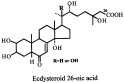
Fig. 5. Formula of ecdysteroid 26-oic acid.

Fig. 6. Formula of juvenile hormone III.
 You have
Access
- 55
- Cited by