1. Introduction
Archosauria is a major vertebrate clade that includes birds, crocodilians and numerous extinct lineages including non-avian dinosaurs, pterosaurs and early pseudosuchian (crocodile-line) taxa such as aetosaurs and ‘rauisuchians’. The timing of origin of Archosauria is well constrained to the Early Triassic (252.3–247.2 Ma: Mundil et al. Reference Mundil, Palfy, Renne, Brack and Lucas2010), based upon extremely rare but diagnostic pseudosuchian material from the Lower Triassic (Olenekian) to lowest Middle Triassic of Germany, Russia, China and the USA (Gower & Sennikov, Reference Gower, Sennikov, Benton, Shishkin, Unwin and Kurochkin2000; Nesbitt, Reference Nesbitt2003, Reference Nesbitt2005; Nesbitt, Liu & Li, Reference Nesbitt2011; Butler et al. Reference Butler, Brusatte, Reich, Nesbitt, Schoch and Hornung2011). Archosaurs subsequently radiated during the Middle Triassic, and by the end of the Triassic Period they were the dominant large-bodied tetrapods in most continental ecosystems (e.g. Benton, Reference Benton1983; Brusatte et al. Reference Brusatte, Benton, Ruta and Lloyd2008, Reference Brusatte, Benton, Desojo and Langer2010b ; Langer et al. Reference Langer, Ezcurra, Bittencourt and Novas2010; Nesbitt, Reference Nesbitt2011). Scientific understanding of the early archosaurian radiation is, however, hampered by the relatively poor early fossil record of the group. As noted above, Early Triassic archosaur fossils are scarce, whereas the early Middle Triassic (Anisian) evolution of the group is highlighted by just a few taxa and fossil assemblages (e.g. Krebs, Reference Krebs1965; Zhang, Reference Zhang1975; Benton & Gower, Reference Benton and Gower1997; Gower & Sennikov, Reference Gower, Sennikov, Benton, Shishkin, Unwin and Kurochkin2000; Nesbitt, Reference Nesbitt2003, Reference Nesbitt2005; Sen, Reference Sen2005; Nesbitt et al. Reference Nesbitt, Sidor, Irmis, Angielczyk, Smith and Tsuji2010; Butler et al. Reference Butler, Brusatte, Reich, Nesbitt, Schoch and Hornung2011).
The Middle Triassic (upper Anisian) Manda beds of southwestern Tanzania potentially provide the most illuminating window on the early archosaurian radiation. Unlike most other early Middle Triassic fossil assemblages, archosaur remains have proved to be abundant within the Manda beds, with extensive material being collected in the 1930s (Stockley, Reference Stockley1932; Haughton, Reference Haughton1932; Nowack, Reference Nowack1937) and early 1960s (Attridge et al. Reference Attridge, Ball, Charig and Cox1964). Although some of these extremely important historical collections of archosaur material were studied by Huene (Reference Huene1938, Reference Huene1939; see also Gebauer, Reference Gebauer2004 and Butler et al. Reference Butler, Barrett, Abel and Gower2009), the majority formed part of the Ph.D. dissertation of A. J. Charig (unpub. Ph.D. thesis, Univ. Cambridge, 1956) and remain largely absent from the published literature. Recently, new expeditions to the Manda beds involving the senior author have led to the discovery and description of abundant new archosaur material (e.g. Nesbitt et al. Reference Nesbitt, Sidor, Irmis, Angielczyk, Smith and Tsuji2010), and have prompted a fresh review of the Manda archosaurs.
One of the few formally described archosaur taxa from the Manda beds is Parringtonia gracilis Huene, Reference Huene1939, described on the basis of a partial maxilla and part of a postcranial skeleton, and largely ignored by later workers. Huene (Reference Huene1939) originally referred Parringtonia gracilis to Pseudosuchia (although the 1939 conception of Pseudosuchia was very different from that of the present day), and noted similarities to Ornithosuchus (an ornithosuchid) and Saltoposuchus (a non-crocodyliform crocodylomorph). However, Huene (Reference Huene1939) concluded that the available material was insufficient to determine if Parringtonia gracilis was closely related to these taxa or instead represented a new pseudosuchian lineage.
Krebs (Reference Krebs1965) noted similarities between the scapulae of Parringtonia gracilis and Erpetosuchus granti. Subsequently, Walker (Reference Walker1970) tentatively linked Parringtonia gracilis with the small pseudosuchian Erpetosuchus granti from the Upper Triassic of Scotland, as well as Dyoplax arenaceus Fraas, Reference Fraas1867 from the Upper Triassic of Germany (currently considered to be a possible ‘sphenosuchian’: Lucas, Wild & Hunt, Reference Lucas, Wild and Hunt1998), within the Erpetosuchidae (see comments in Section 5.c below). Krebs (Reference Krebs and Kuhn1976) provided a formal diagnosis of Erpetosuchidae, retaining within it only Erpetosuchus granti and Parringtonia gracilis. Benton & Walker (Reference Benton and Walker2002) removed Parringtonia gracilis from Erpetosuchidae (which they considered a monospecific family), suggesting that any similarities between Parringtonia gracilis and Erpetosuchus granti were possibly plesiomorphies.
Erpetosuchus granti has often been suggested to be closely related to Crocodylomorpha (e.g. Walker, Reference Walker1968), perhaps forming the sister taxon of this clade (Olsen, Sues & Norell, Reference Olsen, Sues and Norell2000; Benton & Walker, Reference Benton and Walker2002; Brusatte et al. Reference Brusatte, Benton, Desojo and Langer2010a ) and thus potentially a crucial taxon for understanding the evolution of crocodylomorphs. The hypothesis that Parringtonia gracilis and Erpetosuchus granti form a clade, Erpetosuchidae, has never been tested in a phylogenetic analysis, but is potentially crucial for an understanding of the timing of crocodylomorph origins. Moreover, as is also the case for other archosaur material from the Manda beds, a re-examination of Parringtonia gracilis has the potential to shine new light on the early archosaur radiation. For these reasons, we here redescribe the type and only known specimen of Parringtonia gracilis, and explicitly test the phylogenetic position of both Parringtonia gracilis and Erpetosuchus granti in a comprehensive cladistic analysis of early archosaurs.
2. Institutional abbreviations
Repositories for specimens discussed in the text are indicated by the following acronyms: AMNH – American Museum of Natural History, New York, USA; IVPP – Institute of Vertebrate Paleontology and Paleoanthropology, Beijing, China; NHMUK – Natural History Museum, London, UK; NMS – National Museums of Scotland (formerly Royal Scottish Museum, RSM), Edinburgh, UK; PEFO – Petrified Forest National Park, Arizona, USA; PVL – Instituto Miguel Lillo, Tucumán, Argentina; SAM – Iziko South African Museum, Cape Town, South Africa; SMNS – Staatliches Museum für Naturkunde, Stuttgart, Germany; TMM – Vertebrate Paleontology Laboratory, Texas Natural Science Center, Austin, Texas, USA; TTU – Texas Tech University, Lubbock, Texas, USA.
3. Methods
The holotype maxilla of Parringtonia gracilis was scanned using micro-computed tomography (μCT) at NHMUK using a HMX-ST CT 225 System (Metris X-Tek, Tring, UK). Data was reconstructed using CT-PRO software version 2.0 (Metris X-Tek) and rendered using VG Studio MAX 2.0 (Volume Graphics, Heidelberg, Germany). Measurements are given in Appendix 1.
4. Systematic palaeontology
ARCHOSAURIA Cope, Reference Cope1869
Erpetosuchidae Watson, Reference Watson1917 Definition. The most inclusive clade containing Erpetosuchus granti Newton, Reference Newton1894 but not Passer domesticus Linnaeus, Reference Linnaeus1758, Postosuchus kirkpatricki Chatterjee, Reference Chatterjee1985, Crocodylus niloticus Laurenti, Reference Laurenti1768, Ornithosuchus longidens (Huxley, Reference Huxley1877) or Aetosaurus ferratus Fraas, Reference Fraas1877 (new definition). Diagnosis. The following diagnosis is based solely on phylogenetically tested character states; other possible diagnostic character states are discussed below. Erpetosuchidae differs from all other archosauriforms in dentition present only in the anterior half of the maxilla [character 17, state 1], mediolateral length of the posterior portion of the maxilla greater than the dorsoventral length [21–1] and tooth serrations absent [168–1]. Included taxa. Erpetosuchus granti Newton, Reference Newton1894 and Parringtonia gracilis Huene, Reference Huene1939. Notes. A diagnosis of Erpetosuchidae was previously provided by Krebs (Reference Krebs and Kuhn1976, p. 87), based upon the general anatomical characters of Erpetosuchus granti. No phylogenetic definition has been previously provided.
Parringtonia gracilis Huene, Reference Huene1939
-
1939 Parringtonia gracilis n. g. n. sp.; Huene, p. 65, pl. 4.
-
1965 Parringtonia gracilis v. Huene; Krebs, p. 61.
-
1970 Parringtonia; Walker, p. 368.
-
1976 Parringtonia gracilis v. Huene; Krebs, p. 89.
-
2002 Parringtonia gracilis; Benton & Walker, p. 42.
-
2009 Parringtonia gracilis; Butler et al., p. 1022.
Holotype. NHMUK R8646 (field number P.68a): a relatively complete right maxilla, five nearly complete dorsal vertebrae, two dorsal centra, two partial dorsal neural arches, three caudal vertebrae, five osteoderms, left scapula missing the distal end of the blade and a proximal portion of an ?ischium.
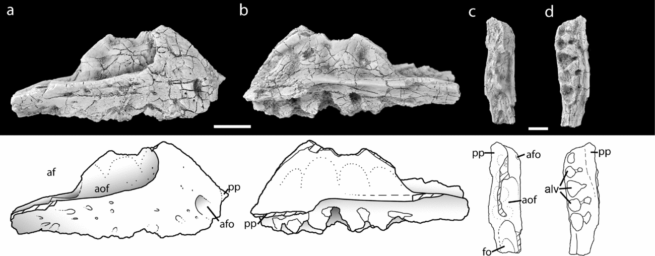
The right maxilla of Parringtonia gracilis (NHMUK R3139) in lateral (a), medial (b), dorsal (c) and ventral (d) views. Abbreviations: af – antorbital fenestra; afo – anterior foramen; alv – alveolus; aof – antorbital fossa; fo – fossa; pp – palatal process. Scales = 1 cm.

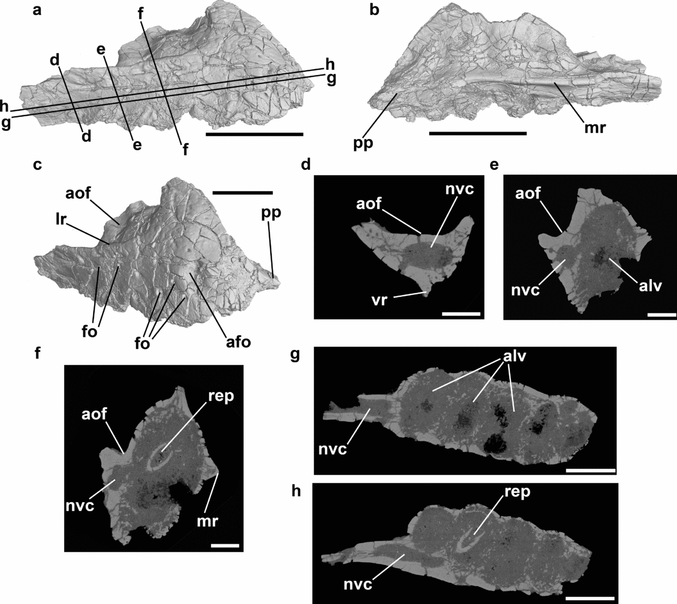
Reconstruction of the maxilla of Parringtonia gracilis (NHMUK R3139) from CT slices in lateral (a), medial (b) and anterolateral (c) views. Location of CT slices in (d–h) are indicated in (a). Abbreviations: afo – anterior foramen; alv – alveolus; aof – antorbital fenestra; fo – foramen; lr – lateral ridge; mr – medial ridge; nvc – neurovascular canal; pp – palatal process; rep – replacement tooth; vr – ventral ridge. Scales = 1 cm.

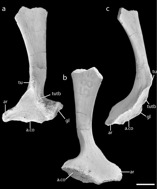
The left scapula of Parringtonia gracilis (NHMUK R3139) in lateral (a), medial (b) and anterior (c) views. Abbreviations: a.co – articulation with the coracoid; ar – acromion process; gl – glenoid; tu – tuber; tutb – tuber for attachment of the m. triceps brachii. Scale = 1 cm.

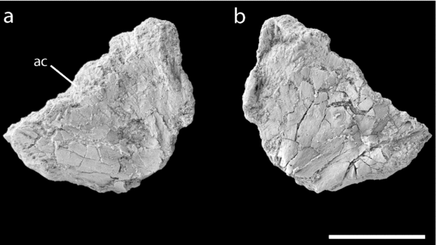
The left ?ischium of Parringtonia gracilis (NHMUK R3139) in lateral (a) and medial (b) views. Abbreviations: ac – acetabulum. Scale = 1 cm.

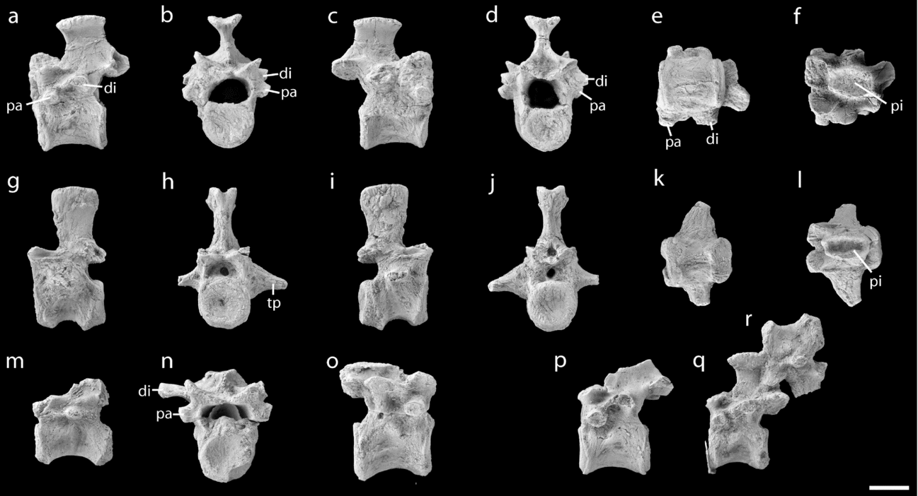
The vertebrae of Parringtonia gracilis (NHMUK R3139). Dorsal vertebra in left lateral (a), anterior (b), right lateral (c), posterior (d), ventral (e) and dorsal (f) views. Anterior caudal vertebra in left lateral (g), anterior (h), right lateral (i), posterior (j), ventral (k) and dorsal (l) views. A posterior caudal vertebra in left lateral (m) view. A dorsal vertebra in anterior (n) and right lateral (o) views. A well-preserved dorsal vertebra in left lateral view (p). A dorsal vertebra in left lateral view (q) attached to a caudal vertebra in right lateral view (r). Abbreviations: di – diapophysis; pa – parapophysis; pi – pit; tp – transverse process. Scale = 1 cm.

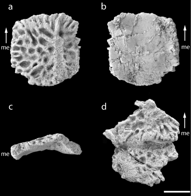
Osteoderms of Parringtonia gracilis (NHMUK R3139). A paramedian and dorsal osteoderm in dorsal (a), ventral (b) and posterior (c) views. Two articulated osteoderms in dorsal (d) view. Abbreviations: me – medial edge. Arrow indicates the anterior direction. Scale = 1 cm.

Type locality and horizon. Field locality B15/1, Mkonoleko/Njalila, northwest of Songea, Ruhuhu Valley, Ruvuma region, southwestern Tanzania (see Stockley, Reference Stockley1932, fig. 1). The specimen was collected by F. R. Parrington from the so-called ‘Upper Bone Bed’ (Huene, Reference Huene1939), now known as the Lifua Member of the Manda beds; however, the exact stratigraphic position is not known. The Manda beds are correlated on the basis of vertebrate biostratigraphy with Subzone C of the Cynognathus Assemblage Zone of the Beaufort Group, South Africa (Gay & Cruickshank, Reference Gay and Cruickshank1999; Catuneanu et al. Reference Catuneanu, Wopfner, Eriksson, Cairncross, Rubidge, Smith and Hancox2005; Sidor et al. Reference Sidor, Angielczyk, Hopson, Nesbitt and Smith2008), which has been dated to the latest Anisian of the Middle Triassic (e.g. Hancox, Reference Hancox2000; Abdala, Hancox & Neveling, Reference Abdala, Hancox and Neveling2005). The locality is number 64085 in The Paleobiology Database (http://paleodb.org).
A. J. Charig (unpub. Ph.D. thesis, Univ. Cambridge, 1956, p. 499) listed rhynchosaur (Stenaulorhynchus sp.: field numbers P.61a, 67, 68b) and indeterminate dicynodont material (field numbers P.57, 61b, 61u, 66, 68u) from the same locality.
Diagnosis. No previous diagnosis has been attempted, with Krebs (Reference Krebs and Kuhn1976) suggesting that Parringtonia gracilis differed from Erpetosuchus granti only in size. Parringtonia gracilis differs from all other archosauriforms except Erpetosuchus granti in the restriction of the maxillary dentition to the anterior portion of the maxilla, the mediolateral expansion of the posterior portion of the maxilla, and a deep anteroposteriorly oriented groove on the dorsal surface of the neural spines. Parringtonia gracilis differs from the holotype specimen of Erpetosuchus granti (NHMUK R3139) in having five maxillary alveoli instead of four, a large foramen on the lateral surface of the anterior portion of the maxilla, the presence of a small tuber on the lateral surface of the scapula distal to the glenoid fossa, and osteoderms that are nearly square instead of being anteroposteriorly longer than wide. Parringtonia gracilis differs from a referred specimen of Erpetosuchus sp. (AMNH 29300) by the presence of five alveoli instead of six alveoli (although possibly nine were present in AMNH 29300 based on nutrient foramina and preserved teeth) and a proportionally shorter ascending (= dorsal) process of the maxilla.
Ontogenetic stage. The holotype of Parringtonia gracilis cannot be aged using typical histological methods because limb bones were not preserved in the specimen. Therefore, we rely on the state of the neurocentral sutures of the vertebrae to estimate skeletal maturity as previously utilized for other archosaurs (Brochu, Reference Brochu1996; Irmis, Reference Irmis2007). All neurocentral sutures in the dorsal vertebrae are clearly visible whereas the neurocentral sutures are closed in the caudal vertebrae thus indicating that the specimen is skeletally immature.
Comments. A. J. Charig (unpub. Ph.D. thesis, Univ. Cambridge, 1956, p. 335) added two small and poorly preserved vertebrae (identified as sacrals by Charig), not described by Huene (Reference Huene1939), to the holotype of Parringtonia gracilis, noting that they were found among large rhynchosaur fragments from the same locality. However, the preservation of these specimens is different from that of the holotype: the vertebrae are articulated (the holotype material is completely disarticulated) and the surface of the bone is different in colour and much more highly fractured. While these elements could represent part of the holotype specimen, this remains uncertain, particularly given that elements of multiple taxa were collected from this locality. In light of this uncertainty and their poor and relatively uninformative preservation we do not discuss these elements further here.
5. Description
5.a. Maxilla (Figs 1, 2)
Most of the body of the maxilla is preserved; the posterior portion and the dorsal portion of the ascending process (= ‘dorsal process’) are missing. The ventral margin is relatively straight in lateral view with small scallops marking the alveoli. Anteriorly, the face of the maxilla arcs medially, forming a rounded anterior surface. This is similar to the condition in Erpetosuchus granti (NHMUK R3139) in which the contact between the maxilla and the premaxilla is pinched medially in ventral view (Benton & Walker, Reference Benton and Walker2002, fig. 2C). There is a large foramen dorsal to the first alveolus that opens anteriorly onto the anterior surface. A similar foramen is also present in Euparkeria (SAM-K-6047), Proterosuchus (Modesto & Sues, Reference Modesto and Sues2004) and Prolacerta (Modesto & Sues, Reference Modesto and Sues2004), but is only rarely present within Archosauria (e.g. Lotosaurus, IVPP V 4880). A similar foramen appears to be absent in Erpetosuchus granti (NHMUK R3139). CT data indicates that this large anterior foramen opens into an elliptical sediment-infilled neurovascular canal that extends posteriorly for the entire length of the element, opening into the antorbital fossa (see below). The neurovascular canal arches dorsally above the first two alveoli and then extends lateral to the alveoli and immediately ventral to the ventral surface of the antorbital fossa. The main neurovascular canal is fully surrounded by bone in some places, but elsewhere it communicates medially with the alveoli.
Two tiny nutrient foramina are present immediately posterior to the large anterior foramen. A further intermediate sized foramen is present, dorsal to the second alveolus. A longitudinal row of several foramina is present just ventral to the ridge that forms the ventral margin of the antorbital fossa. All of the foramina communicate with the main neurovascular canal, either directly or via short canals.
The ridge forming the ventral margin of the antorbital fossa runs subparallel to the oral margin above alveoli four and five. Dorsal to alveolus three the ridge begins to arc dorsally and then posterodorsally, immediately before its termination (as a result of breakage of the ascending process). Anteriorly, the antorbital fossa is not recessed medial to this ridge, unlike the condition in Erpetosuchus granti (NHMUK R3139). Posteriorly, the fossa is recessed medial to the ridge, and some of the internal surface of the fossa is therefore hidden in lateral view. In addition, the ridge expands laterally as it extends posteriorly. As a result, the ridge progressively creates an increasingly well-developed overhang over the ventral portion of the maxilla, with the lateral surface of the posterior part of the maxilla facing ventrolaterally rather than strictly laterally. A similar condition is present in Erpetosuchus granti (NHMUK R3139). The internal surface of the antorbital fossa is corrugated, with several large dorsoventrally extending furrows; this corrugated appearance is probably an external reflection of the alveoli, and is similar to the condition in Riojasuchus (PVL 3829).
Medially, the maxilla bears a sharp longitudinal ridge that bisects the element and that expands transversely at its anterior end to form the palatal process. This process projects a short distance anterior to the main body of the maxilla and is horizontally oriented with a broken medial margin. Because of the breakage it is unclear if the palatal process met its antimere on the midline. The surface dorsal to the ridge and palatal process is corrugated (see above). The five alveoli are positioned ventrolateral to the longitudinal ridge. There are apparently only four teeth in Erpetosuchus granti (NHMUK R3139) and possibly nine in a specimen referred to Erpetosuchus sp. (AMNH 29300: Olsen, Sues & Norell, Reference Olsen, Sues and Norell2000). Each alveolus has a subcircular outline and is separated from adjacent alveoli by thin sheets of bone and unfused interdental plates. Small foramina lie at the dorsal apices of the spaces between the interdental plates. The first two alveoli are significantly smaller than the remaining three. The tooth row is very gently arched anteromedially towards its anterior end. A sharp longitudinal ridge extends from the posterior edge of the fifth alveolus and proceeds posteriorly, forming the ventral margin of the edentulous portion of the element.
In dorsal view, the maxilla is transversely expanded relative to maxillae of other archosaurs. A transversely concave, dorsally facing surface is located posterior to the anterior margin of the antorbital fenestra, and bears a posteriorly opening foramen that represents the exit of the neurovascular canal described above. This concave surface is separated by a weak U-shaped ridge from the rest of the dorsal surface of the element.
A single maxillary tooth was discovered in the fourth alveolus when the specimen was CT scanned. The small tooth appears to lack serrations, is round in cross-section, and slightly posteriorly recurved (Fig. 2). Similar teeth are present in both the holotype of Erpetosuchus granti (NHMUK R3139) and a referred specimen of Erpetosuchus sp. (AMNH 29300).
5.b. Scapula (Fig. 3)
The left scapula is nearly complete and is only missing the distal portion of the blade. The scapula is described with the blade oriented vertically. The element consists of an expanded ventral plate and a blade that bends slightly anteriorly along its length and expands in anteroposterior width at its distal end. The blade has a relatively straight posterior margin and a concave anterior margin, with the result that the distal expansion is asymmetrical, being developed most strongly anteriorly. At its base the blade is constricted anteroposteriorly; the distal expansion (as preserved) of the blade is more than twice the width of the basal constriction. At the proximal constriction the blade has a subcircular cross-section, but distally the blade is transversely compressed. The heavy waisting of the proximal scapula blade in Parringtonia gracilis is similar to that of Erpetosuchus granti and was noted by Krebs (Reference Krebs1965, Reference Krebs and Kuhn1976) and used as a character state to unite the two taxa (Krebs, Reference Krebs and Kuhn1976). The distal portion of the scapula blade is more broadly expanded in Parringtonia gracilis in comparison with Erpetosuchus granti (NHMUK R3139).
The lateral surface of the blade bears two small yet distinct tubera: the first is positioned just dorsal to the glenoid on the posterior margin of the base of the blade, and the second is positioned anteriorly on the lateral surface of the scapula blade at the point of its proximal constriction. The more proximally located tuber is possibly the attachment site of m. triceps brachii and is well developed, as in Erpetosuchus granti and other pseudosuchians such as the loricatan pseudosuchian Batrachotomus (Gower & Schoch, Reference Gower and Schoch2009). The second, more distally placed, tuber is the more pronounced of the two and is unique to Parringtonia gracilis. A sharp and narrow ridge extends from the anteroventral corner of this tuber and forms the anterior margin of the prominent acromion process.
The glenoid faces posterolaterally, similar to the plesiomorphic condition in archosauriforms (Nesbitt, Reference Nesbitt2011). The large acromion process is distinctly raised above the concave proximal surface of the scapula, a character that supports the position of Parringtonia gracilis as an archosaur (Nesbitt, Reference Nesbitt2011). A short, low, anteroposteriorly extending ridge is present on the lateral surface of the proximal end, ventral to the acromion.
In anterior view, the scapula is strongly arched laterally along its length, with the point of maximum curvature occurring at the junction between the proximal plate and the blade. The scapula blade itself is relatively straight in anterior view along most of its length. When the sutural surface for the coracoid is held horizontally, the blade of the scapula blade is directed slightly anterodorsally rather than strictly dorsally.
5.c. Ischium (Fig. 4)
Huene (Reference Huene1939) identified a bone fragment as the proximal portion of a left ischium, but this identification was considered doubtful by A. J. Charig (unpub. Ph.D. thesis, Univ. Cambridge, 1956). Here, we follow the identification of Huene (Reference Huene1939) because it appears the bone has an expanded proximal portion and possibly an acetabular rim. Unfortunately, all of the edges, with the exception of the dorsal edge, are broken. Therefore, it is unclear whether, and how, the ischium contacted the pubis and ilium (contra Huene, Reference Huene1939). The dorsal edge is the thickest portion of the preserved element and is rounded. The medial surface is gently concave and the proximal portion tapers posteriorly.
5.d. Dorsal vertebrae (Fig. 5)
The centra are spool shaped, with lateral and ventral surfaces that are gently concave anteroposteriorly; however, several vertebrae (interpreted as more anterior dorsals) bear a weak midline keel on the ventral surface. The lengths of the centra are typically about 150% of their dorsoventral heights. The weakly amphicoelous articular surfaces are subcircular in outline with gently rounded and slightly thickened rims. The lateral surfaces of the centra lack any kind of lateral fossa immediately ventral to the neurocentral suture, unlike the condition in Euparkeria (SAM-K-6047), suchians (e.g. Revueltosaurus, PEFO 34561) and dinosauromorphs (e.g. Marasuchus, PVL 3870). There are also no lateral fossae in presacral 9 of Erpetosuchus granti (NHMUK R3139).
The neural spines lie over the posterior half of the centrum. They are short and only slightly taller dorsoventrally than the centrum. They are transversely and anteriorly narrowest at mid-height, and they expand weakly anteroposteriorly and very strongly transversely at their apex. In dorsal view, this apical expansion has convex lateral margins and flattened anterior and posterior margins. The dorsal surface is roughly textured and transversely concave, forming a median pit that is also present in Erpetosuchus granti (NHMUK R3139). The dorsal expansions of the neural spines are similar to those present in Euparkeria (SAM-K-6049B) and Erpetosuchus granti (NHMUK R3139). The lateral expansions of the dorsal vertebrae of Erpetosuchus granti (NHMUK R3139) are more restricted anteroposteriorly than Parringtonia gracilis giving them a rectangular shape with a longer mediolateral side. The anterior and posterior surfaces of the spines are formed by sharp ridges.
The large prezygapophyses face dorsomedially at about 30° to the horizontal and have articular surfaces that are gently concave transversely. They do not extend beyond the anterior margin of the centrum. The postzygapophyses face ventrolaterally at a similar angle to the prezygapophyses and overhang the posterior end of the centrum. A deep median fossa located at the base of the neural spine separates the postzygapophyses. Hypantrum–hyposphene accessory vertebral articulations are absent.
The spacious neural canal is relatively large compared to that of the centrum size. Anteriorly, it is broader than deep, with a nearly rectangular outline. The two isolated dorsal centra demonstrate that the neural canal was deeply excavated into the dorsal surface of the centrum. The short parapophyses lie lateral to the mid-point of the neural canal, well above the neurocentral suture and broadly separated from the diapophyses. Their articular surfaces are flattened and face laterally and slightly ventrally. The diapophyses are broken in nearly all of the preserved vertebrae, with one exception. The diapophysis of this vertebra extends posterolaterally and slightly dorsally, and has subparallel anterior and posterior margins in dorsal view. Its distal end is slightly expanded dorsoventrally to form the convex articular surface. In some, but not all, of the dorsals there is a low ridge connecting the dorsal margin of the parapophysis to the anterior margin of the diapophysis. A shallow concavity is present beneath the diapophysis; however, deep fossae and well-developed vertebral laminae are absent.
5.e. Caudal vertebrae (Fig. 5)
Three caudal vertebrae, one anterior, one anterior–mid and one mid–posterior, are represented in the material. The caudal vertebrae have centra that are all approximately the same length. Neurocentral sutures are indistinguishably fused in all three vertebrae. The anterior caudal has dorsoventrally deep, laterally directed transverse processes that have a triangular outline in dorsal and anterior views. The centrum has subcircular and gently concave articular faces; in lateral view the centrum is longer than dorsoventrally deep with constricted lateral surfaces that are concave anteroposteriorly and dorsoventrally. A sharp break-in-slope (paramedian keel) separates the lateral and ventral surfaces of the centrum; the ventral surface is flattened with a weakly developed median groove. The ventral margin of the centrum is bevelled at anterior and posterior ends (this is most obvious in lateral view), with the posterior bevelled surface being the larger of the two. This surface, which articulated with the chevron, is subdivided by a subtle median break-in-slope.
The spacious neural canal of the anterior caudal has a subrectangular outline, being wider transversely than dorsoventrally deep. It is widest at its anterior end and decreases in transverse width posteriorly. The prezygapophyses extend only very slightly beyond the anterior margin of the centrum; they face dorsomedially at approximately 15° above the horizontal. Their articular faces are gently concave transversely. The postzygapophyses face ventrolaterally at a similar angle. A small median fossa is present between the postzygapophyses in posterior view. The neural spine lies over the posterior half of the centrum and its height is 1.5 times greater than the height of the centrum. It is narrowest in anteroposterior and transverse dimensions immediately above the postzygapophyses, and it expands anteroposteriorly and transversely towards its apex. At its apex, the transverse expansion of the spine is marked, but does not occur to the same degree as in the presacral vertebrae. Among early archosaurs, the presence of transverse expansions of the anterior caudal neural spines is rare and only occurs to our knowledge in some aetosaurs (Walker, Reference Walker1961, fig. 10a–d). The dorsal surface of the spine bears a deep anteroposteriorly extending pit surrounded by a distinct rim, as present in the dorsal vertebrae. The anterior and posterior margins of the spine are formed by sharp ridges that birfurcate dorsally to form the lateral margins of the dorsal expansion. The lateral surfaces of the spine are gently convex anteroposteriorly.
The neural spine of the anterior–mid caudal is incomplete. It differs from the anterior caudal primarily in possessing a much less well-developed transverse process. On the ventral surface of the centrum there is a sharp midline ridge or keel, in addition to the paramedian keels seen in the anterior caudal. The mid–posterior caudal (also lacking its neural spine, and with damaged zygapophyses) retains a short and dorsoventrally shallow transverse process. The centrum is nearly as elongated as the more anterior caudal vertebrae, but is substantially reduced in height.
5.f. Osteoderms (Fig. 6)
Five osteoderms, one of which is nearly complete, are preserved. The osteoderms are nearly square with rounded corners. This is in contrast to the osteoderms of Erpetosuchus granti, which are distinctly longer than wide (NHMUK R3139). There is no pointed process on the anterior edge, unlike the condition in paracrocodylomorphs (Nesbitt, Reference Nesbitt2011). The osteoderms are distinctly bent into an L-shape, with an anteroposteriorly extending rounded ridge (which is positioned at two-thirds of the transverse distance from the medial margin of the element marking the point of the bend). The anterior edge of each osteoderm does not have a smooth surface devoid of sculpturing for articulation with the preceding osteoderm like that of aetosaurs (e.g. Lucasuchus; TMM 31100-361), but instead has a sculptured anterior facet that indicates that the osteoderms were imbricated in life, as in Erpetosuchus granti (Benton & Walker, Reference Benton and Walker2002). Deep pits and grooves cover the dorsal surface of the osteoderm and radiate from the midpoint of the anteroposteriorly extending ridge as in Erpetosuchus granti (NHMUK R3139). The presence of deep sculpturing in small osteoderms is rare among early archosaurs, with most small early archosaur osteoderms having relatively smooth surfaces (e.g. Aetosaurus ferratus; SMNS 18554). The ventral surface is smooth and convex anteroposteriorly. The anterior, posterior and lateral edges taper, whereas the medial edge is slightly thickened, suggesting that it contacted its antimere at the midline. It is not clear if Parringtonia gracilis had any osteoderms lateral to the paramedian row, as occurs in one specimen referred to Erpetosuchus granti (Benton & Walker, Reference Benton and Walker2002)
It is not clear how the osteoderms were arranged. However, it is likely that there was only a single pair of paramedian rows (two rows) and there was only one set per vertebrae given the length of each osteoderm relative to each presacral vertebra. This pattern is present in Erpetosuchus granti (NMS 1966.4.3; Benton & Walker, Reference Benton and Walker2002).
6. The phylogentic relationships of Parringtonia gracilis
6.a. Methodology
We assessed the phylogenetic position of Erpetosuchus granti and Parringtonia gracilis by modifying the most comprehensive early archosaur phylogeny to date (Nesbitt, Reference Nesbitt2011). A total of 412 characters and 79 taxa were used in the analysis, with the rhynchosaur Mesosuchus used to root the most parsimonious trees (MPT). The matrix was analysed in PAUP* ver. 4.0b10 using a heuristic search subjected to 1000 random addition replicates with tree bisection and reconnection branch swapping. Branches were collapsed if they had a minimum length of zero. All characters were equally weighted. Characters 32, 52, 121, 137, 139, 156, 168, 188, 223, 247, 258, 269, 271, 291, 297, 328, 356 and 399 were ordered because they represent a range with intermediate stages between two extremes. Parringtonia gracilis was scored from the only known specimen and Erpetosuchus granti was scored from the holotype (NHMUK R3139) and referred specimens detailed in Benton & Walker (Reference Benton and Walker2002) (see Appendix 2 for scores). The North American specimen of Erpetosuchus sp. (Olsen, Sues & Norell, Reference Olsen, Sues and Norell2000) was not used to score this taxon because the scores are identical to that of Erpetosuchus granti.
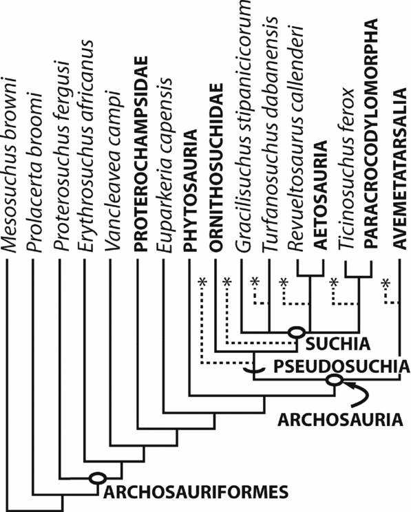
Our results (MPT = 1710, tree length (TL) = 1304, consistency index (CI) = 0.369, retention index (RI) = 0.772) show that Parringtonia gracilis and Erpetosuchus granti form a well-supported clade (Bremer support = 3) within Archosauria. The results of this analysis are largely congruent with those of Nesbitt (Reference Nesbitt2011). However, the addition of Parringtonia gracilis and Erpetosuchus granti decreases resolution at the base of Archosauria, with an unresolved polytomy containing Parringtonia gracilis + Erpetosuchus granti, Avemetatarsalia, Ornithosuchidae, Aetosauria + Revueltosaurus, Ticinosuchus + Paracrocodylomorpha, Gracilisuchus and Turfanosuchus (Fig. 7) present in the strict consensus tree. A combination of missing data and character conflict (e.g. the presence of a dorsally elongated quadratojugal (a character of Rauisuchidae + Crocodylomorpha) combined with the presence of a lateral expansion of the distal end of the neural spines in the dorsal vertebrae (a primitive archosauriform character)) in Parringtonia gracilis and Erpetosuchus granti appears to cause the low resolution. An Adams consensus tree (Adams, Reference Adams1972) places Parringtonia gracilis + Erpetosuchus granti in a polytomy with Ornithodira and Pseudosuchia, whereas the relationships within Pseudosuchia are highly similar to those of Nesbitt (Reference Nesbitt2011). Reduced consensus (Wilkinson, Reference Wilkinson2003; carried out using REDCON 3.0, Wilkinson, Reference Wilkinson2001) confirmed that Parringtonia gracilis and Erpetosuchus granti act as ‘wildcard’ taxa in the analysis. A posteriori deletion of Parringtonia gracilis and Erpetosuchus granti yields a reduced component consensus tree that is highly similar to the strict consensus tree presented by Nesbitt (Reference Nesbitt2011), although relationships at the base of Pseudosuchia are slightly less resolved.
Phylogenetic positions of Erpetosuchidae (Erpetosuchus granti + Parringtonia gracilis) among early archosaurs in the analysis of Nesbitt (Reference Nesbitt2011). A strict consensus of the early archosaur phylogeny (MPT = 1710, TL = 1304, CI = 0.369, RI = 0.772) results in a large polytomy including Avemetatarsalia, Ornithosuchidae, Aetosauria + Revueltosaurus, Ticinosuchus + Paracrocodylomorpha, Gracilisuchus and Turfanosuchus. Asterisks mark the possible phylogenetic positions of Erpetosuchidae. Taxa in bold and all capitals have been collapsed for brevity but were originally included in the analysis as species-level taxa (see Nesbitt, Reference Nesbitt2011).

This poorly resolved result also appears to be linked to the unresolved phylogenetic positions of Gracilisuchus stipanicicorum and Turfanosuchus dabanensis at the base of Suchia. If these taxa are a priori removed and the analysis rerun, the resultant strict consensus tree (MPT = 90, TL = 1281, CI = 0.376, RI = 0.775) has relationships that are identical to those of Nesbitt (Reference Nesbitt2011) and places the clade Parringtonia gracilis + Erpetosuchus granti as the sister taxon of aetosaurs + Revueltosaurus, although this relationship is weakly supported (Bremer support = 1) by only the following character states: posterior portion of the maxilla ventral to the antorbital fenestra expands dorsoventrally (27–2: CI = 0.182) and two paramedian pairs of osteoderms (four osteoderms per segment) (406-1: CI = 0.5).
6.b. Parringtonia gracilis and Erpetosuchus granti
Our results indicate that Parringtonia gracilis and Erpetosuchus granti form a clade, Erpetosuchidae (see definition and diagnosis in Section 4 above). Within the numerical phylogenetic analysis conducted, the two taxa are united by three unique unambiguous optimized synapomorphies: dentition present only in the anterior half of the maxilla (17-1), mediolateral length greater than dorsoventrally length of the posterior portion of the maxilla (21-1) and tooth serrations absent (168-1). These three characters states have a high consistency index in this analysis (CI = 0.7–1) indicating that they essentially only occur in Parringtonia gracilis and Erpetosuchus granti (see Nesbitt, Reference Nesbitt2011). Our analysis also identifies one other unambiguously optimized character state that unites Parringtonia gracilis and Erpetosuchus granti: paramedian osteoderms with a distinct longitudinal bend near the lateral edge (404-1). However, given the uncertainty of the phylogenetic position of Erpetosuchidae, and that this character state has a low consistency index (= 0.25), this optimization is most likely an artefact given that many early archosaur groups with osteoderms also have this character state (e.g. crocodylomorphs; Nesbitt, Reference Nesbitt2011).
Parringtonia gracilis and Erpetosuchus granti also share many character states not yet sampled in the cladistic analysis. The following shared character states appear to represent synapomorphies of the two taxa, but this must be considered tentative prior to their incorporation into future phylogenetic analyses. Parringtonia gracilis and Erpetosuchus granti share a sharp ridge on the lateral margin of the maxilla that marks the ventral extent of the antorbital fossa. Ventral to this ridge, the external surface of the maxilla is inclined medially towards the alveolar margin. This medially inclined external surface of the maxilla continues posteriorly onto the jugal in Erpetosuchus granti, effectively exposing much of the external surface of the jugal in ventral view. This morphology represents a character state that unites the North American and European specimens of Erpetosuchus with Parringtonia gracilis.
Both Parringtonia gracilis and Erpetosuchus granti bear a hypertrophied tuber hypothesized for the attachment of the m. triceps brachii on the posterolateral surface of the proximal scapula blade. Although other archosauriforms have a small tuber in the same location (Nesbitt, Reference Nesbitt2005, Reference Nesbitt2011), the size of the tuber in Parringtonia gracilis and Erpetosuchus granti is exceptionally large relative to the overall size of the scapula (Fig. 3). Similarly, the extreme waisting of the proximal portion of the scapula blade is rare among non-dinosaurian archosauromorphs. Krebs (Reference Krebs and Kuhn1976) hypothesized that the presence of an anteriorly inclined scapular blade unites Parringtonia gracilis and Erpetosuchus granti. The scapula blade does slightly arc anteriorly in both taxa; however, the amount of curvature in the element in Parringtonia gracilis and Erpetosuchus granti is not dissimilar to that of other archosaurs (e.g. Postosuchus kirkpatricki, TTU P9000).
The transversely expanded distal ends of the neural spines bear median pits on their dorsal surfaces in the presacral vertebrae (vertebrae 4–12) of Erpetosuchus granti, and similar median pits occur in the posterior presacral vertebrae and caudal vertebrae (exact positions unknown) of Parringtonia gracilis (Fig. 5). Many pseudosuchians (e.g. the aetosaur Desmatosuchus; Parker, Reference Parker2008) and taxa just outside Archosauria (e.g. Euparkeria, SAM-K-6047) have laterally expanded distal neural spines (= spine tables), but the dorsal surface is either convex or nearly flat. The deep pits in Parringtonia gracilis and Erpetosuchus granti are only present in these two taxa, as far as we have observed.
Benton & Walker (Reference Benton and Walker2002) challenged the assignment of Parringtonia gracilis to Erpetosuchidae by Walker (Reference Walker1970) and Krebs (Reference Krebs and Kuhn1976), suggesting that most of the similarities between Parringtonia gracilis and Erpetosuchus granti are plesiomorphic for Archosauria. Of the six autapomorphies of Erpetosuchus granti listed by Benton & Walker (Reference Benton and Walker2002), three of the character states (characters 1, 2 and 6) are present on the maxilla, an element that is present in both Parringtonia gracilis and Erpetosuchus granti. Parringtonia gracilis and Erpetosuchus granti share a reduced maxillary tooth row (4–5 teeth) that is restricted to the area anterior to the midpoint of the antorbital fenestra (autapomorphy 1 of Benton & Walker, Reference Benton and Walker2002). In the discussion of this character state, Benton & Walker (Reference Benton and Walker2002) stated that only the anterior part of the maxilla of Parringtonia gracilis is preserved and therefore that it was not clear if Parringtonia gracilis had an anteriorly restricted maxillary dentition. However, there is a clear absence of alveoli in the preserved posterior portion of maxilla (Figs 1, 2). Additionally, Parringtonia gracilis and Erpetosuchus granti share large antorbital fenestra set in a deep fossa whose margins are marked by distinct sharply angled ridges (autapomorphy 2 of Benton & Walker, Reference Benton and Walker2002) and teeth oval in cross-section, lacking distinct anterior and posterior carinae and lacking marginal serrations (autapomorphy 6 of Benton & Walker, Reference Benton and Walker2002), as discussed in Section 4 above.
6.c. The phylogenetic position of Erpetosuchidae
Parringtonia gracilis and Erpetosuchus granti form a clade historically referred to as Erpetosuchidae (Walker, Reference Walker1970; Krebs, Reference Krebs and Kuhn1976) and phylogenetically defined for the first time here. Unfortunately, the phylogenetic position of this clade within Archosauriformes is poorly resolved in the results of our early archosaur phylogenetic analysis. The high number of autapomorphic features in the skull of Erpetosuchus granti (see Benton & Walker, Reference Benton and Walker2002), the uncertainty in determining cranial homologies between Erpetosuchus granti and other early archosaurs (e.g. the ‘overhanging’ squamosal; see discussion in Nesbitt, Reference Nesbitt2011), genuine character conflict (see above) and the absence of information on the anatomy of the braincase, pelvis and hindlimb all contribute to the poor resolution of the relationships of the clade. Minimally, however, Erpetosuchidae can be assigned to the Archosauria based on the presence of palatal processes of the maxilla that are in contact along the midline (32-1), an antorbital fossa that is present on the posterodorsal portion of the maxilla (137-2) and a distinctly raised acromion process (220-1) of the scapula. The cranial and postcranial skeletons of Erpetosuchidae share no unambiguous synapomorphies with most of the clearly monophyletic groups of Triassic archosaurs (e.g. Aetosauria, Dinosauromorpha, Pterosauria).
Previously, Erpetosuchus granti was incorporated into a small phylogenetic analysis including an aetosaur, Gracilisuchus, Postosuchus and early crocodylomorphs by Olsen, Sues & Norell (Reference Olsen, Sues and Norell2000). The subsequent analyses of Benton & Walker (Reference Benton and Walker2002) and Brusatte et al. (Reference Brusatte, Nesbitt, Irmis, Butler, Benton and Norell2010a ) placed Erpetosuchus granti into more comprehensive early archosaur phylogenies with increased taxon sampling, but continued to recover Erpetosuchus granti as the sister taxon to Crocodylomorpha. In all three previous analyses (Olsen, Sues & Norell, Reference Olsen, Sues and Norell2000; Benton & Walker Reference Benton and Walker2002; Brusatte et al. Reference Brusatte, Benton, Desojo and Langer2010a ), the sister-taxon relationship between Erpetosuchus granti and Crocodylomorpha was supported by three character states: the absence of a postfrontal, the complete fusion of the parietal, and an anterodorsally sloped quadrate and quadratojugal. Nevertheless, accurate recognition of the absence of both a postfrontal and an interparietal suture relies upon the ability to observe clear sutures between skull elements. Unfortunately, the only available skull material of Erpetosuchus granti that includes the relevant parts of the skull roof is preserved as a natural mould, as is the case for most of the other Lossiemouth reptiles (Benton & Walker, Reference Benton and Walker1981, Reference Benton and Walker1985). Frequently, sutures cannot be clearly identified in fossils preserved in this manner, and this is particularly true for specimens from the Lossiemouth Sandstone Formation. Therefore, the presence or absence of a postfrontal and an interparietal suture cannot be confirmed with the available material, and these characters were scored as unknown for Erpetosuchus granti in our analysis. Furthermore, it is clear that some early crocodylomorphs (e.g. Dromicosuchus grallator, Sues et al. Reference Sues, Olsen, Carter and Scott2003) have a clear interparietal suture. The anterodorsally sloping quadrate and quadratojugal may represent a shared character of crocodylomorphs and Erpetosuchus granti; however, this character should be scored cautiously given that the accurate assessment of the orientation of the quadrate and quadratojugal in life requires exquisite preservation and the absence of crushing. Crocodyliforms clearly have a similarly sloping quadrate and quadratojugal, but the orientation of these elements in early crocodylomorphs such as Dibothrosuchus elaphros (Wu & Chatterjee, Reference Wu and Chatterjee1993) and Dromicosuchus grallator (Sues et al. Reference Sues, Olsen, Carter and Scott2003) remain uncertain.
Given these scoring differences, we examined the evidence for a possible sister-taxon relationship of Erpetosuchidae and Crocodylomorpha in our analysis. Even if Erpetosuchus granti is scored as lacking both a postfrontal and an interparietal suture, Erpetosuchidae is still recovered in a polytomy at the base of Archosauria, and is not recovered as the sister taxon of Crocodylomorpha. A sister-taxon relationship between Erpetosuchidae and Crocodylomorpha requires 13 extra steps in our analysis. If Erpetosuchus granti is solely used to represent Erpetosuchidae (i.e. if Parringtonia gracilis is a priori excluded), it is recovered as the sister taxon of aetosaurs + Revueltosaurus, but this position is poorly supported (Bremer support = 1).
Our results illustrate the difficulties involved in accurately reconstructing the phylogenetic position of Erpetosuchidae within Archosauria. Consequently, the discovery of new material of erpetosuchids is likely required to fully resolve the relationships of the enigmatic clade.
At the moment, we restrict Erpetosuchidae to only include Erpetosuchus and Parringtonia gracilis. However, the poorly understood Dyoplax arenaceus (SMNS 4760) from the Carnian Stuttgart Formation (Lucas, Wild & Hunt, Reference Lucas, Wild and Hunt1998; Kozer & Bachmann, Reference Kozer and Bachmann2010) has been hypothesized to be a member of the group (Walker, Reference Walker1961, Reference Walker1968, Reference Walker1970; Romer, Reference Romer1966; Carroll, Reference Carroll1988) but has also been considered to be an aetosaur (Zittel, Reference Zittel1890; Huene, Reference Huene1902; McGregor, Reference McGregor1906), a protosuchid crocodyliform (Benton, Reference Benton, Fraser and Sues1994) or a sphenosuchian (Lucas, Wild & Hunt, Reference Lucas, Wild and Hunt1998; Benton & Walker, Reference Benton and Walker2002). The broad array of taxonomic assignments is largely the result of poor preservation of the anatomical details of the specimen: the specimen is preserved as a natural cast in sandstone (Fraas, Reference Fraas1867). Clark, Sues & Berman (Reference Clark, Sues and Berman2000) questioned the recent assignment to a crocodylomorph by Lucas, Wild & Hunt (Reference Lucas, Wild and Hunt1998), noting that proposed cranial similarities between Dyoplax and crocodylomorphs are impossible to verify on the cast.
The assignment by Walker (Reference Walker1961) of Dyoplax arenaceus to Erpetosuchidae was based solely upon gross similarity. Although the skeleton of Dyoplax arenaceus is largely complete, the elements that can be compared between Erpetosuchus granti, Parringtonia gracilis and Dyoplax arenaceus are limited. The body of Dyoplax arenaceus appears to be an external cast but it is not clear if the skull is an internal or external cast; if the skull is an internal cast, comparisons with the external skull cast of Erpetosuchus granti is impossible. As preserved, the skull of Dyoplax arenaceus appears to lack the mediolaterally expanded posterior portion of the maxilla that occurs in Erpetosuchus granti and Parringtonia gracilis. Furthermore, Dyoplax arenaceus lacks the ventrally deflected jugal and narrow frontal and parietal of Erpetosuchus granti (Benton & Walker, Reference Benton and Walker2002). Postcranially, little is visible in Dyoplax arenaceus. The osteoderms of Dyoplax arenaceus are superficially similar to those of Erpetosuchus granti and Parringtonia gracilis, but the common character states have a wide distribution among pseudosuchians (Nesbitt, Reference Nesbitt2011).
Dyoplax arenaceus cannot be shown to pertain to Erpetosuchidae at this time, although we cannot exclude the possibility that future discoveries and future analyses may show it to be the sister taxon of Erpetosuchus granti and Parringtonia gracilis. We refrain from including it into a phylogenetic analysis at this time, but note that the taxon can be clearly assigned to Archosauria based on the presence of an antorbital fenestra with a fully circumscribing antorbital fossa. The presence and arrangement of the dorsally placed osteoderms is suggestive of a pseudosuchian assignment (see Nesbitt, Reference Nesbitt, Liu and Li2011).
7. Discussion
Our redescription of the anatomy and reassessment of the phylogenetic relationships of Parringtonia gracilis demonstrates the taxon is the oldest unambiguous member of Erpetosuchidae: this result has several implications for the understanding of this poorly known archosaur clade. For over 150 years, the enigmatic Erpetosuchus granti was little more than a curiosity, with few clues to its evolutionary history. Now, we know that Erpetosuchus granti from Scotland has a nearly indistinguishable sister taxon in the Upper Triassic of North America (Olsen, Sues & Norell, Reference Olsen, Sues and Norell2000) and a very close relative from the Middle Triassic of Africa. Erpetosuchidae minimally consists of members stretching from the Anisian to the Norian that are broadly distributed in northern and southern Pangaea. Thus, this fragmentary fossil record hints at a possibly widespread and diverse group of archosaurs that lived for much of the Triassic. Furthermore, the distinct morphology of the anterior portion of the skull of Erpetosuchus granti appears to have been present in Parringtonia gracilis, thus demonstrating that restriction of maxillary teeth to the anterior portion of that element and the deflection of the maxilla and jugal was present early in the history of the group. These two features may relate to a specialist dietary strategy, suggesting that erpetosuchids may have had a similar diet for much of their evolutionary history.
The early Middle Triassic age of Parringtonia gracilis also has important implications for previous hypotheses about the timing of origin of Crocodylomorpha. Formerly, the Late Triassic Erpetosuchus granti was hypothesized to be the sister taxon of Crocodylomorpha (Olsen, Sues & Norell, Reference Olsen, Sues and Norell2000; Benton & Walker, Reference Benton and Walker2002; Brusatte et al. Reference Brusatte, Benton, Desojo and Langer2010a ), a group that also has its first appearance in the Late Triassic (Clark, Sues & Berman, 2000). The sister-taxon relationship between Erpetosuchus granti and Crocodylomorpha implied that the inferred temporal origin of Crocodylomorpha was consistent with direct evidence from the fossil record (Brusatte et al. Reference Brusatte, Benton, Desojo and Langer2010a ). However, in this analysis, we do not find a close relationship between Erpetosuchidae and Crocodylomorpha.
Clarification of the phylogenetic position of Parringtonia gracilis provides important data on the archosaurian assemblage of the Manda beds. Thus, the earliest erpetosuchid is added to an exceptionally diverse archosaur assemblage that contains either the first or early appearances of multiple major archosaur clades including poposauroids (Nesbitt, Reference Nesbitt2003; Butler et al. Reference Butler, Barrett, Abel and Gower2009), loricatans (Huene, Reference Huene1938; Charig, Reference Charig1957) and avemetatarsalians (Nesbitt et al. Reference Nesbitt, Sidor, Irmis, Angielczyk, Smith and Tsuji2010). Erpetosuchidae, as demonstrated by its co-occurrence with early members of other archosaurian subgroups, was a group present during the early radiation of Archosauria.
Acknowledgements
We thank Paul Barrett, Angela Milner and Sandra Chapman for access and collections help at NHMUK. We thank Stig Walsh and Richard Abel (NHMUK) for assistance with CT data, and Phil Crabb (NHMUK) for photography. Reviews from Hans-Dieter Sues and Grzegorz Niedźwiedzki improved the manuscript. Funding was provided to SJN by NSF grant no. 1024036. RJB was supported during the completion of this work by the DFG Emmy Noether Programme (BU 2587/3-1) and an Alexander von Humboldt Postdoctoral Research Fellowship.
Appendix 1. Measurements
Maxilla, anteroposterior length as preserved: 30.4 mm
Maxilla, maximum height as preserved: 13 mm
Scapula, maximum height as preserved: 34 mm
Scapula, maximum anteroposterior expansion, proximal end: 18 mm
Scapula, minimum anteroposterior width of shaft: 4.7 mm
Scapula, maximum anteroposterior expansion of distal shaft as preserved: 10.7 mm
Ischium, dimensions: 21 by 13.5 mm
Complete osteoderm, anteroposterior length: 14 mm
Complete osteoderm, transverse width: 13.7 mm
Dorsal vertebra, labelled ‘7’, centrum length, width, height (both anterior), total height of vertebra: 12 mm, 13.3 mm, 9 mm, 8.3 mm, 22.4 mm
Dorsal centrum, labelled ‘1’, same measurements: 9.6 mm, 7.7 mm, 6.8 mm, NA
Dorsal centrum, labelled ‘2’, same measurements: 9.7 mm, 8 mm, 6.7 mm, NA
Dorsal vertebra, labelled ‘3’, same measurements: 10.1 mm, 7.6 mm, 7 mm, 16.5+ mm
Dorsal vertebra, labelled ‘4’, same measurements: 10.2 mm, 7.8 mm, damaged, 15+ mm
Dorsal vertebra, labelled ‘5’, same measurements: 10.6 mm, 7.2 mm, 8.1 mm, 21.1 mm
Dorsal vertebra, labelled ‘6’, same measurements: 11 mm, 8 mm, 7.6 mm, 20 mm
Anterior caudal, same measurements: 9.8 mm, 7.8 mm, 7.7 mm, 21 mm
Anterior-mid caudal, same measurements: 10.5 mm, 6.4 mm, 5.6 mm, 14+ mm
Mid caudal, same measurements: 9.6 mm, 5 mm, 4.8 mm, 10.8+ mm
Appendix 2. Taxon scores and terminal taxa information
Scores for taxa incorporated into Nesbitt (Reference Nesbitt2011)
Parringtonia gracilis

Erpetosuchus granti

Erpetosuchus granti Newton, Reference Newton1894
Age. late Carnian–Norian, Late Triassic.
Occurrence. Lossiemouth Sandstone Formation, exact locality unknown (from either Lossiemouth East Quarry or Spynie Quarries), close to Elgin, Scotland.
Holotype. NHMUK R3139, a nearly complete skull and mandible, cervical and anterior dorsal vertebrae, shoulder girdle, forelimbs and osteoderms.
Referred Material. NMS 1966.43.4A, B, dorsal vertebrae, ribs and osteoderms; NMS 1992.37.1, cervical vertebrae and osteoderms; NHMUK R4807, articulated vertebrae.
Revised Status. Valid, Archosauria.
Key references. Newton (Reference Newton1894); Olsen, Sues & Norell (Reference Olsen, Sues and Norell2000); Benton & Walker (Reference Benton and Walker2002).