Introduction
The surgical management of congenital mitral valve disease in paediatric patients presents significant challenges due to the varied anatomical configurations and growth considerations. Reference Brown, Fiore, Ruzmetov, Eltayeb, Rodefeld and Turrentine1,Reference Iddawela, Joseph and Ganeshan2 These challenges are especially pronounced in neonates and infants, who have the most severe lesions, have smaller annuli, fragile tissues, and require more complex postoperative care. Preserving growth potential in this group is a major problem. Reference Isaacson, Lucjak and Johnson3 Reintervention becomes inevitable as the child grows, but avoiding valve replacement during infancy is crucial due to the high mortality rate—up to 41%—and the high rate of reintervention. Reference Caldarone, Raghuveer and Hills4–Reference Beierlein, Becker and Yates6 Mitral valve repair in children generally shows good outcomes, with low mortality and an acceptable reoperation rate. Reference Hetzer, Walter and Hübler7–Reference Shi, Xu and Yan11 However, some studies indicate that infants undergoing mitral valve repair are at a higher risk of mortality Reference Isaacson, Lucjak and Johnson3,Reference Sughimoto, Konstantinov, d’Udekem, Brink, Zannino and Brizard12 and have a higher rate of reoperation within five years. Reference Isaacson, Lucjak and Johnson3 Very few studies Reference Geoffrion, Pirolli and Pruszynski13 have explored the long-term consequences of mitral valvuloplasty in neonates and infants. Our objective was to evaluate the outcomes of patients with congenital mitral valve disease who underwent mitral valvuloplasty or replacement during their first year of life.
Methods
Study design
This study is a retrospective review of our institution’s database. We identified all patients who underwent mitral valve repair or replacement at one year of age or younger between January 2001 and March 2023. The inclusion criteria comprised patients with primary mitral stenosis, mitral regurgitation, or a combination of both, who underwent surgical intervention on the mitral valve. Patients with supravalvular mitral membrane, atrioventricular septal defect, mitral cleft, or single ventricle physiology were excluded. We retrospectively reviewed their clinical, echocardiographic, and surgical records, considering the surgical indication and findings from the most recent preoperative clinical and echocardiographic assessments. Associated left heart obstruction lesions included coarctation of the aorta, aortic valve stenosis (valvular or subvalvular), and, less frequently, hypoplastic left ventricle. Shone syndrome was defined as the association of two or more left-sided obstructive lesions. Operative reports were reviewed to identify the type of procedure performed; no conversions from repair to replacement were reported, although undocumented cases cannot be excluded. Transthoracic echocardiography was performed preoperatively, before discharge, and during each cardiologist consultation. The mean gradient across the mitral valve was measured using mitral inflow continuous wave Doppler, and significant mitral stenosis was defined as a mean gradient greater than 5 mmHg. Follow-up was determined from the most recent available data. The outcomes assessed included early mortality (within 30 days), late mortality, the need for reintervention, and residual symptoms at the last follow-up. Adverse events, including postoperative complications, were also documented. Data collection was performed using the institution’s electronic medical records. This study has been registered in the General Data Protection Register of the APHP under the registration number: 2024 1024121250. This retrospective, single-centre study follows MR004 guidelines for data reuse (CNIL-France). Informational notices are available upon request, and GDPR compliance is overseen locally by the Data Protection Officer.
Surgical procedure
Surgery is always based on a transesophageal echocardiography evaluation performed before the incision. The gradient (with a description of stenotic sites) and regurgitation (its grade, location and mechanisms) are assessed, and the mitral annulus diameter is compared to normal. Leaflet motion is also observed. In all patients, the mitral valve was evaluated for potential repair before considering valve replacement. The decision between repair and replacement was made intraoperatively based on valve morphology and the likelihood of achieving a durable repair. The key feature was the presence of two functional commissures and the possibility of reconstructing a subvalvular apparatus for the anterior leaflet (including techniques such as leaflet detachment, papillary muscle incision, transposition, or the use of artificial chordae).
All surgeries were performed via a median sternotomy using cardiopulmonary bypass. Trans-septal or interatrial groove incisions were used, leaving the choice to the surgeon. In patients younger than 6 months, the trans-septal approach was preferred. The repair strategy depended on the observed lesions, aiming to restore every aspect of a functional mitral valve as needed:
Papillary muscles and chordae
At least two independent papillary muscles were preserved, with chordae connecting the two leaflets. When indicated (single papillary muscle or attachment to the free wall), the papillary muscles were split between them and laterally towards the free wall of the left ventricle. In cases of near-complete absence of chordae, the muscles were incised vertically, almost reaching the leaflet.
Leaflet mobility
Both leaflets needed to have acceptable mobility. Any anomalous fibrotic tissue localised on the leaflets—especially on the atrial side or at the commissure level—was excised. Accessory chordae were sectioned to improve mobility, especially for the mural leaflet.
Coaptation surface
To improve the coaptation surface, an anterior leaflet extension was commonly performed in these infants. This was done with an almost circular patch (slightly oval) with a height two-thirds of the initial diameter of the mitral annulus. Treated autologous patches were used for nearly all patients. At redo surgery, two patients presented with recurrent stenosis related to calcification and rigidity of the previously glutaraldehyde-treated pericardial patch. These patients underwent re-repair using untreated autologous pericardium, which was considered sufficiently thick for use at systemic pressure due to the previous surgery. One patient required concomitant enlargement of the mural leaflet with commissure detachment to avoid a gutter effect.
Annulus diameter restoration
Restoring a normal annulus diameter was achieved by plication of the posterior annulus from the posterior to the anterior commissure. This was done using a polytetrafluoroethylene (PTFE) band, tailored in a 0.6 mm patch to match the annulus’s shape. The band was cut in the middle to allow for future growth.
Prolapse repair
In cases of leaflet prolapse, excessive mobility was corrected by transferring chordae, shortening them, or using artificial chordae.
Regurgitation management
Large indentations causing localised regurgitation were closed. We avoided any commissure closure that could induce a significant gradient.
Final evaluation
The final repair was assessed by a water test to objectively measure minimal regurgitation and verify the coaptation surface created by native tissue. The valve’s opening was examined directly using tissue forceps. Calibrating with a Hegar dilator can risk tearing the tissue. After coming off bypass, a transesophageal echocardiography was performed to confirm the repair’s quality.
Statistical analysis
Continuous variables are presented as means (± standard deviations) or medians (interquartile range Q1–Q3), while categorical variables are reported as frequencies and percentages. Univariable analysis was performed using either the Chi-squared test or Fisher’s exact test for categorical variables and the Student’s t-test or Mann–Whitney test for continuous variables. Survival plots were generated using the Kaplan–Meier method, and survival functions were compared using the log-rank test. Patients who died intraoperatively were excluded from survival analyses, as no postoperative follow-up time could be recorded. Associations between potential risk factors and overall survival were analysed using Cox regression analysis. A p-value of less than 0.05 was considered statistically significant. All data analyses were performed using GraphPad Prism Software (GraphPad Software, La Jolla, CA, USA).
Results
Demographics
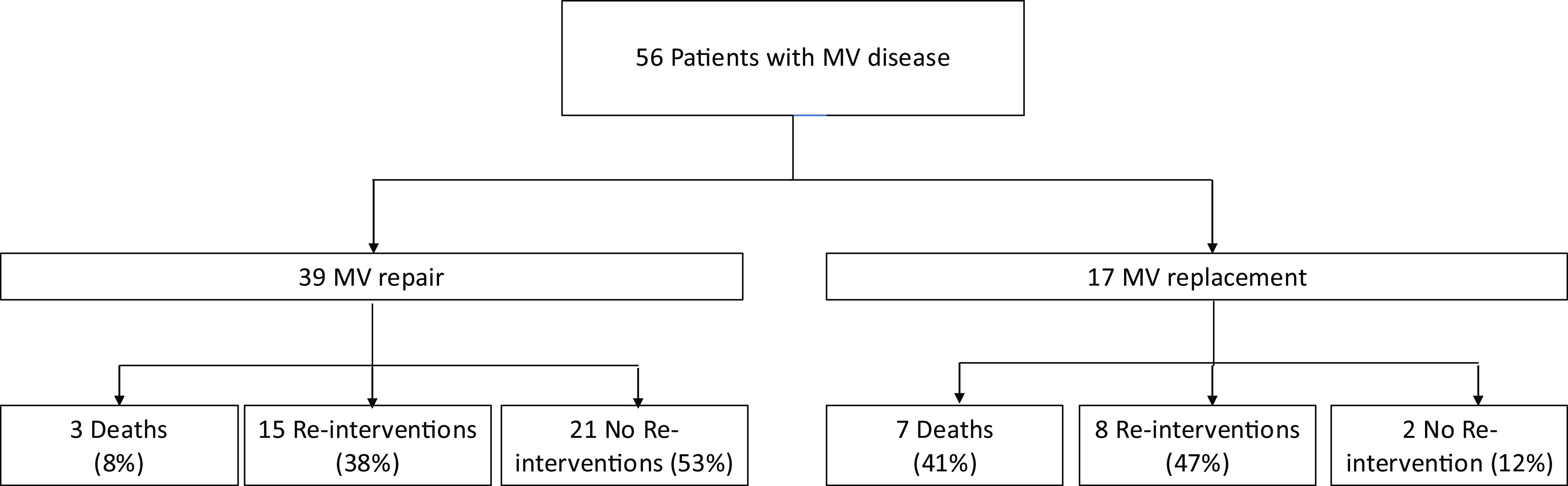
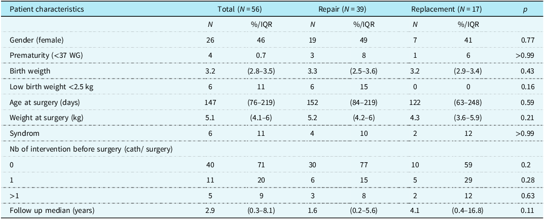
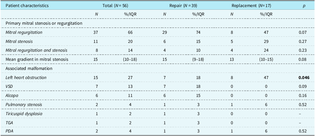
Eighty-five infants who underwent mitral surgery were identified in our database during the study period, with fifty-six patients meeting the inclusion criteria, each included only once based on their initial mitral valve procedure (Figure 1). Patient demographic details are shown in Table 1. The median birth weight was 3.2 kg (interquartile range 2.8–3.5). The median age at surgery was 147 days (interquartile range 76–219), and the median weight at surgery was 5.1 kg (interquartile range 4.1–6). Comissural fusion was the most frequent malformation (50%). Sixteen patients (29%) had previous interventions, either surgery or catheterisation, most commonly for left heart obstruction (coarctation repair, aortic valvuloplasty, or balloon dilation) or for coronary reimplantation in ALCAPA. Left heart obstruction lesions included coarctation of the aorta, aortic valve stenosis (valvular or subvalvular), and, less frequently, hypoplastic left ventricle. Shone syndrome was defined as the association of two or more left-sided obstructive lesions. The median follow-up time was 2.9 years (interquartile range 0.3–8.1). Thirty-nine patients (70%) underwent mitral valve repair, and seventeen underwent mitral valve replacement. There was no significant difference between the populations who underwent mitral valve repair and those who had mitral valve replacement. Cardiac preoperative characteristics are described in Table 2. Thirty-seven patients (66%) had mitral regurgitation, eleven (20%) had mitral stenosis, and eight (10%) had both mitral regurgitation and stenosis. In cases of mitral stenosis, the mean gradient was 15 mmHg (interquartile range 10–18). Associated cardiac malformations were common, including left heart obstruction (27%), ventricular septal defect (13%), and ALCAPA (11%). Left heart obstruction was significantly more frequent in patients who underwent mitral valve replacement (p = 0.046).
Flowchart.

Cohort demographics

Mitral valve characteristics

Surgeries
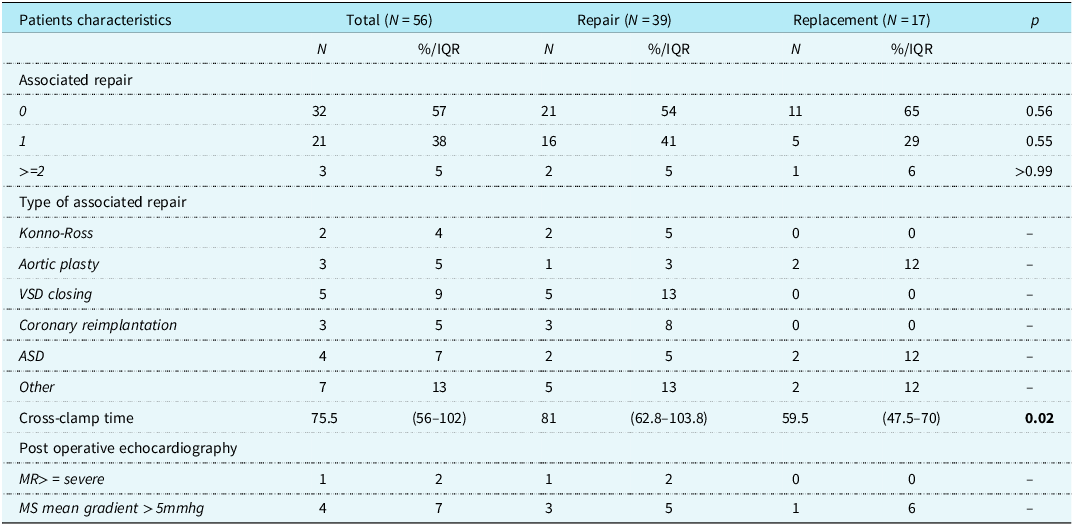
Surgical characteristics are presented in Table 3. Twenty-four patients (43%) underwent additional procedures, such as ventricular septal defect closure (9%), aortic valvuloplasty (5%), and Konno-Ross procedures (4%). Associated surgical procedures were performed in both groups, with no statistically significant difference in their frequency..The median cross-clamp duration was 75.5 minutes (interquartile range 56–102). Cross-clamp time was significantly shorter in patients who underwent mitral valve replacement (p = 0.016). Postoperative echocardiography revealed moderate mitral regurgitation in seven patients (13%), severe mitral regurgitation in one patient and a residual mean gradient in four patients (7%).
Surgical characteristics

Early outcomes
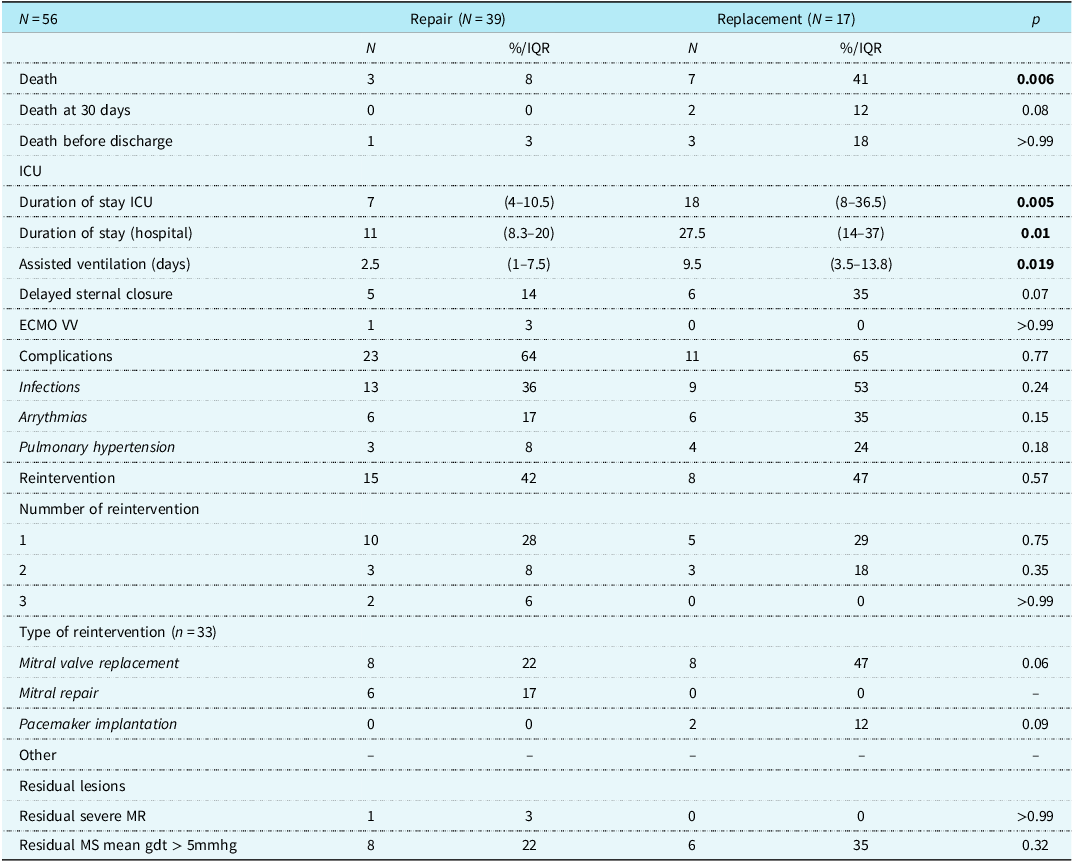
Outcomes are detailed in Table 4. The 30-day mortality rate was 12% for patients who underwent mitral valve replacement and 0% for those who had mitral valve repair. Deaths in the repair group were mainly due to chronic respiratory failure or occurred at home from presumed cardiac causes, whereas in the replacement group, mortality was primarily related to postoperative complications, infection, pulmonary hypertension, or bronchospasm. Patients with mitral valve replacement had significantly longer ICU and hospital stays, and ventilation time compared to those with mitral valve repair (p = 0.005, p = 0.01, and p = 0.019, respectively). The most frequent adverse events were infections and arrhythmias. However, there was no significant difference in the number of complications between the two groups (p = 0.77).
Outcomes

Long-term outcomes
Survival
The Kaplan–Meier plots for overall survival are shown in Figures 2 and 3. Overall survival rates were 96% at 30 days, 86% at 1 year, and 77% at 5, 10, and 15 years; survival was significantly higher in patients who underwent mitral valve repair compared to those who had mitral valve replacement (p = 0.039). In the mitral valve repair group, overall survival rates were 100% at 30 days, 93% at 1 year, and 89% at 5, 10, and 15 years. In contrast, in the mitral valve replacement group, survival rates were 88% at 30 days, 74% at 1 year, and 60% at 5, 10, and 15 years. Additionally, overall survival did not show a significant difference in patients with Shone’s syndrome (Figure 3). In the Cox regression analysis, mitral valve replacement was the only significant risk factor for mortality compared to mitral valve repair (hazard ratio 9.0, 95% confidence interval 1.3–94.8, p = 0.038).
( a ) Kaplan–Meier plot for overall survival stratified by mitral valve repair or replacement. ( b ) Kaplan–Meier plot for re-intervention-free survival stratified by mitral valve repair or replacement.

Kaplan–Meier plot for overall survival stratified by Shone syndrome.

Reintervention
Twenty-three patients (41%) required reintervention: sixteen underwent mitral valve replacement, six had mitral valve repair, and two required pacemaker implantation. One patient died after reintervention (mitral valve replacement). Among patients who required reintervention after mitral repair (n = 15), three (20%) underwent early reoperation within the first three postoperative days (two replacements and one repeat repair). Among the remaining cases, the median time to reintervention was 1.1 years (interquartile range 0.45–6.6), and five patients (41%) underwent redo surgery before the age of one year. Kaplan–Meier curves for reintervention-free survival are shown in Figure 3. In the repair group, reintervention-free survival rates were 81%, 65%, 46%, and 9% at 1, 5, 10, and 15 years, respectively. In the replacement group, corresponding rates were 74%, 54%, 54%, and 38%. Reintervention-free survival did not differ significantly between the repair and replacement groups (log-rank test, p = 0.31). At the last follow-up, one patient had residual severe mitral regurgitation, and fourteen had mitral stenosis with a gradient greater than 5 mmHg.
Discussion
Mitral valve replacement and repair in infants are uncommon procedures, with limited data available on this specific population. This large retrospective single-centre cohort describes both techniques in terms of mortality and reintervention. The two patient groups are not directly comparable, as surgical choice depends on lesion complexity, the surgeon’s expertise, and the acceptable risk of technical failure. While some patients could have undergone either intervention, this study evaluates outcomes based on the selected approach rather than comparing two equivalent strategies.
Population
The study population was comparable to those reported in recent series, Reference Isaacson, Lucjak and Johnson3,Reference Geoffrion, Pirolli and Pruszynski13 with a median age at surgery of 4 months. The distribution of mitral pathologies was similar, with 66% having mitral regurgitation, 20% stenosis, and 14% a combination of both. The cross-clamp time for mitral valvuloplasty was also consistent with published data.
Mortality
Overall mortality in our cohort was 22%, with the majority occurring within the first 2 years following mitral valve repair or replacement. Recent studies across paediatric age groups similarly emphasise a high risk of mortality in children under 2 years old, particularly during the first postoperative year. Reference Sughimoto, Konstantinov, d’Udekem, Brink, Zannino and Brizard12 Beierlein et al. Reference Beierlein, Becker and Yates6 found a 30-day mortality rate of 42% and a 10-year survival rate of only 33% in patients aged 2 years or younger, identifying operative mortality as the key factor contributing to poor long-term survival. A recent study Reference Brancaccio, Trezzi and Croci14 showed that infants and neonates have a threefold risk for death compared to children. In contrast, some authors, Reference Rafii, Davies, Carroll, Quaegebeur and Chen15 have reported no significant differences in survival between younger and older children undergoing mitral valve replacement, with a 10-year survival rate of 82% in patients under 2 years and a 40% freedom from reoperation.
In our study, mortality in the replacement group was very high (41%), consistent with Beierlein Reference Beierlein, Becker and Yates6 findings. Our results differ from those reported by Rafii et al. Reference Rafii, Davies, Carroll, Quaegebeur and Chen15 who found comparable survival between children under and over 2 years of age (median age 1.2 years). The younger median age in our cohort (147 days) likely accounts for the higher early mortality observed after mitral valve replacement, as younger infants are at increased operative risk. The mitral valve repair group showed a lower mortality rate of 8%, aligning with other studies Reference Isaacson, Lucjak and Johnson3 who reported a 10.9% mortality at discharge in neonates undergoing valvuloplasty and a 22% mortality on follow-up. This mortality rate is notably higher than that observed in older children; in the same study, children older than 1 year had a 0% discharge mortality and a 10% mortality at 5 years. Although surgical practices evolved over the 22-year study period, with an increased proportion of mitral repairs after 2012, the overall trends remained consistent, with repair demonstrating lower mortality compared to replacement.”
Regarding mortality risk factors, mitral valve repair versus replacement was the only significant risk factor for survival identified in our cohort. Other risk factors, such as Shone’s syndrome or concurrent surgeries, have been described in the literature Reference Sughimoto, Konstantinov, d’Udekem, Brink, Zannino and Brizard12 but were not significant in our analysis, likely due to the small sample size. These findings underline the current limitations of prosthetic valve replacement in infancy, where mortality often results from postoperative complications and haemodynamic instability. Recent advances in partial heart transplantation, reported for aortic and pulmonary valves, Reference Overbey, Aykut and Kucera16 may open promising avenues for valve replacement by providing living tissue capable of growth and adaptation.
Overall, mitral valve repair demonstrates superior mortality outcomes compared to replacement in this infant population.
Reintervention
Data on reintervention following mitral valvuloplasty in infants are limited.
In older children, the rate of reintervention is relatively low (23%). Reference Kalfa, Vergnat and Ly8 In children under 2 years, cumulative incidence of reoperation at 6 years is higher 38.6%. Reference Mayr, Vitanova and Burri10 In the specific group of infants, Geoffrion et al. Reference Geoffrion, Pirolli and Pruszynski13 observed a high reintervention rate, with a 10-year reintervention-free survival rate of 48.9% following mitral repair or replacement.
In our study, the reintervention rate was high (41% of the cohort) and comparable across both groups. Among those undergoing mitral valve repair, reintervention-free survival was 65% at 5 years, 46% at 10 years. For the mitral valve replacement group, the reintervention-free survival was 54% at both 5 and 10 years and 38% at 15 years. The recent study by Isaacson et al. Reference Isaacson, Lucjak and Johnson3 found no significant difference in reintervention rates between infants and older children following mitral valve repair, with a 5-year reintervention rate of 22% in both groups; however, the study did not extend beyond this period. In our cohort, the majority of reinterventions occurred after 5 years, which may explain the differences observed between the two studies. Of the fifteen patients who underwent reintervention following an initial mitral repair, 6 (40%) had a subsequent mitral repair, indicating that valve replacement can be avoided in some cases.
Surgical aspects
Patch and anterior enlargement
The need for patch enlargement was frequent in our experience. It allows for a reasonable surface of coaptation after splitting the papillary muscles, especially in Hammock valves. This splitting is usually necessary to achieve a large opening of the mitral valve and some mobility of the leaflets. Our first choice is to enlarge the anterior leaflet, as the “gutter effect” is not a concern in this technique, even when the commissures are not fully detached.
Patch nature
The decision to use glutaraldehyde-treated autologous pericardium (treated for 10 minutes) is mainly based on the physical properties obtained after treatment, which are close to those of the native valve. This material also offers the possibility of achieving an immediate good result (with no significant gradient or regurgitation). However, the risk of calcification and subsequent stenosis is high, leading to a significant proportion of failures. In such cases, whenever possible, the patch has been replaced with fresh pericardium, which is slightly more resistant after redo surgery. None of these patients experienced early recurrent failure.
Patch geometry
Most of the patches were tailored with an oval shape rather than a crescent geometry. This allows for a significant surface of coaptation, which is made up of the entire native anterior leaflet. The oval shape often prevents commissural regurgitation, even when the anterior valve has been detached slightly beyond the commissures.
Annuloplasty
Almost all patients underwent posterior plication of the mitral annulus to reduce the initial diameter. We attempted to allow for potential future growth by using two independent strips of PTFE, which are sometimes (for a very dilated annulus) attached together with a resorbable stitch.
Very early failure
Two patients experienced tears in the week following the repair, both of which were located near the commissure on the leaflet. We now systematically use fresh pericardium mattress stitches in this area.
Addressing leaflet prolapse
We generally addressed prolapse by chordae transfer or shortening or by closing the indentations. When these options are not available, we do not hesitate to use artificial PTFE chordae as a second choice.
Repair versus replacement
Our practice has evolved over time, and we are now more aggressive in choosing repair for most patients. We recognise that, for some, this is a palliative approach, but the high risks associated with mechanical valves in very young infants likely make this strategy advantageous. Delaying valve replacement is technically beneficial and has long-term advantages, such as enabling the use of a larger prosthesis and reducing valve-related complications.
Balancing repair and replacement: a need for a more conservative approach?
Our findings suggest that repair should be prioritised whenever feasible, even if it means accepting residual lesions. Given the high mortality associated with early mitral valve replacement, delaying replacement offers key advantages: it allows for patient growth, enabling the use of a larger prosthetic valve, and reduces long-term complications linked to mechanical valves, such as thromboembolism and anticoagulation risks. While not all valves can be repaired, our results reinforce the idea that pushing the limits of valvuloplasty is often preferable to early replacement, especially in borderline cases. The variability in the literature likely stems from selection bias—severe cases naturally require replacement—but the consistently poor outcomes in this group highlight the need for a more conservative approach to valve replacement in infancy. Future progress in infant mitral valve surgery will likely depend on early referral, precise anatomical evaluation, and concentration of expertise in specialised centres.
Limitation
This study has several limitations, primarily due to its retrospective, single-institution design. Our cohort included a heterogeneous population with diverse congenital mitral pathologies, and varied repair techniques were utilised. The relative rarity of mitral valve repair or replacement in infants, combined with the small sample size, limits the statistical power. Some clinically relevant differences did not reach statistical significance for this reason. Another limitation is the relatively short median follow-up (2.9 years) compared to the study period, which may lead to underestimation of late mortality and reintervention rates. The mitral valve replacement cohort has an extended follow-up period, as these procedures were performed during an earlier timeframe. Additionally, the small sample size was insufficient to adequately adjust for potential confounders, such as disease severity. The repair and replacement groups are not directly comparable, as valve replacement was generally indicated in patients with more complex anatomy, for whom repair was deemed unlikely to provide lasting function.”
Conclusion
Mitral valve surgery in infants is associated with significant risk and a high rate of re-intervention Although mitral repair is associated with improved mortality outcomes, it often delays but does not always prevent the need for eventual valve replacement.
Availability of data and materials
All data generated or analysed during this study are included in this published article.
Funding statement
The authors declare that they have no specific funding for this research.
Competing interests
The authors declare that they have no competing interests
Ethical standard
This study has been registered in the General Data Protection Register of the APHP under the registration number: 2024 1024121250. This attests to its compliance with the General Data Protection Register. This retrospective, single-centre study follows MR004 guidelines for data reuse (CNIL-France), where written consent is not required. Informational notices are available upon request, and GDPR compliance is overseen locally by the Data Protection Officer.