Introduction
In recent years, the B12-based ternary compound AlMgB14 has roused great attention owing to its excellent properties such as extremely high hardness, low density, excellent electrical conductivity, good wear resistance, and oxidation resistance [Reference Russell, Cook, Harringa and Lewis1, Reference Cook, Harringa, Lewis and Russell2, Reference Lei, Meng, Zhuang, Chen and Dai3, Reference Tian, Bastawros, Lo, Constant, Russell and Cook4, Reference Zhoua, Xu, Zhang, Baib, Huang, Wu and Luo5]. It was reported [Reference Bahadur and Ahmed6] that the maximum microhardness value of AlMgB14 added with sub-micro TiB2 particles could reach 46 GPa, which is barely less than that of diamonds and cubic nitride boride (c-BN). Ahmed et al. [Reference Ahmed, Bahadur, Cook and Peters7] discovered that the hardness of AlMgB14–TiB2 was increased from 28.2 to 36.8 GPa when 0–70 wt% TiB2 particles were added; moreover, its fracture toughness was enhanced from 2.97 MPa m1/2 to 4.07 MPa m1/2 correspondingly. Because of its excellent properties and lower cost [Reference Kevorkijan, Škapin, Jelen, Krnel and Meden8], AlMgB14 ceramic matrix composites have great potential applications in cutting, grinding, polishing operations, and self-lubricating coating. To improve its brittle nature and enlarge its practical application, AlMgB14 ceramic matrix composites are joined to metal materials such as steel and Ti alloys. Up to now, multiple methods of joining ceramics or ceramic matrix composites to metals have been developed, including brazing [Reference Sciti, Bellosi and Esposito9], diffusion bonding [Reference Kim, Dong, Lim and Kim10], transient liquid bonding [Reference Zhang, Cai, Chen and Xu11], and self-propagation high-temperature synthesis [Reference Yang and Mu12]. However, only a few literatures reported the joining characteristics of AlMgB14 materials. Cook et al. [Reference Russell, Cook, Harringa and Lewis1] found that AlMgB14 could be joined to Fe when heating at 1473 K for 2 h. Zhuang et al. [Reference Zhuang, Lei, Chen, Hu and Meng13] successfully associated AlMgB14–30 wt% TiB2 with Nb and Mo plates by the field-assisted diffusion bonding method. Active metal brazing, as we all know, is a widely used technique for joining ceramics (such as diamond, c-BN, and Al2O3) to metals because of its convenience, cost-effectiveness, and high-quality process [Reference Ahmed, Bahadur, Cook and Peters7]. However, research about the brazing performance of AlMgB14 or AlMgB14 ceramic matrix composites has not been reported to date. In this article, AlMgB14–30 wt% TiB2 (denoted as AMBT) ceramic was brazed to Ti6Al4V alloy (TC4 for convenience) using inactive AgCu eutectic alloy. There are two reasons for using TC4: First, its coefficient thermal expansion (CTE) (8.6 × 10⁻⁶ K−1) is relatively close to that of AlMgB14 (9 × 10⁻⁶ K−1) [Reference Russell, Cook, Harringa and Lewis1], and, second, the AgCu eutectic alloy of lower cost has been successfully used in joining ceramic to titanium alloy. Several studies demonstrated [Reference Zhao, Wang, Cao, Song, Tang and Feng14, Reference Barrena, Matesanz and Salazar15, Reference Zhao, Song, Hu, Zhang, Cao, Fu and Feng16] that the spreading and wettability of AgCu melt on ceramic substrates can be significantly improved when more Ti atoms from TC4 dissolved and diffused into this melt under higher brazing temperature. This process triggered the interfacial reaction between the filler alloy and ceramic substrate. Therefore, AgCu filler alloy foil was used to braze AMBT to TC4 at different temperatures, and the effects of active Ti element in TC4 on the interfacial microstructure and mechanical properties of the AMBT/TC4 joints were investigated.
Results and discussion
Joint microstructure characterization
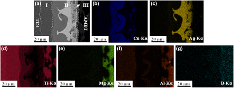
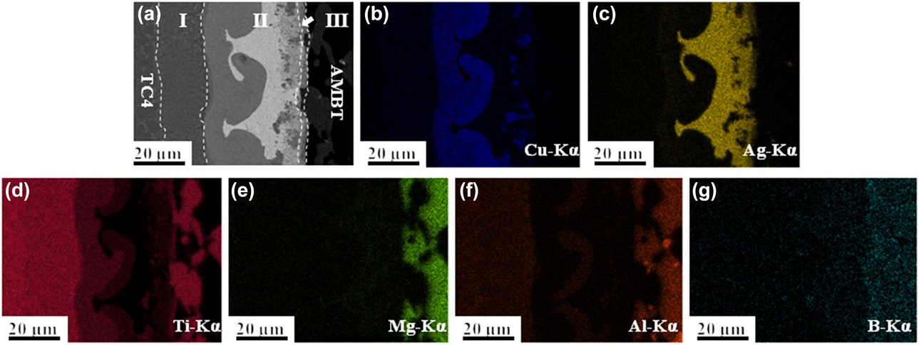
Figure 1 displays the microstructure and the energy-dispersive spectrometer (EDS) compositional maps of the TC4/AMBT joint brazed with AgCu filler alloy at 910 °C for 10 min, in which the brazed joint can be divided into three regions: region I (diffusion zone next to TC4 substrate); region II (the brazing beam); region III (an interfacial reaction layer adjacent to AMBT substrate). In Fig. 1(a), a sound joint without any crack or void can be seen, indicating a good wetting and close contact between TC4 and AMBT. Besides, an array of whiskers perpendicular to AMBT substrate also can be seen. In Figs. 1(b)–1(g), region I mainly consists of Ti element. In the brazing beam, the gray reaction layers bordering region II were primarily composed of Ti and Cu, the remaining phases were the white Ag-rich matrix phase and some gray blocky Cu-rich phase as well as the Ti- rich whisker phase. Region III was a thin reaction layer exhibiting a high Ti character. It needs to be pointed out that the Ti content in this layer is higher than that in the whisker phase.
Typical microstructure and corresponding element distribution images of the TC4/AMBT joint brazed with AgCu filler at 910 °C for 10 min: (a) microstructure; (b–g) element map distributions of Mg, Al, Ti, Ag, B, and Cu.
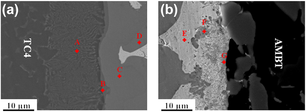
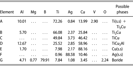
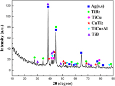
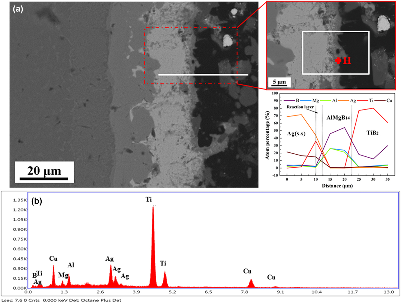
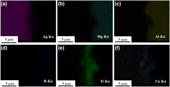
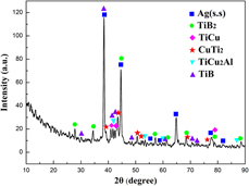
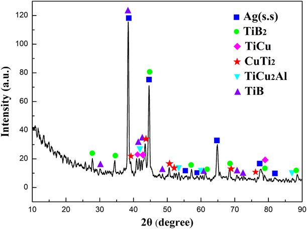
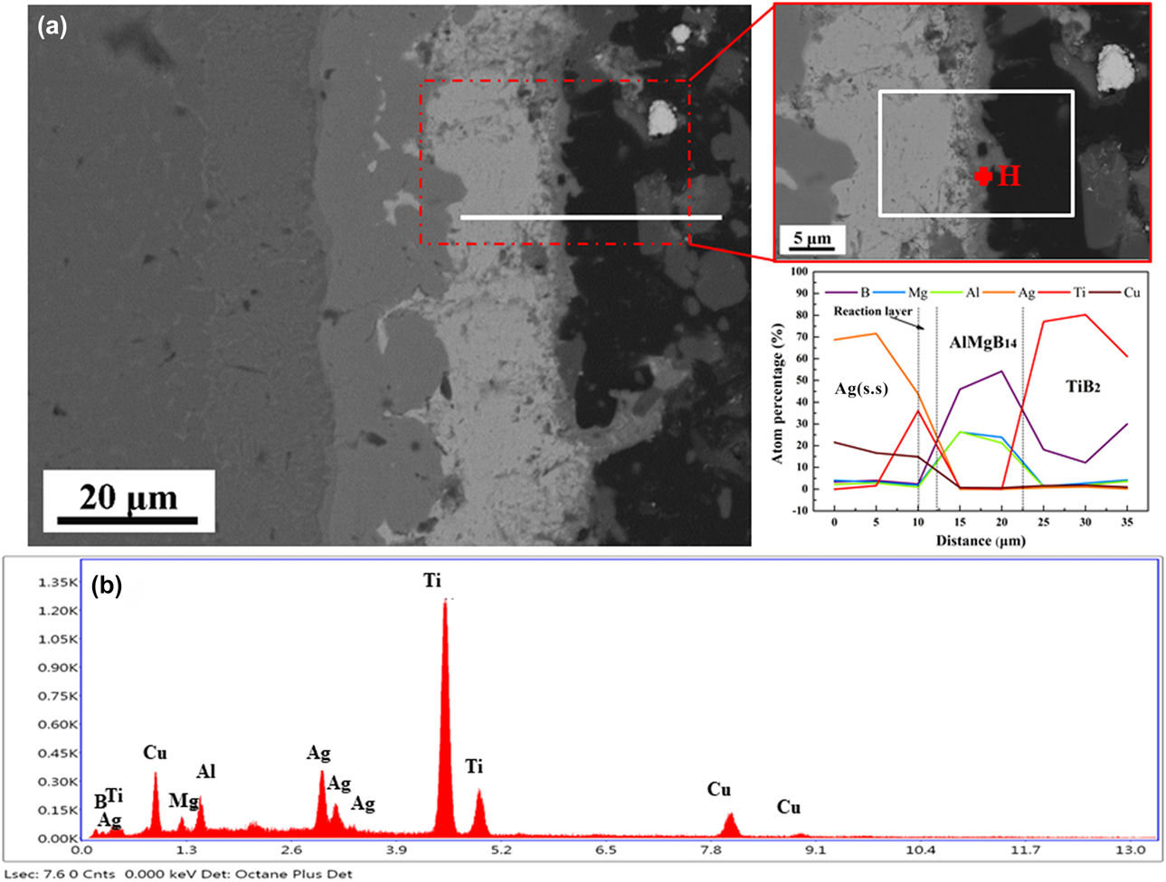
To ascertain the microstructure of the brazed joints, magnified images of a typical bonding interface are displayed in Figs. 2(a) and 2(b). The EDS analysis results of marked points in Fig. 2 are listed in Table I. Combined with chemical compositions in Table I and the Ti–Cu–Al ternary phase diagram [Reference Villars, Prince and Okamoto17], the eutectoid phases in region I were determined as Ti(s.s) + Ti2Cu; the formation of these eutectoid phases was attributed to the decrease in the eutectoid transformation temperature of Ti caused by the diffusion of a little Cu from the AgCu filler into the TC4 substrate [Reference Russell, Cook, Harringa and Lewis1, Reference Qiu, Wang, Yang and Wang18]. The dark layers marked by B–D were confirmed as Ti2Cu, TiCu, and TiCu2Al in sequence, which is in agreement with the literature reported by Qiu et al. [Reference Qiu, Wang, Yang and Wang18], in which formation of these Ti–Cu compound layers was explained by the differences in the Gibbs free energies at different temperatures. The white Ag-rich matrix phase marked by E and the blocky Cu-rich gray phase marked by F are Ag-based solid solution (Ag(s.s)) and Cu-based solid solution (Cu(s.s)), respectively. Region III was the key factor to evaluate the brazing performance of the AMBT to TC4 by the AgCu eutectic filler. The EDS results showed that this reaction layer marked G consisted of B (81.71 at.%), Ti (8.02 at.%), Al (4.73 at.%), and Cu (3.54 at.%). By taking into account the high percentage of the detected B elements, this layer might be deduced as boride. Although the B element is the so-called ultra-light element, quantitative analysis of the element by EDS is difficult. X-ray diffractometer (XRD) analysis of this reaction layer by grounding layer by layer from the TC4 side is given in Fig. 3. The results confirmed that the TiB phase can be detected apart from the formation of Ag(s.s), TiB2, TiCu2Al, and Ti–Cu compounds. Thus, it is important to determine whether the whisker phase or the reaction layer is the TiB phase. Our study found that the reaction layer can be generated at a brazing temperature of 880 °C, whereas the whiskers perpendicular to the reaction layer cannot be detected, as displayed in Fig. 4(a). Elemental distribution of the enclosed white rectangle area [right side of Fig. 4(a)] seen in Fig. 5 indicates that the reaction layer mainly composed of Ti and trace Cu, and free of Al, Mg, and Ag elements can be found in this layer. Distribution of the B element cannot be clearly distinguished in this region, but the elemental composition of this layer marked point H was detected as B (64.64 at.%), Mg (0.55 at.%), Al (1.99 at.%), Ag (2.70 at.%), Ti (23.97 at.%), and Cu (6.15 at.%) by EDS; the corresponding energy spectrum can be seen in Fig. 4(b), which is significantly different from that of the detected AlMgB14 (B 93.84 at.%, Mg 3.41 at.%, and Al 2.75 at.%). According to the atomic ratio of Ti and B and the Ti–B binary phase diagram [Reference Panda and Ravi Chandran19], this reaction can be preliminarily deduced as the TiB2 phase. This deduction of TiB2 formation can be further confirmed by line scanning analysis in Fig. 4(a). The changing trend of Ti and B elements in the reaction layer was similar to that in the gray TiB2 phase which was added into the AlMgB14 matrix as a strengthening phase when neglecting the interference of the Ag element. In addition, the existence of trace Cu could be explained as residual Cu from the brazing filler. For the formation Ti–Cu compounds, Ti atoms in the brazing seam might prefer to react with B in the AMBT substrate because the mixing enthalpy of B–Ti (−42 kJ/mol) is much lower than that of Cu–Ti (−17 kJ/mol) [Reference Fu, Wang, Zhang and Hu20]; as a result, the initially affiliated trace Cu atoms might stay at the reaction region where Ti participated. The formation of TiB2 layer also can be speculated from the perspective of thermodynamic analysis. The possible reactions between Ti and B as well as the Gibbs free energies of each reaction at 880 °C were analyzed by means of an HSC chemistry 6 software, the results of which are illustrated in the following reactions:
 $${\rm{Ti}} + {\rm{Ti}}{{\rm{B}}_2} = 2{\rm{TiB}} \cdot \Delta G = - 52.1\;{{{\rm{kJ}}} / {{\rm{mol}}}}\quad ,$$
 $${\rm{Ti}} + {\rm{B}} = {\rm{TiB}} \cdot \Delta G{\rm{\ }} = - 165.4\;{{{\rm{kJ}}} / {{\rm{mol}}}}\quad ,$$
 $${\rm{Ti}} + 2{\rm{B}} = {\rm{Ti}}{{\rm{B}}_{\rm{2}}} \cdot \Delta G{\rm{\ }} = - 260.8\;{{{\rm{kJ}}} / {{\rm{mol}}}}\quad .$$
High magnification BSE images of the interfacial microstructure of the TC4/AMBT joint: (a) TC4–braze alloy interface; (b) braze alloy–AMBT interface.

Chemical compositions (at.%) and possible phases of each spot marked in Fig. 4.

XRD pattern of the reaction layer adjacent to AMBT.
EDS analysis of the reaction layer of the joint brazed at 880 °C: (a) microstructural morphology of the TC4/AMBT joint and line scanning result of the reaction; (b) EDS pattern of marked point H.
Elemental distribution of the enclosed rectangle region in Fig. 4(a). (a) Al; (b) Mg; (c) Al; (d) B; (e) Ti; (f) Cu.

It can be seen that owing to its lowest negative Gibbs free energies, reaction (3) is the most possible to occur than reaction (1) at 880 °C. According to Le Chatelier’s principle, reaction (1) might also take place because of its negative Gibbs free energies when Ti content is in excess. The TiB phase existing like whiskers has been widely reported as a result of the reaction between the diboride phase and Ti [Reference He, Yang, Lin, Jia, Feng and Liu21, Reference Yang, Wang, Wang, Zhang, Wang and Feng22]; the at.% of Ti in TiB is lower than that in TiB2. Based on this discussion, it can be concluded that the reaction layer is the TiB2 phase and the whiskers formed in the joint when brazing at 910 °C is TiB, which can be detected by XRD. Above all, the microstructure of the TC4/AMBT joint brazed with the AgCu eutectic filler alloy at 910 °C for 10 min can be determined as TC4/Ti(s.s) + Ti2Cu/Ti2Cu/TiCu/TiCu2Al/Ag(s.s) + Cu(s.s)/TiB whiskers/TiB2 reaction layer/AMBT.
Evolution of joint interlayer microstructure
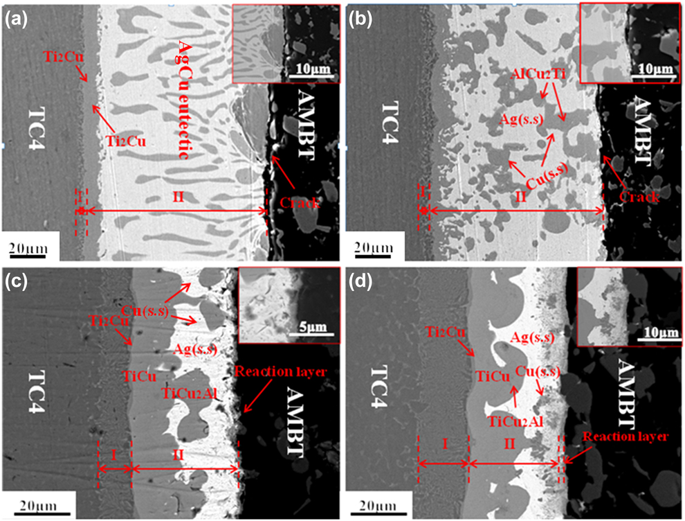
Figure 6 shows the interlayer microstructure of the TC4/AMBT joints brazed at different temperatures for 10 min. It can be seen in Fig. 6(a) that the brazing seam (region II) mainly presented a typical AgCu eutectic structure except Ti2Cu: TiCu intermetallic layers with a total width of 6.7 μm were formed bordering region I when brazing at 820 °C. The thicknesses of regions I and II were about 3.3 μm and 103.3 μm, respectively. In addition, it is noteworthy that a visible crack along the interface between the AMBT and filler alloy can be seen in Fig. 6(a), indicating the poor wettability of the AMBT substrate by the filler alloy. This phenomenon can be explained as follows: Because the partial enthalpy of Ti solution in the molten Cu (−10 kJ/mol) was lower than that of Ag (39 kJ/mol) [Reference Qiu, Wang, Yang and Wang18], Ti had a strong tendency to affiliate and react with Cu rather than Ag. Because the quantity of dissolved and diffused active Ti in TC4 was limited at lower brazing temperatures, only a part of Cu in the molten AgCu diffused toward TC4 and reacted with Ti. As a result, few Ti–Cu compound layers were generated bordering region I. The rest of the molten liquid metal formed AgCu eutectic structure in the seam during cooling. Because of no more active Ti in the molten alloy, the AMBT cannot be wetted by the AgCu alloy, and it is difficult to achieve a metallurgical bonding between AMBT and filler alloy [Reference Yang, Zhang, Chen, Qi, He and Feng23]. Besides, the high residual stresses yielded by a significant difference in the CET mismatch of the AgCu alloy (18.5 × 10⁻⁶ K−1) [Reference Asthana and Singh24] and AMBT (8.64 × 10⁻⁶ K−1) might have contributed to the formation of the crack.
Microstructures of the TC4/AMBT joints brazed at different temperatures for 10 min: (a) 820 °C; (b) 850 °C; (c) 880 °C; and (d) 910 °C.
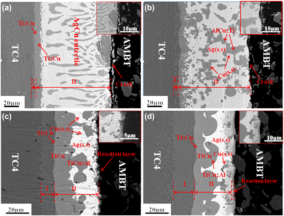
When the brazing temperature rose to 850 °C [Fig. 6(b)], the brazing seam (region II) was mainly composed of distributed blocky Cu(s.s), darker TiCu2Al over Cu(s.s), and a white Ag(s.s) matrix. The thickness of region I increased from 3.3 to 7.5 μm, whereas that of region II slightly decreased from 103.3 to 94.5 μm; the same crack existed as shown in Fig. 6(a). It can be deduced that almost all of the Ti atoms from TC4 were consumed by reacting with Cu in the AgCu molten liquid phase, although the quantity of Ti atoms that diffused into the molted AgCu alloy increased at 850 °C. As a result, the quantity of Ti atoms that could promote the wettability of the AMBT was inadequate and the blocky TiCu2Al and Cu(s.s) precipitated from the molten liquid phase during the cooling process as described by the following reaction [Reference Liu, Liu, Zhang, Tao, Zhang and Qi25]:
 $$L = {\rm{Cu}}\left( {{\rm{s}}{\rm{.s}}} \right) + {\rm{TiC}}{{\rm{u}}_2}{\rm{Al}}\quad .$$
When the brazing temperature was further increased to 880 °C, the brazing seam was made up of three gray Ti–Cu compound layers (in the sequence of Ti2Cu, TiCu, and TiCu2) and a white Ag (s.s) matrix with a few blocky TiCu2Al and Cu(s.s) over it, as displayed in Fig. 6(c). It was noted that a continuous TiB2 layer with a thickness of 1.1 μm formed at the interface of the brazing filler and AMBT substrate, and no crack was found. Region I was obviously thickened from 6.29 μm in Fig. 6(b) to 11.45 μm, and the share of Ti–Cu compound layers in region II was boosted significantly, whereas the thickness of region II was remarkably decreased from 94.5 to 39.5 μm. With improving the brazing temperature, a great number of Ti atoms dissolved and diffused into the molten filler to react with Cu, which resulted in large amounts of Ti–Cu compound layers gathering at the border of region I [Reference Zhao, Wang, Cao, Song, Tang and Feng14, Reference Barrena, Matesanz and Salazar15, Reference Qiu, Wang, Yang and Wang18]. Besides, because of the lower negative mixing enthalpy of Ti–B, Ti atoms might attach and react with the AMBT substrate. Consequently, the wettability and spreading of the molten alloy on AMBT remarkably improved; the width of the brazing seam dramatically decreased because the massive molten filler was squeezed out of the brazing seam [seen in Fig. 8(b)]. A few blocky TiCu2Al and Cu(s.s) were precipitated in Ag(s.s) during the cooling process as mentioned earlier.
When brazing at 910 °C, the morphology of the brazing seam was generally the same as that in Fig. 6(c), except that a few Cu(s.s) were detected over Ag(s.s). The thickness of region I increased from 11.45 to 17.39 μm, and the share of Ti–Cu compound layers in the brazing beam further increased accordingly. Only a few Cu(s.s) precipitated at Ag(s.s) during the cooling process because a great number of Ti and Al atoms diffused toward both substrate sides. The TiB2 reaction layer did not change but formed an array of TiB whiskers with the length of about 10 μm.
To conclude, the microstructure of AMBT/TC4 joints can be controlled by the dissolution and diffusion of active Ti elements in TC4 at different brazing temperatures. When the brazing temperature is above 880 °C, the wettability of the AMBT substrate can be remarkably enhanced because of the affinity and reaction of much Ti from TC4 with the AMBT substrate. Additionally, Ti was also a crucial factor in the formation of a TiB2 reaction layer, realizing the metallurgical combination between the filler alloy and AMBT substrate.
Joint mechanical properties
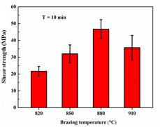
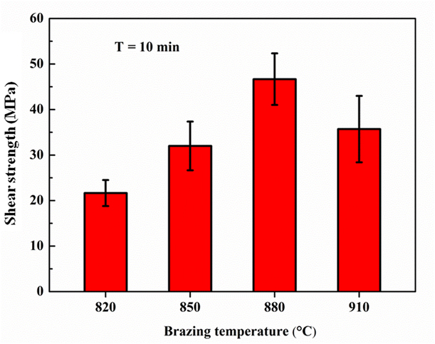
It is essential to investigate the effect of brazing temperatures on the strength of TC4/AMBT joints because the microstructures of brazed joints are considered to be decisive for the strength of the joints. Figure 7 depicts the relationship between the average shear strength and the brazing temperature for the TC4/AMBT joints brazed with AgCu filler alloy. It can be seen that the shear strength of TC4/AMBT joints increased from 21.16 to 46.67 MPa with the brazing temperature increased from 820 °C to 880 °C, and then dropped to 35.7 MPa when brazing at 910 °C. The obtained maximum shear strength was 46.67 MPa at 880 °C for 10 min.
Effect of brazing temperature on the shear strength of TC4/AMBT joints brazed for 10 min.
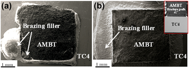
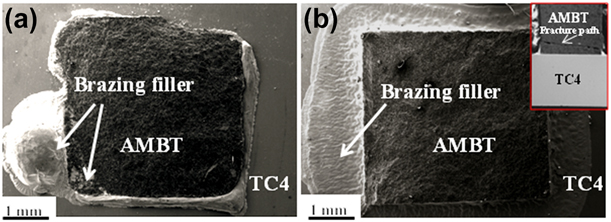
The variation in shear strength is highly correlated with the microstructure and fracture morphology of the brazed joints. When brazing was carried out at 820 and 850 °C, almost no active Ti from TC4 diffused onto the surface of the AMBT substrate, which resulted in the AMBT not being wetted thoroughly by the brazing filler, as seen in Fig. 8(a). The crack [in Figs. 6(a) and 6(b)] existing between the brazing filler and AMBT substrate was detrimental to the joint strength because the fracture was initiated at this unbounded interface and extended into the AMBT substrate under the loading force, as shown in Fig. 8(a), in which the brazing filler was detected on the fracture surface. However, when the brazing temperature was increased to 880 °C, the formation of much brittle Ti–Cu intermetallic layers in the brazing seam was accompanied with the generation of a TiB2 layer at the interface of the AMBT and brazing filler. Formation of this layer could realize a metallurgical combination between the filler alloy and AMBT substrate. In Fig. 8(b), the fracture morphology and path of brazed joints show that the fracture merely happened at the AMBT substrate rather than at the brittle layers in the seam, which indicates that the strength of the AMBT–brazing filler interface is higher than that of the AMBT substrate. Therefore, the joint with the maximum shear strength of 46.67 MPa was obtained. When the brazing temperature was further increased to 910 °C, the fracture morphology and position of the brazed joint were the same as that of the joint brazed at 880 °C, but the shear strength dropped to 35.7 MPa. This phenomenon can be explained by the residual stress. The fracture propagated inside the AMBT substrate, which indicates that the residual stress in the joint lowered the property of the AMBT near the brazing seam [Reference Mendez, Park and Eagar26]. Moreover, the plastic deformation ability of the brazing seam degraded when Ti–Cu intermetallic layers became thicker and the width of the brazing seam further dropped at higher brazing temperatures [Reference Shiue, Wu and Chen27, Reference He, Zhang, Sun and Liu28, Reference Wang, Zhang, Liu and Wang29]; thus, the brazed joint strength dropped because the residual stress could not be controlled by plastic deformation.
Fracture morphologies of TC4/AMBT joints brazed at different temperatures for 10 min: (a) 820 °C; (b) 880 °C.
Conclusions
AMBT was successfully brazed to TC4 using active Ag72–28Cu (wt%) filler alloy, and the influence of brazing temperature on the microstructure and mechanical properties of the AMBT/TC4 joints were studied. Primary conclusions can be drawn as follows:
(1) The typical microstructure of the TC4/AMBT joint is TC4/Ti(s.s) + Ti2Cu/Ti2Cu/TiCu/TiCu2Al/Ag(s.s) + Cu(s.s)/TiB whiskers/TiB2 reaction layer/AMBT.
(2) With increasing the brazing temperature, the diffusion region of the TC4 became thicker, whereas the width of the brazing seam dropped significantly for the dissolution and diffusion of active Ti from the TC4 substrate. When the brazing temperature was over 880 °C, Ti–Cu intermetallic compound layers in the sequence of Ti2Cu, TiCu, and TiCu2Al, and a few distributed Cu(s.s) among Ag(s.s) formed in the brazing seam. Meanwhile, a continuous TiB2 reaction layer was generated between the brazing filler and AMBT substrate.
(3) The shear strength of TC4/AMBT joints increased first and then dropped when the brazing temperature was above 880 °C. The maximum strength of the brazed joint was 46.7 MPa at the brazing temperature of 880 °C for 10 min.
Experimental
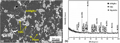
Commercial AlMgB14–TiB2 ceramic was provided by Quanzhou Qi Jin New Materials Co., Ltd., Quanzhou, China. AlMgB14 (mean particle size 2.1 μm, >95% purity) and 30 wt% TiB2 (mean particle size 3–5 μm, >99% purity) powders were mixed and subsequently hot-pressed at 1400 °C for 15 min under the pressure of 50 MPa in a vacuum furnace. The relative density of the prepared AlMgB14–TiB2 ceramic was 87%. Figure 9 shows the microstructure and XRD pattern of the AMBT, and the black matrix and gray-white particles are AlMgB14 and TiB2, respectively. Besides, a few pores existed in the AMBT. By using wire electro-discharge machining, AMBT and TC4 were cut into pieces with the dimension of 4 mm × 4 mm × 4 mm and 10 mm × 5 mm × 3 mm, respectively. The AMBT and TC4 pieces were ground with 800-grit SiC papers and polished with diamond pastes. A commercially inactive Ag72Cu28 (wt%) alloy foil with a thickness of 100 µm was tailored into the same size of AMBT pieces and sandwiched between the AMBT and TC4. A normal load of 1.5 KPa was applied to keep the surface of the couples in close contact. Before assembling, all of the specimens were ultrasonically cleaned in acetone for 20 min and dried in air. Brazing experiments were carried out in the temperature range of 820–910 °C for 10 min with a vacuum of 3.0 × 10⁻³ Pa. The specific joining process was as follows: The assembly was heated to 710 °C at 20 °C/min and kept for 10 min; then further heated to the brazing temperature at 5 °C/min and isothermally held for 10 min; and subsequently cooled down to 200 °C at 5 °C/min. The interfacial microstructure of the brazed joints was characterized by a field-emission scanning electron microscope (FESEM, ZEISS) equipped with an energy-dispersive spectrometer (EDS, EDAX, Oberkochen, Germany). The formed phases in the joints were identified by an X-ray diffractometer (XRD, DX-2700, Dandong Hao Yuan Instrument Co., Ltd., Dandong, China) with Cu Kα radiation. The bonding properties of the brazed joints were evaluated by a universal testing machine (Instron 5967, Instron Corporation, Norwood) at a replacement speed of 0.5 mm/min. At least five samples were tested to determine the joint shear strength under the same conditions.
Characterization of the AMBT sample: (a) secondary electron image; (b) XRD diffraction pattern.

Acknowledgments
This project is supported by the National Natural Science Foundation of China (Grant Nos. 51775138, U1537206, and U1737205) and the Key Research & Development program of Shandong Province (No. 2017GGX40103).