Introduction
Cornudiscoides Kulkarni, Reference Kulkarni1969 is a small monogenoid belonging to Ancylodiscoidinae Gusev, Reference Gusev1976, Dactylogyridae Bychowsky, 1933, from locations in India, Sri Lanka, Malaysia and Thailand (Lim et al., Reference Lim, Timofeeva and Gibson2001). According to the classification of Kritsky & Boeger (Reference Kritsky and Boeger1989) Dactylogyridae comprised nine subfamilies including Ancylodiscoidinae. Lim et al. (Reference Lim, Timofeeva and Gibson2001) raised Ancylodiscoidinae to family level as Ancylodiscoididae supported by Bychowsky & Nagibina (Reference Bychowsky and Nagibina1978) (Šimková et al., Reference Šimková, Plaisance, Matejusová, Morand and Verneau2003). Later, Mendoza-Palmero et al. (Reference Mendoza-Palmero, Balsco-Costa and Scholz2015) suggested the revision of the taxonomic status of Ancylodiscoidinae and Ancylodiscoididae, due to the lack of phylogenetic support. Cornudiscoides was established by Kulkarni along with three species, C. heterotylus (type species), C. microtylus and C. megalorchis, from Mystus tengara (Hamilton, 1822) at Hyderabad, Andhra Pradesh. This parasite extensively parasitizes the members of Bagridae, especially Mystus Scopoli, 1777 and Sperata Holly, 1939. According to the IUCN Red List, Mystus (Ng, Reference Ng2010) and Sperata (Rema & Raghavan, Reference Rema and Raghavan2013) are widely utilized, both as food and as ornamental fish.
In 1976, Gusev described two new species of Cornudiscoides – C. proximus and C. geminus – from Mystus vittatus and transferred Ancylodiscoides jaini Gusev, 1963 to Cornudiscoides as C. jaini. Much later Dubey et al. (Reference Dubey, Gupta and Agarwal1992) recorded C. geminus and reported two new species, C. raipurensis and C. vittati. Agrawal & Vishwakarma (Reference Agrawal and Vishwakarma1999) added six new species, viz. C. tukarami, C. gussevi, C. bleekerai, C. susanai, C. gomtai and C. agarwali, along with re-description of C. proximus and C. geminus. Agrawal & Vishwakarma (Reference Agrawal and Vishwakarma1999) re-examined C. raipurensis, which differed in body ratio and size of dorsal anchors compared with C. proximus, and confirmed that it was C. proximus, along with synonymization of Neomurraytrema shuklai (Agrawal and Singh, 1985) and Neomurraytrema lucknowensis (Agrawal and Sharma,1988) as C. proximus and C. geminus, respectively. Lim et al. (Reference Lim, Timofeeva and Gibson2001) validated 17 species of Cornudiscoides in which 11 species (including C. raipurensis synonymized as C. proximus by Agrawal & Vishwakarma, Reference Agrawal and Vishwakarma1999) were described from India and six from Malaysia. Devak & Pandey (Reference Devak and Pandey2007) listed 19 known species of Cornudiscoides. However, Pandey & Agrawal (Reference Pandey and Agrawal2008), in their monograph based on traditional morphological observations, recorded only 12 Indian species. Subsequently, Chaudhary & Singh (Reference Chaudhary and Singh2011), while establishing the phylogeny of C. proximus using partial sequences of 28S rDNA, listed 14 Indian species.
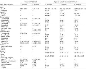
Important diagnostic features, such as sclerotized structures of the haptor and copulatory complex, may show variations in their morphology depending upon the age of the parasite, season, locality and geographic distribution of the host, proving that morphological studies alone are not completely reliable (Gilmore et al., Reference Gilmore, Cone, Lowe, King, Jones and Abbot2012). In the present work, the authors studied three species of Cornudiscoides – C. proximus, C. geminus and C. agarwali – which are very common compared to other known species. The haptoral region is more or less similar in all three species (present study) but the male copulatory complex has significant variations (comparative measurements are given in table 1). The inter-specific differentiation on the basis of the male copulatory complex is common in species of Ancylodiscoidinae (Gusev, Reference Gusev1976; Lim et al., Reference Lim, Timofeeva and Gibson2001; Wu et al., Reference Wu, Zhu, Xie, Wang and Li2007). No molecular study has been performed apart from the involvement of 28S rDNA for molecular characterization of C. proximus (Chaudhary & Singh, Reference Chaudhary and Singh2011). Sequences of large ribosomal subunit have been used successfully to study the phylogenetic relationships of monogenoideans at higher levels (Mollaret et al., Reference Mollaret, Jamieson, Adlard, Hugall, Lecointre, Chombard and Justine1997, Reference Mollaret, Jamieson and Justine2000; Jovelin & Justine, Reference Jovelin and Justine2001; Olson & Littlewood, Reference Olson and Littlewood2002). Meanwhile, other researchers have shown that the 18S rRNA gene is very useful in molecular taxonomy and phylogenetic studies of parasites (Matejusová et al., Reference Matejusová, Koubková, Amelio and Cunniungham2001) along with the usefulness of its secondary structure to establish phylogeny (Cunningham et al., Reference Cunningham, Aliesky and Collins2000). The inclusion of two ribosomal subunit sequences complemented very efficiently the traditional morphometric analyses and phylogenetic study of C. proximus, C. geminus and C. agarwali. Also, it is worth mentioning here that establishing the distinction of congeners using two molecular markers together is a first endeavour in freshwater monogenoideans.
Comparative measurements of three species of Cornudiscoides; L, length; W, width.

Methods
Collection of monogenoideans
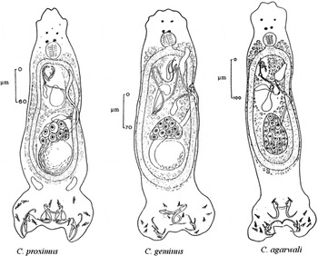
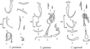
The three species of Cornudiscoides were collected from the gills of Mystus vittatus (Hamilton, 1822). Hosts were caught during April–May 2015 from the River Gomti (a tributary of the Ganges) and purchased from a local fish market at Lucknow (26°8′N 80°9′E). The fish specimens were identified using Fish base (Froese & Pauly, Reference Froese and Pauly2015) and Jayaram (Reference Jayaram1955). Out of 79 examined hosts, 49 were infected with worms. Live hosts were sacrificed, gills excised and transferred into Petri dishes containing water. Living worms were studied under a phase-contrast microscope (Olympus CX41, Tokyo, Japan). The parasites were identified with the help of Pandey & Agrawal (Reference Pandey and Agrawal2008). Comparative figures and measurements of the three species are shown in figs 1 and 2, and table 1. A total of 98 C. proximus, 78 C. geminus and 59 C. agarwali were collected and stored in 100% alcohol for the molecular work.
Whole mounts of three species of Cornudiscoides (Agrawal & Vishwakarma, Reference Agrawal and Vishwakarma1999).

Hard parts of three species of Cornudiscoides. A, Dorsal anchor with patch; B, ventral anchor; C, copulatory complex; D, dorsal bar; E, ventral bar; F, hooks; G, vaginal aperture (Agrawal & Vishwakarma, Reference Agrawal and Vishwakarma1999).

Molecular analysis
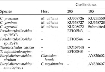
Genomic DNA was extracted from ethanol-preserved specimens using DNeasy Tissue Kit (Qiagen, Hilden, Germany) according to the manufacturer's protocol. The polymerase chain reaction (PCR) was used to amplify the partial ribosomal DNA from the 28S region, using the primer Ancy55F forward (5′-GAGATTAGCCCATCACCGAAG-3′) and LSU1200R reverse (5′-GCATAGTTCACCATCTTTCGG-3′) (Plaisance et al., Reference Plaisance, Littlewood, Olson and Morand2005) and universal primers forward (5′-ACCCGCTGAATTAAGCAT-3′) and reverse (5′-CTCTTCAGACTTTTCAAC-3′) (Shrivastava et al., Reference Shrivastava, Agrawal and Upadhyay2013). The 18S region was amplified using the forward primer worm A (5′-ACGAATGGCTCATTAAATCAG-3′) and reverse primer worm B (5′-CTTGTTACGACTTTTACTTCC-3′) (Plaisance et al., Reference Plaisance, Littlewood, Olson and Morand2005). A PCR reaction volume of 25 μl contained 1 × PCR buffer (Invitrogen, California, USA), 1.5 mm of MgCl2 (Invitrogen), 200 μm of each deoxynucleoside triphosphate (dNTP) (Promega, Wisconsin, USA), 0.4 μm of each primer, 2.5 U of Taq polymerase (Invitrogen) and 5 μl of genomic DNA. The thermal cycle started with 3 min at 94°C for initial denaturation; followed by 35 cycles of 30 s at 94°C, 30 s at 52°C for annealing, and 2 min at 72°C (extension); and a final extension at 72°C for 10 min followed by cooling at 4°C. PCR products were examined on 1% agarose gel, stained with ethidium bromide and visualized on a gel documentation system. The PCR products were purified and sequenced by Xcelris Labs Limited, Ahmadabad, using Big Dye Terminator version 3.1 Cycle sequencing Kit (Applied Biosystems, California, USA). Six new sequences were deposited to the GenBank database (table 2). Sequences of two genera, Pseudancylodiscoides Yamaguti, 1963 and Thaparocleidus Jain, 1952 (table 2), of the same subfamily, were also retrieved for the present analysis, as data for both the subunits were available in GenBank.
GenBank accession numbers of three species of Cornudiscoides and other species of Dactylogyridae included in this study.

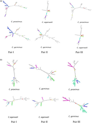
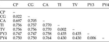
BLASTn (National Center for Biotechnology Information; NCBI) was performed for partial sequences of both 28S and 18S rDNA to uncover the degree of resemblance between species. Separate alignments for two regions were executed with Clustal Omega software (Sievers et al., Reference Sievers, Wilm, Dineen, Gibson, Karplus, Li, Lopez, McWilliam, Remmert, Söding, Thompson and Higgins2011) using the default option. The ATGC count was calculated with the help of ATGC calculator. Motif-based comparison and alignments have been computed for both larger and smaller subunits to find best sequence motifs, common in two species, using the Freiburg RNA Tool (Smith et al., Reference Smith, Heyne, Richter, Will and Backofen2010) (fig. 3a and b). Phylogenetic analysis was conducted with the help of MEGA version 6.06 software (Tamura et al., Reference Tamura, Stecher, Peterson, Filipski and Kumar2013). Each dataset was analysed through neighbour joining (NJ), minimum evolution (ME), unweighted pair group method with arithmetic mean (UPGMA) and estimated p-distance value methods. Substitution, including transitions, transversions, gaps and missing data, were decimated. In the analysis the codon positions (first, second and third) and non-coding sites were also included. Bootstrap values were calculated on the basis of 1050 replicates for 18S and 28S rDNA molecular datasets. The pairwise comparisons were performed using the MEGA version 6.06 tool and the genetic distance was estimated with the help of p-distance (table 3) method for both the genes.
Secondary structures from the ExpaRNA output showing regions of exact pattern matches (EPMs) with the same colour for pairs of species; (a) 28S and (b) 18S regions.

A comparison of sequence differences (%) of species of Ancylodiscoidinae for large subunits.

CP, C. proximus; CG, C. geminus; CA, C. agarwali; TI, Thaparocleidus infundibulovagina; TV, Thaparocleidus varicus; PY3, Pseudoancylodiscoides sp. 3; PY4, Pseudoancylodiscoides sp. 4.
Results
Molecular characterization
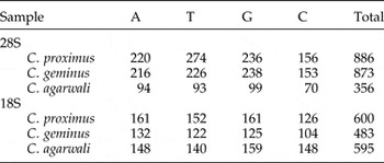
The analyses of two different regions were computed for three species of Cornudiscoides. Approximately 985 bp and 650 bp of large subunit and small subunit sequences, respectively, were amplified for C. proximus, C. geminus and C. agarwali. Nucleotide BLAST for the 28S gene of C. proximus, C. geminus and C. agarwali revealed the highest similarity (91%) with Bifurcohaptor indicus (JX852710). Similarly, BLASTn (NCBI) for the 18S gene of both C. proximus and C. geminus expressed 94% proximity with Euryhaliotrematoides triangulovagina (AY820608) whereas, C. agarwali showed 95% identity with Haliotrema macasarensis (EU836228). The newly sequenced single stranded ribosomal DNA (ssrDNA) alignment with other ssrDNA sequences displayed an homologous region in DNA and expressed similarity among them, while insertions and deletions exposed inter-specific genotypic differences. In the 28S region sequence alignment of 278 positions, 243 sites showed homology while 35 sites were variable and no deletions were observed. In the 490 aligned sites of the 18S region of Cornudiscoides, 380 similar sites along with 98 variable sites and 12 deletions were found. The nucleotide variability of 28S and 18S rDNA within the three species is shown in table 4.
Composition of nucleotide sequences in partial 28S and 18S rDNA of three species of Cornudiscoides.

Secondary structures
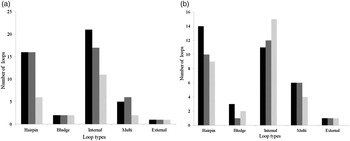
rRNA secondary structures contain a variety of loops, such as stem-loop or hairpin loop, multi-loop, internal loop, external loop, stacked base pair and external bases (Lyngsø et al., Reference Lyngsø, Zuker and Pedersen1999). The types of loops present in 28S and 18S regions of C. proximus, C. geminus and C. agarwali are shown in fig. 4. Pairwise motif identification showed conserved regions between species of Cornudiscoides. ExpaRNA analyses identified exact pattern matches (EMPs) between species pairs, displayed in different colours (fig. 3a and b). Small ribosomal subunits have more EMPs than large ribosomal subunits, besides these observations the secondary structure of the three species in both regions (18S and 28S) have some common folding patterns. All these observations give a clear-cut interrelationship among the three species.
Types of loops present in (a) 28S and (b) 18S regions of Cornudiscoides proximus (black bars), C. geminus (dark grey bars) and C. agarwali (light grey bars).

Phylogenetic analysis
Six sequences of Cornudiscoides along with six sequences retrieved from GenBank (table 2) were used to evaluate their phylogenetic relationship with respect to species of Thaparocleidus and Pseudoancylodiscoides, because these two species are very similar to Cornudiscoides. Thaparocleidus differs from Cornudiscoides in terms of a divided ventral bar or one connected by a thin median ligament and one pair of long marginal hooks, while the presence of modified needle-like hooks in Cornudiscoides distinguished it from Pseudoancylodiscoides (Lim et al., Reference Lim, Timofeeva and Gibson2001; Wu et al., Reference Wu, Zhu, Xie, Wang and Li2007).
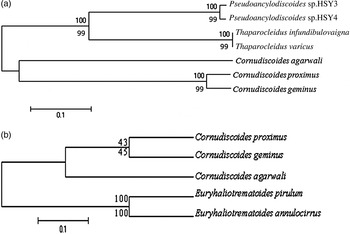
The phylograms for 28S and 18S rDNA regions have similar topology according to NJ, ME and UPGMA methods. The bootstrap values are different for the two regions; in the case of 28S, the values are 99/100, which are of high significance, but the values 43/45 for 18S are poorly significant (bootstrap values greater than 50% are well supported). The phylogenetic tree for the 28S region shows that all three species of Cornudiscoides analysed herein cluster together, forming a sister group with species of Thaparocleidus and Pseudoancylodiscoides (all being members of the Ancylodiscoidinae) (fig. 5a). For phylogenetic analysis of the 18S region, Euryhaliotrematoides species behave as an outgroup with respect to Cornudiscoides species, clustering in a clade (fig. 5b).
Tree topology for rDNA for members of the Ancylodiscoidinae to show (a) 28S and (b) 18S regions, using NJ and ME methods. Bootstrap values support 1050 replicates below the nodes for NJ and above for ME.

Discussion
The partial sequences of 28S and 18S rRNA genes were used to assess the genetic differentiation of three species of Cornudiscoides and their phylogenetic relationship among other groups of monogenoidean parasites of siluriform fish. The large and small ribosomal subunits are extremely useful to explain the phylogeny of monogenoideans at the level of family and subfamily (Šimková et al., Reference Šimková, Matejusová and Cunniungham2006), and the nucleotide sequences of monogenoideans have sufficient phylogenic information to decode the relationship among them (Cunningham et al., Reference Cunningham, Aliesky and Collins2000).
In the present study, all the three species of Cornudiscoides are very similar regarding haptoral structures (bilobed haptor, dorsal anchor, ventral anchor, dorsal bar, paired ventral bar and two types of marginal hooks) but dissimilar regarding the male copulatory complex and median ligament (present only in C. proximus) (figs 1 and 2).
For the first time, we have attempted the comprehensive analyses of three species of Cornudiscoides, using 28S and 18S rDNA partial sequences to show the genetic variation among them. Different sets of primers amplified a 28S region of 886 bp, 873 bp and 356 bp of C. proximus, C. geminus and C. agrawali, respectively, and an 18S region of 600 bp, 483 bp and 595 bp of C. proximus, C. geminus and C. geminus, respectively. The dispersion of ATGC bases (table 4) for the two regions among C. proximus, C. agarwali and C. agarwali deviated from species to species and exhibited clear-cut intra-specific differences. The BLASTn search disclosed the obscure relationship of these parasites with other species. The inter-specific BLASTn search demonstrated the large alignment lengths of base pairs, and more ambiguous sites between C. proximus and C. agarwali than C. geminus. Alignment of the three species for 18S rDNA demonstrated a more conserved region than 28S, and facilitated the calculation of inter-specific relatedness, revealing that C. proximus and C. geminus are more closely related to each other than C. agarwali. The alignment data were further supported by pairwise distance estimation for the 28S region, which showed 0.022, 0.697 and 0.705 values between C. proximus–C. geminus, C. proximus–C. agarwali and C. geminus–C. agarwali pairs, respectively (table 3). These results revealed the proximity of C. proximus and C. geminus rather than C. agarwali. In secondary structures of 28S and 18S regions for paired species (fig. 3a and b), the C. geminus–C. proximus pair exhibited more EMPs than other pairs. In the computed phylogenetic tree for both 28S and 18S regions, the three species of Cornudiscoides cluster together forming a single clade, showing a close relationship with each other and confirming the distinction from dactylogyrids (Thaparocleidus, Pseudoancylodiscoides and Euryhaliotrematoides) (fig. 5a and b).
Summing up all the results, we conclude that use of both the markers seemed to be successful in genetic characterization and proving the existence of three different species of Cornudiscoides in nature, and confirmed the morphometric analyses. As in previous molecular studies conducted on monogenoidean parasites, the present results showed that the 18S gene is a better potential marker than the 28S gene (Blair & Barker, Reference Blair and Barker1993; Cunningham et al., Reference Cunningham, McGillivary and Mackenze1995; Zhu et al., Reference Zhu, Gasser and Chilton1998; Matejusová et al., Reference Matejusová, Koubková, Amelio and Cunniungham2001), but in this study we could not get significant results from either one (18S or 28S marker). So, according to our findings, both the markers are useful for the characterization of parasites. In future, apart from the 28S and 18S regions, other genetic markers, such as internal transcribed spacer (ITS), mitochondrial cytochrome oxidase I (COI), mitochondrial nicotinamide adenine dinucleotide (NAD) and inter-genic spacer (IGS) regions, may provide a clear picture of the systematics of other Cornudiscoides species. We are, therefore, convinced that the molecular markers strongly demonstrate the genetic delineation and confirm the validation of C. proximus, C. geminus and C. agarwali.
Financial support
The authors are thankful for the facilities developed from UGC-SAP (DRS-I and II) and under the priority area ‘Helminth Taxonomy’ and DST-PURSE programmes of the Department of Zoology, University of Lucknow, utilized for the present work. We also acknowledge UGC for the award of Junior Research Fellowship (JRF) to J.V. (F25-1/2013-14 (BSR)/7-109/2007 (BSR). For study leave, the author A.K.V. is indebted to Jamia Millia Islamia, New Delhi.
Conflict of interest
None.