


Introduction
The genus Sporopodium, one of the most conspicuous taxa of Ectolechiaceae, is widely distributed in tropical and subtropical rainforests, with most species being foliicolous (Lücking Reference Lücking2008; Sérusiaux et al. Reference Sérusiaux, Lücking and Lumbsch2008). The 25 species currently recognized are characterized by campylidial conidiomata, typically drop-shaped conidia, muriform ascospores, large and often pruinose apothecia with an irregular margin, and farinose thalli with a complex chemistry including chlorinated xanthones (Santesson Reference Santesson1952; Sérusiaux Reference Sérusiaux1986; Aptroot & Sipman Reference Aptroot and Sipman1993; Aptroot et al. Reference Aptroot, Diederich, Sérusiaux and Sipman1997, Reference Aptroot, de Souza, Santos, Oliveira-Junior, Barbosa and Cáceres2022; Lücking Reference Lücking1999, Reference Lücking2008; Lücking & Lumbsch Reference Lücking and Lumbsch2001; Lücking & Kalb Reference Lücking and Kalb2002; Øvstedal & Elix Reference Øvstedal and Elix2007; Sérusiaux et al. Reference Sérusiaux, Lücking and Lumbsch2008; Singh & Pinokiyo Reference Singh and Pinokiyo2008; Lücking et al. Reference Lücking, Hodkinson and Leavitt2017).
However, in the past 20 years, little progress has been made in the taxonomy and systematics of this genus, with only one new species described (Aptroot et al. Reference Aptroot, de Souza, Santos, Oliveira-Junior, Barbosa and Cáceres2022). In addition, morphological and chemical characters have been the main tools for the delimitation of species within Sporopodium, with no comprehensive DNA-based phylogeny thus far available.
Based on collections from China, Thailand and Malaysia, we report here on our latest discoveries of Sporopodium in subtropical and tropical Asia. The molecular phylogenetic reconstruction of the material studied, along with a detailed analysis of morphological, anatomical and chemical characters, supports the distinction of five new species.
Materials and Methods
The study was principally based on a larger number of specimens collected by the authors in China, Thailand and Malaysia. The materials are deposited in the herbaria of the Institute of Microbiology of the Chinese Academy of Sciences, China (HMAS-L), Ramkhamhaeng University, Thailand (RAMK), and the Botanischer Garten und Botanisches Museum Berlin, Germany (B). We provide up to two numbers for specimens cited in this study, a collection number followed by a herbarium accession number, for example KYW0585 (RAMK 31610), although some individual specimens have only a collection number or an accession number.
Morpho-anatomical analysis and TLC were performed as described in Wang et al. (Reference Wang, van den Boom, Sangvichien and Wei2020a), using the equipment as described in that study. Both thalli and apothecia were used in TLC analysis. Atranorin and norstictic acid (substances in Lethariella cladonioides (Nyl.) Krog) were used as controls.
HPLC analysis
Approximately 20–30 mg of dried thallus and apothecia were scraped from each thallus, placed into 2 ml tubes, and subjected to ultrasonic extraction in acetone for 2 h. After being dissolved in 300 μl acetone, the sample solutions were filtered through a 0.4 mm syringe membrane prior to HPLC analysis. HPLC was performed in a Shimadzu liquid chromatograph (Prominence Modular HPLC LC-20A; Shimadzu, Kyoto, Japan). The run conditions were as follows: column, YMC-Pack ODS-A 150 × 4.6 mm I.D., S-5 μm, 12 nm (YMC Co., Ltd, Kyoto, Japan); solvent system, MeOH:H2O:phosphoric acid with 80:20:1 ratio; flow rate, 1.0 ml min−1; column temperature, 40 °C; detector, SPD-M20A, range 180–700 nm. The retention times and UV-spectra of detected peaks were compared with those of known data (Yoshimura et al. Reference Yoshimura, Kinoshita, Yamamoto, Huneck and Yamada1994; Huneck & Yoshimura Reference Huneck and Yoshimura1996).
DNA extraction, PCR amplification and sequencing
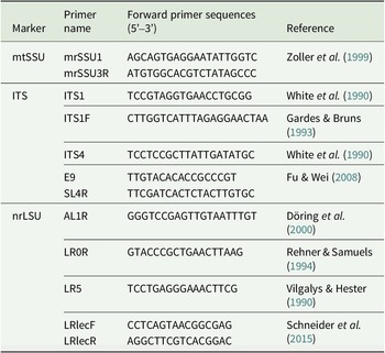
Primers used in this study are listed in Table 1. Total DNA extraction, PCR cycling parameters, PCR product purification and sequencing were performed as described in Wang et al. (Reference Wang, van den Boom, Sangvichien and Wei2020a). The success rate of PCR for Sporopodium is relatively low; the reason is unknown but is possibly due to the complex chemistry interfering in the PCR. We had to repeat the procedure multiple times or use different primers to obtain successful sequences (Tables 1 & 2).
Primers used in this study.
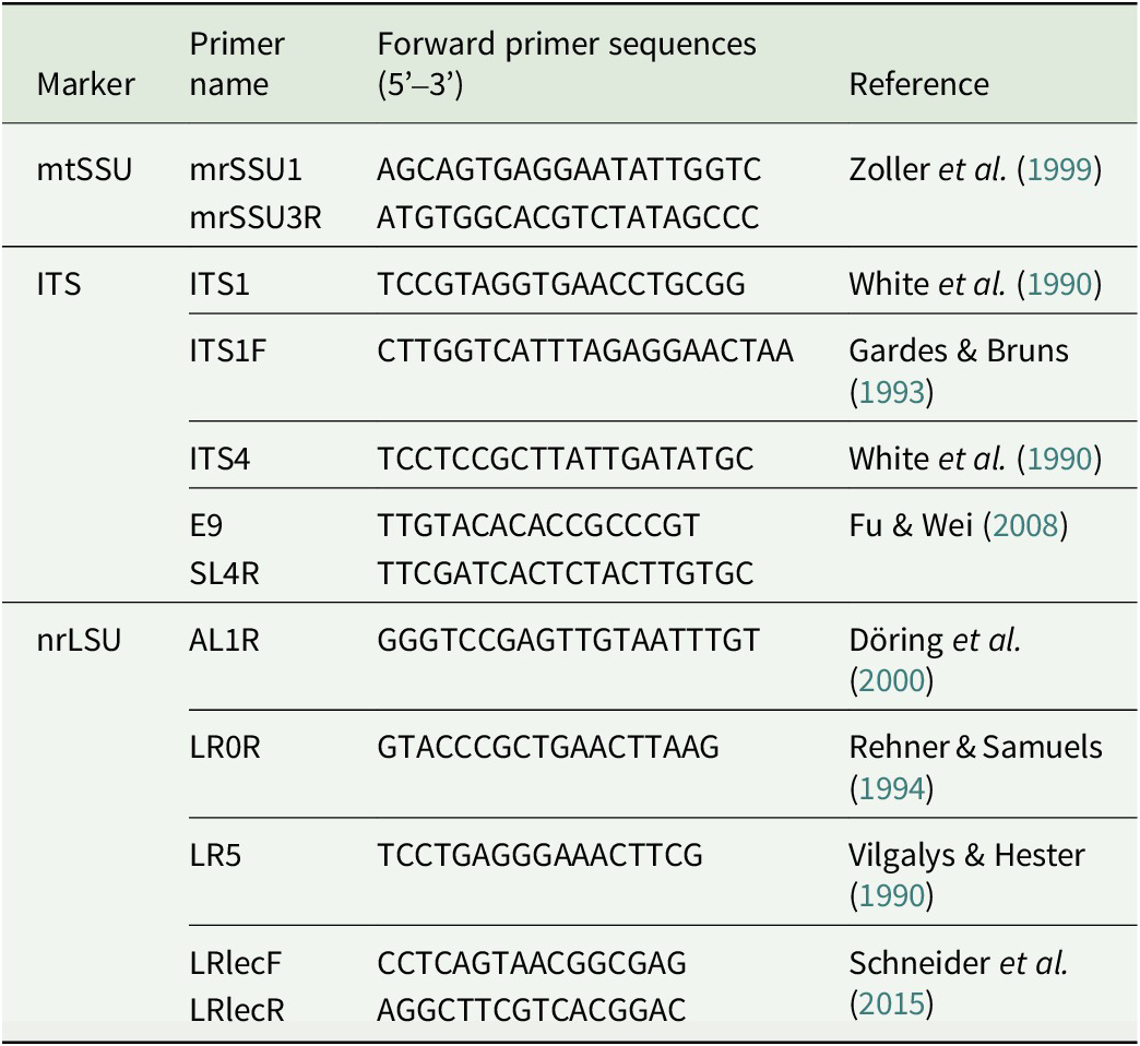
Table 1. Long description
The table is organized into four columns: Marker, Primer name, Forward primer sequences (5 prime to 3 prime), and Reference.
* mt S S U Marker:
- Primer mr S S U 1: A G C A G T G A G G A A T A T T G G T C. Reference: Zoller et al. 1999.
- Primer mr S S U 3 R: A T G T G G C A C G T C T A T A G C C C. Reference: Zoller et al. 1999.
* I T S Marker:
- Primer I T S 1: T C C G T A G G T G A A C C T G C G G. Reference: White et al. 1990.
- Primer I T S 1 F: C T T G G T C A T T T A G A G G A A C T A A. Reference: Gardes and Bruns 1993.
- Primer I T S 4: T C C T C C G C T T A T T G A T A T G C. Reference: White et al. 1990.
- Primer E 9: T T G T A C A C A C C G C C C G T. Reference: Fu and Wei 2008.
- Primer S L 4 R: T T C G A T C A C T C T A C T T G T G C. Reference: Fu and Wei 2008.
* nr L S U Marker:
- Primer A L 1 R: G G G T C C G A G T T G T A A T T T G T. Reference: Döring et al. 2000.
- Primer L R 0 R: G T A C C C G C T G A A C T T A A G. Reference: Rehner and Samuels 1994.
- Primer L R 5: T C C T G A G G G A A A C T T C G. Reference: Vilgalys and Hester 1990.
- Primer L R lec F: C C T C A G T A A C G G C G A G. Reference: Schneider et al. 2015.
- Primer L R lec R: A G G C T T C G T C A C G G A C. Reference: Schneider et al. 2015.
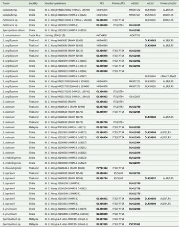
Specimens of Sporopodium and outgroup species used in the phylogenetic analyses (Figs 1–3) with voucher information and GenBank Accession numbers. New sequences and new species are in bold.
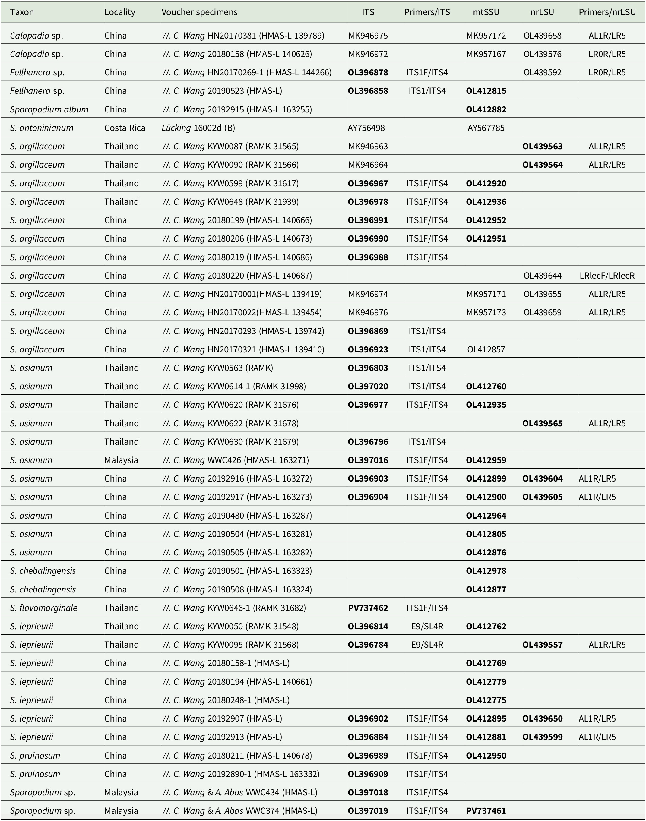
Table 2. Long description
The table contains 8 columns: Taxon, Locality, Voucher specimens, I T S, Primers forward slash I T S, mt S S U, nr L S U, and Primers forward slash nr L S U.
Key entries include:
* Calopadia species from China (HMAS-L 139789) with I T S MK946975 and nr L S U OL439658.
* Fellhanera species from China (HMAS-L 144266) with I T S OL396878 and nr L S U OL439592.
* Sporopodium album from China (HMAS-L 163255) with mt S S U OL412882.
* Sporopodium argillaceum specimens from Thailand and China, with various I T S accessions like MK946963 and OL396967, and nr L S U accessions like OL439563.
* Sporopodium asianum (new species) from Thailand, Malaysia, and China, with multiple new sequences including I T S OL396803 and mt S S U OL412760.
* Sporopodium chebalingensis from China with mt S S U OL412978.
* Sporopodium flavomarginale from Thailand with I T S PV737462.
* Sporopodium leprieurii from Thailand and China with sequences such as I T S OL396814 and nr L S U OL439557.
* Sporopodium pruinosum from China with I T S OL396989.
Primers listed include I T S 1 F forward slash I T S 4, E 9 forward slash S L 4 R, and A L 1 R forward slash L R 5 for nr L S U. Bold text in the original table indicates new sequences and new species.
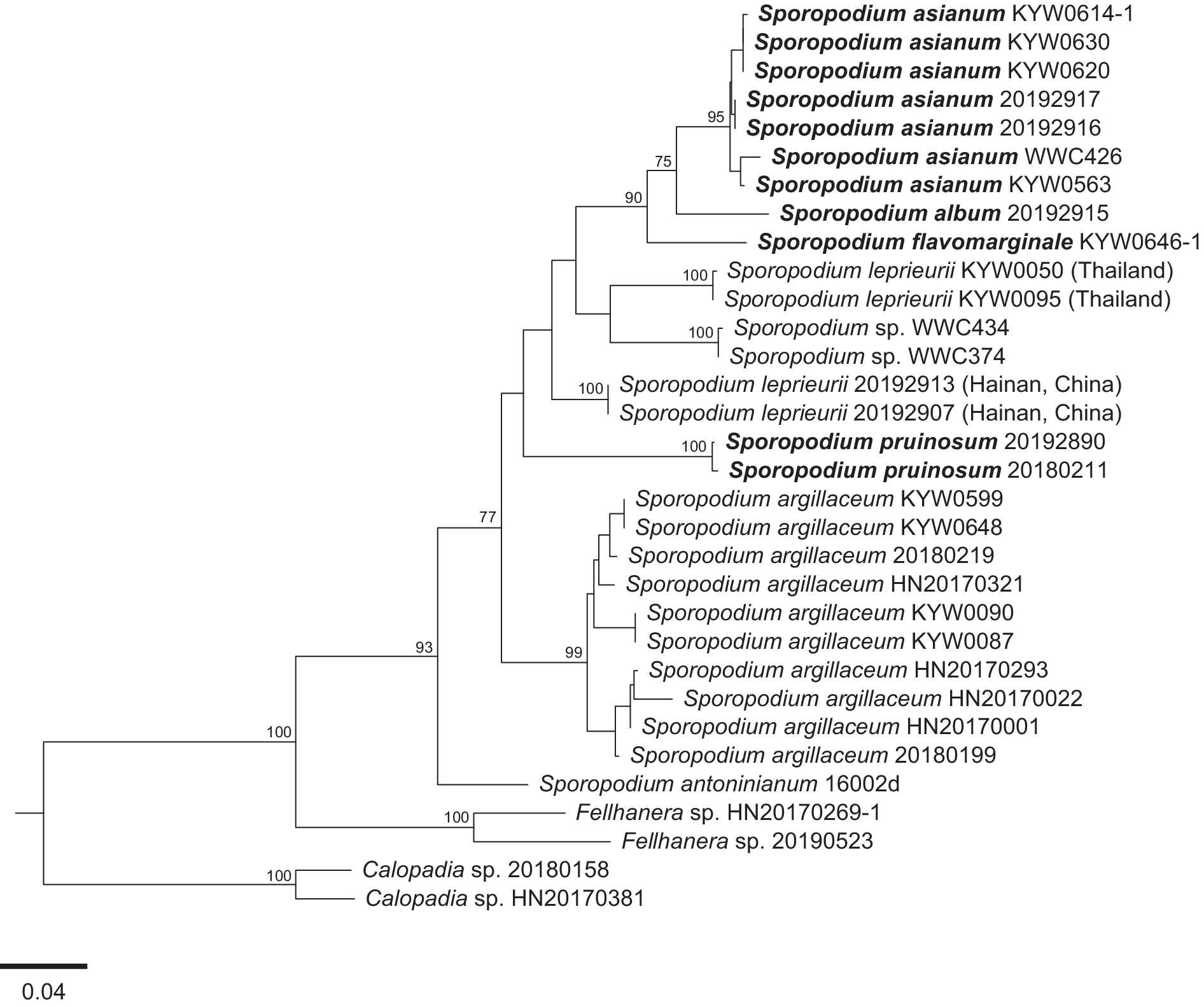
Phylogram of Sporopodium species using maximum likelihood (ML) inferred from the ITS data set. Bootstrap values ≥ 75% are indicated at the branches. The tree was rooted using Calopadia sp. Newly described species are marked in bold. Scale = 0.04 substitutions per site.
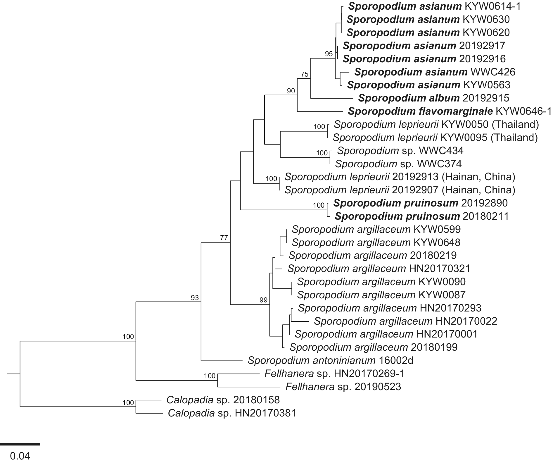
Figure 1. Long description
A maximum likelihood phylogram with a scale bar of 0.04 substitutions per site at the bottom left. The tree is rooted at the bottom with a clade containing Calopadia sp. 20180158 and Calopadia sp. H N 20170381. Moving upward, the next branch leads to a clade of Fellhanera sp. H N 20170269-1 and Fellhanera sp. 20190523. The main Sporopodium group is divided into two large clusters.
* The lower cluster contains Sporopodium antoninianum 16002d and a large sub-clade of Sporopodium argillaceum specimens including K Y W 0 5 9 9, K Y W 0 6 4 8, 20180219, H N 20170321, K Y W 0 0 9 0, K Y W 0 0 8 7, H N 20170293, H N 20170022, H N 20170001, and 20180199.
* The upper cluster branches into several groups. From bottom to top:
- Sporopodium pruinosum 20192890 and 20180211.
- Sporopodium leprieurii specimens from Hainan, China (20192913, 20192907).
- A group containing Sporopodium sp. W W C 4 3 4 and W W C 3 7 4, and Sporopodium leprieurii from Thailand (K Y W 0 0 5 0, K Y W 0 0 9 5).
- Sporopodium flavomarginale K Y W 0 6 4 6-1.
- The uppermost clade includes Sporopodium album 20192915 and several Sporopodium asianum specimens (K Y W 0 6 1 4-1, K Y W 0 6 3 0, K Y W 0 6 2 0, 20192917, 20192916, W W C 4 2 6, and K Y W 0 5 6 3).
Bootstrap values are labeled at nodes, with values such as 100, 99, 93, 90, 77, and 75 indicating branch support.
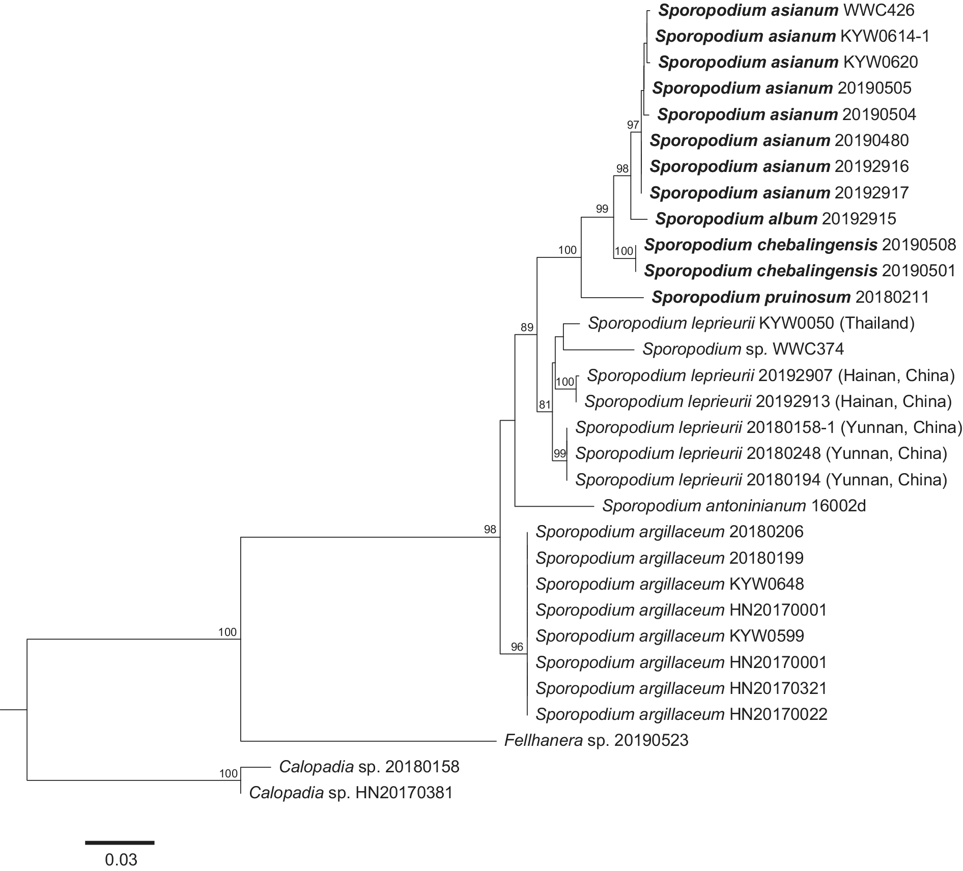
Phylogram of Sporopodium species using maximum likelihood (ML) inferred from the mtSSU data set. Bootstrap values ≥ 75% are indicated at the branches. The tree was rooted using Calopadia sp. Newly described species are marked in bold. Scale = 0.03 substitutions per site.
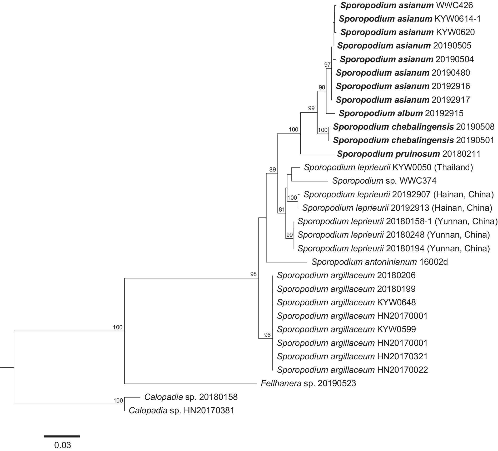
Figure 2. Long description
The phylogram originates from a root on the left, splitting into two primary lineages.
* The bottom lineage is the outgroup, containing Calopadia species 20180158 and H N 20170381 with a bootstrap value of 100.
* The main upper lineage branches into a clade containing Fellhanera species 20190523 and a large cluster of Sporopodium species.
* The Sporopodium argillaceum clade contains eight specimens (e.g., 20180206, H N 20170022) with a bootstrap support of 96.
* A sister clade to S. argillaceum, supported by a bootstrap of 98, contains several subgroups:
- Sporopodium antoninianum 16002d sits at the base of this subgroup.
- A clade of Sporopodium leprieurii specimens from Thailand, Hainan, and Yunnan, China, with internal bootstrap values ranging from 81 to 100.
- A top-most clade containing Sporopodium pruinosum 20180211, Sporopodium chebalingensis (bolded), Sporopodium album 20192915, and a large cluster of Sporopodium asianum (bolded) specimens.
Bootstrap values are indicated at each node, with many reaching 99 or 100. A scale bar at the bottom left indicates 0.03 substitutions per site.
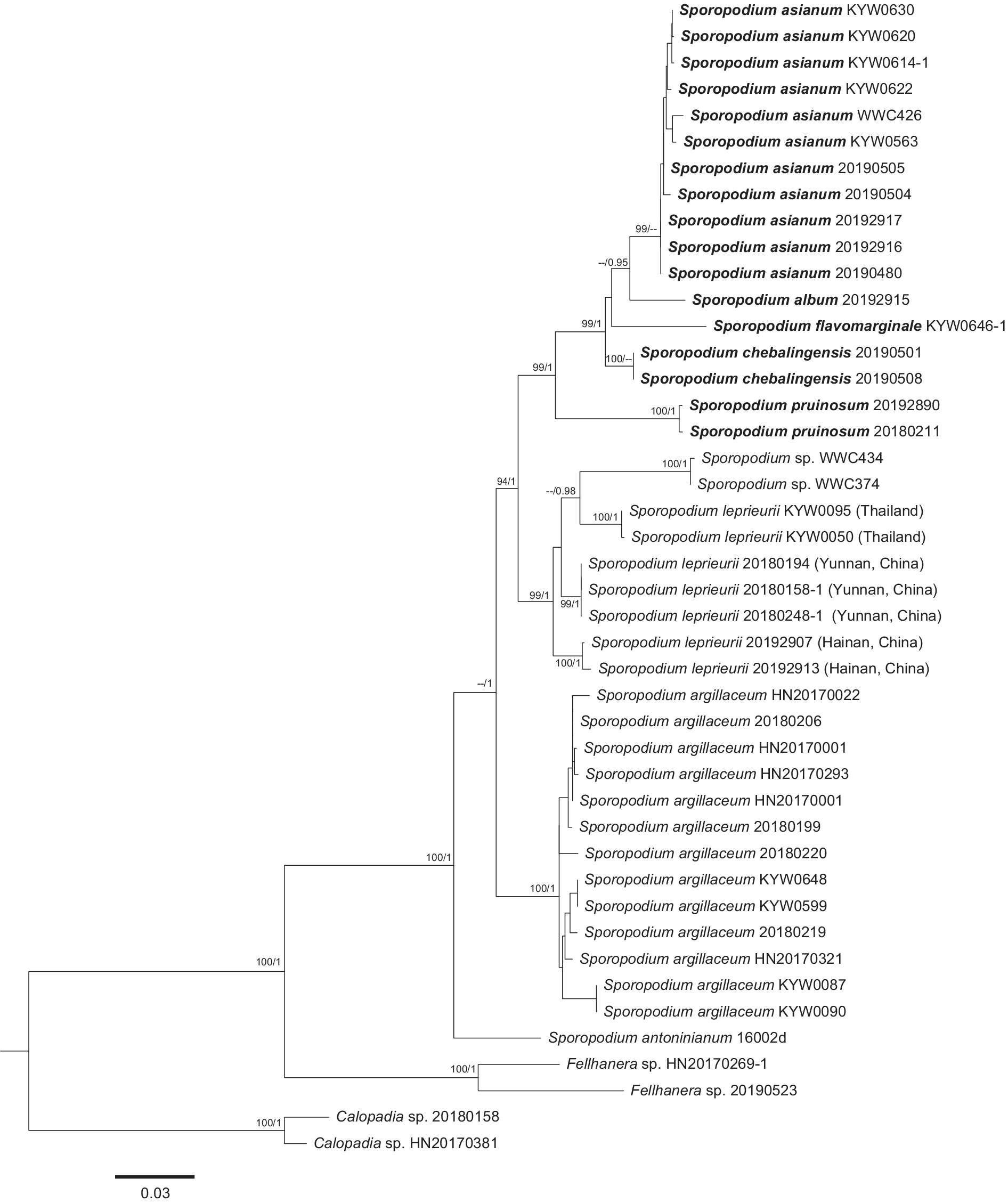
Phylogram of Sporopodium species using maximum likelihood (ML) inferred from a concatenated data set of ITS + mtSSU + nrLSU. Bootstrap values (BS) ≥ 75% and posterior probabilities (PP) ≥ 0.95 are indicated at the branches (BS/PP). The tree was rooted using Calopadia sp. Newly described species are marked in bold. Scale = 0.03 substitutions per site.
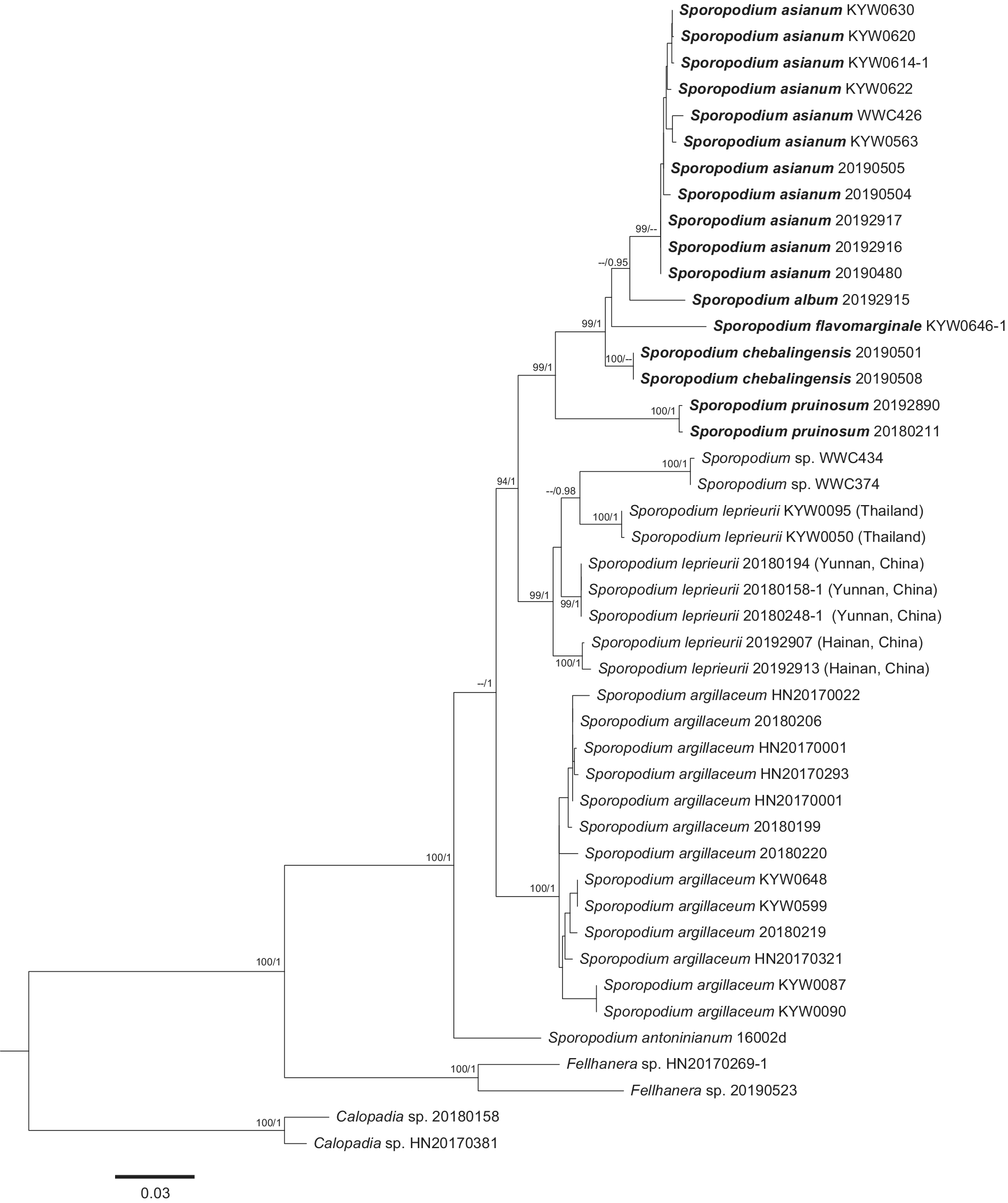
Figure 3. Long description
A phylogenetic tree with a scale bar of 0.03 substitutions per site at the bottom left. The tree is rooted by a basal clade containing Calopadia species 20180158 and H N 20170381 with support values of 100 forward slash 1. Moving up the tree, the next major branch leads to a clade of Fellhanera species and Sporopodium antoninianum. The largest section of the tree contains several distinct Sporopodium clades.
* The bottom-most major clade in this group consists of 13 specimens of Sporopodium argillaceum, supported by values of 100 forward slash 1.
* Above this is a clade containing Sporopodium leprieurii specimens from Thailand, Yunnan China, and Hainan China.
* Further up are smaller sister clades including Sporopodium species W W C 434 and W W C 374, Sporopodium pruinosum, and Sporopodium chebalingensis.
* The top-most section features a large cluster of Sporopodium asianum specimens, including K Y W 0630, K Y W 0620, and others, with Sporopodium album and Sporopodium flavomarginale branching off just below them.
Support values are written as B S forward slash P P at each node. Bolded names indicate newly described species, including Sporopodium asianum, Sporopodium album, Sporopodium flavomarginale, Sporopodium chebalingensis, Sporopodium pruinosum, and Sporopodium argillaceum.
Sequence alignment and phylogenetic analysis
Geneious v. 6.1.2 (Biomatters Ltd, Auckland, New Zealand) was used to assemble and edit primary sequences. A total of 64 newly generated sequences were aligned together with 12 sequences retrieved from GenBank (Table 2). Two specimens of Calopadia were chosen as outgroup based on previous phylogenetic analysis (Wang et al. Reference Wang, Sangvichien, Wei and Wei2020b).
The assembled sequences were aligned online using MAFFT v. 7.511 (https://mafft.cbrc.jp/alignment/server/?tdsourcetag=s_pcqq_aiomsg; Katoh et al. Reference Katoh, Asimenos and Toh2009). In advanced settings, we chose auto mode (progressive method depends on data size); our MAFFT alignments were conducted using the L-INS-i method, and ‘try to align gappy regions’. Other parameter settings were as follows: scoring matrix for nucleotide sequences = 200PAM/κ = 2; gap opening penalty = 1.53; offset value = 0; mafft-homologs = use UniRef50. The results were exported in fasta format.
The program Gblocks v. 0.91b (Castresana Reference Castresana2000) was used to delimit ambiguous regions, implementing the options for a less stringent selection (http://molevol.cmima.csic.es/castresana/Gblocks_server.html), which yielded final alignments of 608 bp (ITS), 820 bp (mtSSU), and 833 bp (nrLSU), respectively. Parameter settings were as follows: minimum number of sequences for a conserved position = 50% of the number of sequences + 1; minimum number of sequences for a flank position = 50% of the number of sequences + 1; maximum number of contiguous non-conserved positions = 8; minimum length of a block = 2; allowed gap positions = all. Alignments were concatenated in Geneious v. 6.1.2 for the multilocus phylogenetic analysis.
Maximum likelihood (ML) analysis and Bayesian inference (BI) were used to infer phylogenetic trees based on the single gene loci ITS and mtSSU, and also a combined ITS, mtSSU and nrLSU data set.
ML analysis was performed on the Cipres Science Gateway (http://www.phylo.org; Miller et al. Reference Miller, Pfeiffer and Schwartz2010), using RAxML-HPC Black Box (v. 8.2.12) (Stamatakis Reference Stamatakis2014). Phylogenetic tree inference used maximum likelihood/rapid bootstrapping with the default parameters and the GTRGAMMA model as implemented on the CIPRES portal, NSF XSEDE resource and bootstrap statistics calculated from 1000 bootstrap replicates.
Bayesian analysis was performed using MrBayes v. 3.2.6 (Huelsenbeck & Ronquist Reference Huelsenbeck and Ronquist2001; Ronquist & Huelsenbeck Reference Ronquist and Huelsenbeck2003). The best substitution models were estimated in jModelTest 2.1.10 (Darriba et al. Reference Darriba, Taboada, Doallo and Posada2012), using the Akaike information criterion (AIC). Based on the results, the best-fitting models for ITS, mtSSU and nrLSU were all GTR + I + G. All characters were equally weighted, gaps were treated as missing data, and the default distributions were used for priors. Markov chain Monte Carlo (MCMC) was initiated from a random tree using 5 million generations and sampling every 1000 steps, with the first 25% of trees discarded as burn-in. Stationarity of analysis was determined by examining the standard deviation of split frequencies (< 0.01). A majority-rule consensus tree was constructed from the remaining trees to estimate posterior probability (PP), with values ≥ 0.95 considered as significantly supported. ML and BI trees were visualized with the program FigTree v. 1.4.3 and Adobe Illustrator CC 2023.
Results
The final alignment consisted of 25 ITS, 25 mtSSU and 14 nrLSU sequences newly generated from 42 Ectolechiaceae specimens, plus seven ITS sequences and five mtSSU sequences downloaded from NCBI (Table 2).
Phylogenetic analysis was performed for single loci (ITS, mtSSU) and for the concatenated, multilocus data set (Figs 1–3). In the trees based on the single locus ITS and mtSSU (Figs 1 & 2), all sequences of S. argillaceum (Müll. Arg.) Zahlbr. form a highly supported clade, close to the single sequence of S. antoninianum Elix et al.; in the ITS tree (Fig. 1), the sequences of S. leprieurii Mont. from Thailand and China form two separate clades in a paraphyletic relationship, those from Thailand being an unsupported sister to a clade of two unnamed sequences (WWC374, 434), whereas in the mtSSU tree (Fig. 2), the S. leprieurii sequences fall into a single, supported clade, together with the unnamed taxon but with a substructure indicating the presence of more than one species.
In the mtSSU tree (Fig. 2), four new species from Asia (S. album, S. asianum, S. chebalingensis, S. pruinosum) form a highly supported clade, while in the ITS tree (Fig. 1), S. pruinosum falls outside this clade. Instead, the ITS tree also places the new taxon S. flavomarginale (no mtSSU sequences) into this clade, whereas it lacks S. chebalingensis (no ITS sequences).
In the tree based on the concatenated data set (Fig. 3), all five new species form a strongly supported clade, here named the S. asianum group. Both the S. argillaceum and the S. leprieurii clades are also strongly supported, including material from Thailand and China, but the substructure in the S. leprieurii clade indicates the presence of up to four species, with a strong geographical signal.
Discussion
This is the first broader molecular phylogenetic study of Sporopodium, a largely foliicolous genus in the family Ectolechiaceae, with a focus on tropical Asia. Prior to this study, 25 species were known in this genus (Santesson Reference Santesson1952; Sérusiaux Reference Sérusiaux1986; Aptroot & Sipman Reference Aptroot and Sipman1993; Aptroot et al. Reference Aptroot, Diederich, Sérusiaux and Sipman1997, Reference Aptroot, de Souza, Santos, Oliveira-Junior, Barbosa and Cáceres2022; Lücking Reference Lücking1999, Reference Lücking2008; Lücking & Lumbsch Reference Lücking and Lumbsch2001; Lücking & Kalb Reference Lücking and Kalb2002; Øvstedal & Elix Reference Øvstedal and Elix2007; Sérusiaux et al. Reference Sérusiaux, Lücking and Lumbsch2008; Singh & Pinokiyo Reference Singh and Pinokiyo2008; Lücking et al. Reference Lücking, Hodkinson and Leavitt2017). Our study, although focusing on a limited geographical area, already raises this number to 30 species. Given that the material is from a limited region in South-East Asia, many more species are expected to be discovered based on molecular phylogenetic revision of material from other regions.
Notably, the five new species were all resolved in a single clade, here named the S. asianum group, all so far known only from South-East Asia. Some species appear to have a narrow distribution area, such as S. chebalingensis known so far only from the Guangdong Province of China. Others, such as S. asianum, are more widely distributed in tropical Asia.
Morphologically, the apothecia of these five species are all covered with pruina or contain algal cells, while these characters vary more widely in other regions, such as the Neotropics, where non-pruinose taxa such as S. leprieurii s. lat. are also found. Three of the five new species produce the didymic acid chemosyndrome, a set of dibenzofuranes related to usnic acid which is a newly reported compound for the genus, otherwise known for example from Cladonia.
Unfortunately, of the 25 previously known species of the genus, only three have sequence data available, namely S. antoninianum, S. argillaceum and S. leprieurii, so we cannot comment on the phylogenetic position of the numerous other described species and how clades within this genus will eventually differ from each other. However, it is notable that the sister clade to the S. asianum group, the S. leprieurii group, as well as the early diverging S. argillaceum and S. antoninianum, typically lack pruina on the apothecial discs. Sporopodium argillaceum is one of the species described from Asia and is apparently widely distributed in this region, but not yet known from Africa and the Neotropics. The type material was collected in Vietnam (Santesson Reference Santesson1952). The species is characterized by its dark brown, non-pruinose apothecial disc with a thick and prominent crenulate margin, combined with a greenish grey, verrucose thallus.
Sporopodium leprieurii differs from other species by the combination of oblong-ellipsoid conidia (most Sporopodium species usually have drop-shaped conidia) produced in campylidia with a thickened base and small, brownish hood, as well as the dark brown apothecial discs with a narrow, thin, cream-coloured to greyish margin, combined with a greenish grey, verrucose thallus (Lücking Reference Lücking2008).
Seven specimens identified as S. leprieurii formed an assembly of three subclades; all have oblong-ellipsoid conidia and verrucose thalli but otherwise exhibit subtle morphological differences and so may represent three different taxa. Given that all three are from tropical Asia and the type is from South America (French Guiana), none of them may be genuine S. leprieurii s. str. At present, we lack sequence data for genuine S. leprieurii from the Neotropics, but this clade needs further study; it also encompasses the synonyms currently placed under S. leprieurii, three of which originate from tropical Asia (Lecidea cupulifera Nyl., Sporopodium pycnoblastum Vain., Loflammia demoulinii Sérus.).
Sporopodium antoninianum is a unique species within the genus, easily recognized by its woolly prothallus formed between partially dispersed thallus patches and marginally pilose apothecia. Lücking (Reference Lücking2008) suggested that this species seems to be most related to S. leprieurii, which has a similar thallus morphology and chemistry, although lacks the woolly prothallus and apothecial hairs. The samples WWC374 and 434 were collected from Malaysia; they exhibit a woolly prothallus similar to that of S. antoninianum but appear to be only distantly related to that species (Figs 1–3). Although they were resolved as part of the S. leprieurii clade, this species differs by having a compact and smooth apothecial margin and lacking a woolly prothallus. Due to the limited specimen material and the unresolved taxonomy of the S. leprieurii clade, we refrain from formally describing this material at the current time.
Among foliicolous lichens and also among Ectolechiaceae, Sporopodium is known for its rich secondary chemistry, including a diversity of chlorinated xanthones (Elix et al. Reference Elix, Crook and Lumbsch1992; Lücking & Lumbsch Reference Lücking and Lumbsch2001; Sérusiaux et al. Reference Sérusiaux, Lücking and Lumbsch2008). Notably, xanthones were not detected in any of the five new species of the S. asianum clade; instead, three of the five species produce didymic acid, a dibenzofurane (i.e. related to usnic acid) thus far known, for instance, from Cladonia species. The S. leprieurii clade includes taxa with a pattern of arthothelin, isoarthotheline, asemone, thiophanic acid, argopsin and zeorin (Lücking & Lumbsch Reference Lücking and Lumbsch2001; Sérusiaux et al. Reference Sérusiaux, Lücking and Lumbsch2008). This suggests that secondary chemistry may be an important predictor of phylogenetic relationships in this genus.
Taxonomy
Sporopodium album W. C. Wang & J. C. Wei sp. nov.
Fungal Names No.: FN 570956
Similar to Sporopodium xantholeucum (Müll. Arg.) Zahlbr. but differs in having a green thallus, a dark brown hypothecium, small apothecia with a pruinose disc and white margin, as well as a different chemistry.
Type: China, Hainan Province, Wuzhishan City, Wuzhi Mountain National Forest Park, 18°54′N, 109°41′E, 800 m alt., on leaves, 12 December 2019, W. C. Wang 20193079-1 (HMAS-L 163253—holotype!).
(Fig. 4)
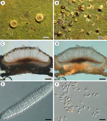
Sporopodium album (holotype, W. C. Wang 20193079-1 HMAS-L). A & B, thalli with apothecia and campylidia. C, thalli with apothecia (Wang 20190503 HMAS-L). D, section of apothecium showing pigmentation. E, muriform ascospores. F, drop-shaped conidia. Scales: A & B = 1 mm; C = 500 μm; D = 200 μm; E & F = 20 μm. In colour online.
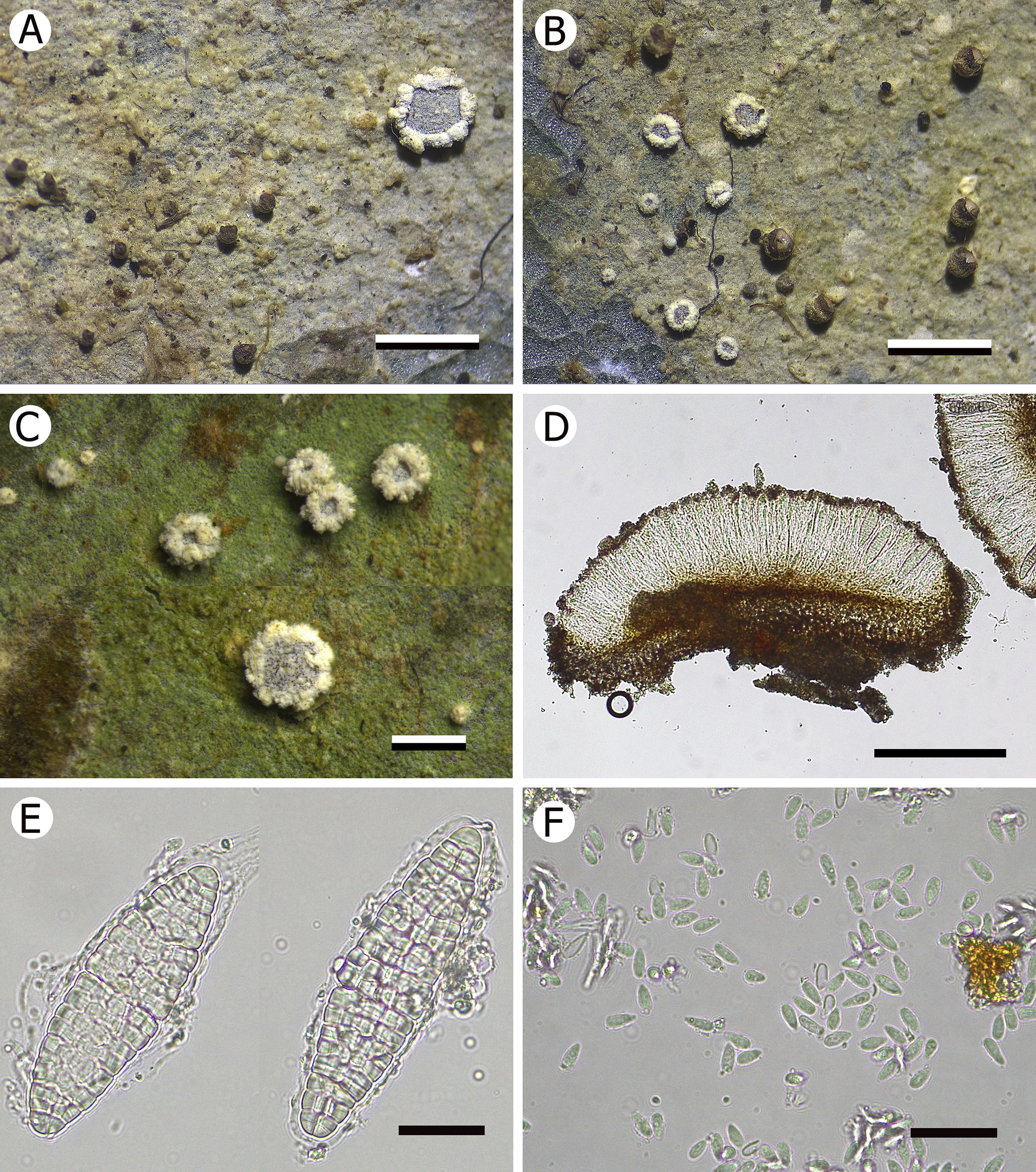
Figure 4. Long description
A six-panel collection of photos labeled A through F.
* Panel A and B: Macroscopic photos of the lichen thallus on a leaf surface. The thallus is pale and granular, featuring circular apothecia with dark centers and thick, white, ruffled margins. Small, dark, rounded campylidia are also scattered across the surface. Scale bars represent 1 mm.
* Panel C: A closer view of the thalli and apothecia on a green leaf substrate. The white margins of the apothecia appear more prominent and textured. Scale bar represents 500 micrometers.
* Panel D: A microscopic cross-section of an apothecium. It shows a dark brown pigmented base and a layer of vertical, tightly packed fungal filaments. Scale bar represents 200 micrometers.
* Panel E: High-magnification view of two muriform ascospores. These are large, elongated, and divided into many internal cells by both transverse and longitudinal walls. Scale bar represents 20 micrometers.
* Panel F: Microscopic view of numerous small, drop-shaped conidia. They are translucent and scattered across the field of view. Scale bar represents 20 micrometers.
Thallus epiphyllous, crustose, continuous, usually small, no more than 1 cm across, marginally irregular, pale green to green or grey-green, occasionally with a grey-blue tinge, surface matt, without or with sparse verrucae. Prothallus not observed. Photobiont a species of Chlorococcaceae; algal cells green, globose, 7.5–10 μm diam.
Apothecia sessile, rounded, distinctly constricted at the base, small, 0.3–0.6 mm diam. and 180–250 μm high; disc flat to slightly convex, strongly convex in old ones, black, but usually covered with white pruina, without epithecial algal cells; margin persistent and prominent, dull, white, irregularly crenulate, farinose, c. 0.06 mm thick. Excipulum 75–125 μm thick, extending to apothecial base, inner part paraplectenchymatous, K−, but small amount of colourless hyphae present in outer part, K−, white under the dissecting microscope, pale brown when examined with the compound microscope, incrusted with numerous pale brown granules which do not dissolve in K (using cross-polarized light); hypothecium c. 45–55 μm high, brown, K−; epithecium colourless or pale brown, containing numerous pale brown granules which do not dissolve in K, c. 12 μm high, K−; hymenium 90–110 μm high, colourless, K−; paraphyses colourless, thin, richly branched and anastomosing. Asci clavate, of the Sporopodium-type, 90–120 × 20–25 μm, with a well-developed, I+ dark blue tholus. Ascospores 1 per ascus, narrowly ellipsoid, strongly muriform, colourless, 80–110 × 16–20 μm, 5–5.5 times as long as wide (n = 60).
Campylidia sessile, upper part dark brown, lower part grey; lobe well developed, c. 0.3 mm wide, mussel-shaped, without inflated socle. Conidia drop-shaped, non-septate, 6–7 × 3 μm, colourless.
Etymology
The epithet ‘album’ refers to its white apothecial margin.
Habitat and distribution
A quite common species, currently known only from China, growing on leaf surfaces in damp understorey or near to a river in rainforests at high altitudes.
Notes
The new species is overall most similar to Sporopodium xantholeucum (Santesson Reference Santesson1952; Lücking Reference Lücking2008), which also has small apothecia with an irregular, farinose margin. However, the latter differs in having a bluish grey thallus containing chlorinated xanthones, pannarin and zeorine, hood-shaped, light grey campylidia, and a pale yellow apothecial margin. Sporopodium flavescens (R. Sant.) Vězda (≡ S. vezdeanum Lumbsch & Elix nom. inval.) from the eastern Paleotropics shares the white apothecial margin with the new species, but its campylidia are bright yellow with an inflated socle and it has a chemistry similar to that of S. xantholeucum. Sporopodium phyllocharis (Mont.) A. Massal. is also similar in the small, pruinose apothecia with white margin, but in addition to the more regular margin, it has a bluish thallus and a brown apothecial disc, as well as a different chemistry (Elix et al. Reference Elix, Lumbsch and Lücking1995). Sporopodium asianum and S. album are closely related phylogenetically and share the same chemistry, but S. asianum has larger (0.6–1 mm diam.), khaki apothecia with a more regular margin and a K+ yellow epithecium.
Additional specimens examined
China: Hainan Province: Wuzhishan City, Wuzhi Mountain National Forest Park, 18°54′N, 109°41′E, 800 m alt., on leaves, 12 xii 2019, W. C. Wang 20193063-1 (HMAS-L 163254); Lingshui County, Diaoluo Mountain National Forest Park, forest trail by the lake, 18°43′31′′N, 109°51′59′′E, 928 m alt., on leaves, 14 xii 2019, W. C. Wang 20192915 (HMAS-L 163255), 20193008 (HMAS-L 163256), 20193011 (HMAS-L 163257), 20193099 (HMAS-L 163258), 20193100 (HMAS-L 163259); Wuzhishan City, Wuzhi Mountain, 700 m alt., on leaves, 27 ix 2008, B. Gao HNF328 (HMAS-L 126374); Ledong County, Jianfeng Ridge, hydrological station, 4 vi 2006, J. C. Wei s. n. (HMAS-L 112789). Guangdong Province: Shixing County, Cheba Ridge National Natural Reserve, roadside from west gate to Triangle Pond, 24.718°N, 114.262°E, 350 m alt., on leaves, 19 iii 2019, W. C. Wang 20190498-1 (HMAS-L 163260), 20190503 (HMAS-L 163261), 20190505-1 (HMAS-L 163262). Yunnan Province: Xishuangbanna City, natural reserve in Tropical Botanical Garden, 14 ix 1987, J. C. Wei 9031 (HMAS-L 118415); Mengyang Town, Wild Elephant Valley, on leaves, 31 i 2018, W. C. Wang 20180221 (HMAS-L 140688). Fujian Province: Yongtai County, Tianmen Mountain scenic spot, 25°49′N, 119°01′E, 500 m alt., on leaves, 9 xii 2021, W. C. Wang & X. L. Wang WWC602-2 (HMAS-L 163263), WWC696 (HMAS-L 163264), WWC703 (HMAS-L 163265), WWC723-1 (HMAS-L 163266), WWC902 (HMAS-L 163267).
Sporopodium asianum W. C. Wang & A. Abas sp. nov.
Fungal Names No.: FN 570957
Similar to S. argillaceum but differs in having pruinose apothecia with a khaki margin, a K+ yellow epithecium and a K+ orange thallus.
Type: Thailand, Nakhon Ratchasima Province, Khao Yai National Park, Pha Kluay Mai Waterfall trail to Haew Suwat Waterfall, 14°23′N, 101°22′E, 800 m alt., on leaves, 5 May 2018, W. C. Wang KYW0630 (RAMK 31679—holotype!).
(Fig. 5)
Sporopodium asianum (holotype, W. C. Wang KYW0630 RAMK). A, thallus with mature apothecia. B, thallus with young apothecia and campylidia. C & D, section of apothecium showing (C) pigmentation in normal light and (D) crystals in exciple and apothecial base (using cross-polarized light). E, muriform ascospore (observed using differential interference contrast microscopy). F, drop-shaped conidia. Scales: A & B = 0.5 mm; C & D = 100 μm; E & F = 10 μm. In colour online.
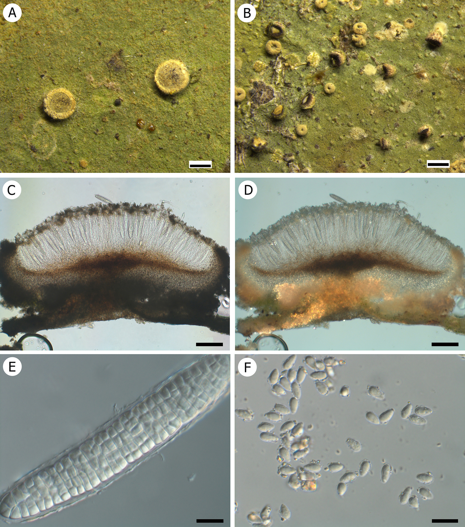
Figure 5. Long description
A collection of six panels labeled A through F.
* Panel A shows a green thallus with two circular, mature apothecia featuring raised margins. A white scale bar at the bottom right represents 0.5 mm.
* Panel B displays a similar green thallus populated with numerous smaller, young apothecia and campylidia. A white scale bar at the bottom right represents 0.5 mm.
* Panel C is a cross-section of an apothecium under normal light, showing a dark, pigmented base and a fan-like arrangement of internal structures. A black scale bar at the bottom right represents 100 micrometers.
* Panel D shows the same cross-section as C but under cross-polarized light, revealing bright, glowing crystals concentrated in the exciple and the base of the apothecium. A black scale bar at the bottom right represents 100 micrometers.
* Panel E is a high-magnification D I C micrograph of a single muriform ascospore, showing a complex grid-like internal cellular structure. A black scale bar at the bottom right represents 10 micrometers.
* Panel F shows a cluster of numerous small, translucent, drop-shaped conidia scattered across the field of view. A black scale bar at the bottom right represents 10 micrometers.
Thallus epiphyllous, crustose, continuous, 1–2 cm across, marginally irregular, grey-green, green or pale green, surface matt, sparsely verrucose; verrucae 0.15–0.2 mm diam., hyphae K+ orange under the compound microscope. Prothallus present, paler than thallus. Photobiont a species of Chlorococcaceae; algal cells green, globose, 7.5–10 μm diam.
Apothecia sessile, rounded, distinctly constricted at the base, 0.6–1 mm diam. and 360–380 μm high; disc flat to slightly convex, brown, but usually covered with khaki to dark khaki pruina, without epithecial algal cells; margin distinct and persistent, dull, khaki, not or slightly crenulate, c. 0.2 mm thick. Excipulum well developed, 100–150 μm thick, extending to apothecial base, distinctly paraplectenchymatous, K−, but small amount of colourless hyphae present in outer surface, K+ yellow, white under the dissecting microscope, but when examined with the compound microscope, outer part dark brown to brownish black, inner part colourless or pale brown, incrusted with numerous pale yellow crystals which dissolve in K; hypothecium narrow, c. 45–60 μm high, brown, K+ yellowish brown to orange; epithecium yellowish brown, containing numerous yellowish brown granules which do not dissolve in K, c. 25 μm high, K+ yellow; hymenium 160–180 μm high, colourless, K−; paraphyses colourless, thin, richly branched and anastomosing. Asci clavate, of the Sporopodium-type, 80–110 × 20–25 μm, with a well-developed, I+ dark blue tholus. Ascospores 1 per ascus, narrowly ellipsoid, strongly muriform, colourless, 70–100 × 12–16 μm, 5.5–6.5 times as long as wide (n = 120).
Campylidia sessile, khaki, the same colour as apothecia, uppermost part black; lobe well developed, 0.3–0.4 mm wide, mussel-shaped; socle present but not inflated. Conidia drop-shaped, non-septate, 6–6.5 × 3 μm, colourless.
Chemistry
Didymic acid (major) and a didymic acid derivative (major) determined by TLC (Fig. 9-1–3) and HPLC.
Etymology
The epithet ‘asianum’ refers to its broad distribution in SE Asia.
Habitat and distribution
A widely distributed species in Asia, currently known from China, Thailand and Malaysia, on leaf surfaces mostly along rivers in lowland tropical or subtropical rainforest.
Notes
Sporopodium asianum is characterized mainly by its khaki apothecia and campylidia, with the apothecia having a K+ yellow epithecium, and a grey-green and K+ orange thallus. It is somewhat similar to S. phyllocharis but differs in the larger apothecia and the chemistry (Santesson Reference Santesson1952; Elix et al. Reference Elix, Lumbsch and Lücking1995; Lücking Reference Lücking2008). Sporopodium argillaceum, another common species in Asia, agrees in the greenish, somewhat verrucose thallus but has a non-pruinose apothecial disc with a prominent, crenulate margin and its thallus often features a marginal reddish prothallus.
Additional specimens examined
Thailand: Nakhon Ratchasima Province: Khao Yai National Park, Pha Kluay Mai Waterfall trail to Haew Suwat Waterfall, 14°23′N, 101°22′E, 800 m alt., on leaves, 5 v 2018, W. C. Wang KYW0585 (RAMK 31610), KYW0613 (RAMK), KYW0615 (RAMK), KYW0616 (RAMK), KYW0617 (RAMK), KYW0618 (RAMK), KYW0619 (RAMK), KYW0620 (RAMK 31676), KYW0621 (RAMK 31677), KYW0622 (RAMK 31678), KYW0624 (RAMK), KYW0625 (RAMK), KYW0626 (RAMK), KYW0627 (RAMK), KYW0628 (RAMK), KYW0629 (RAMK), KYW0631 (RAMK), KYW0455 (RAMK 31961), KYW0585 (RAMK 31610), KYW0614 (RAMK 31998), KYW0638 (RAMK 31909).—Malaysia: Pahang: Fraser Hill, Jeriau Waterfall Trail, 800 m alt., on leaves, 7 ix 2019, W. C. Wang & A. Abas WWC354 (HMAS-L 163268), WWC355 (HMAS-L 163269), WWC360 (HMAS-L 163270), WWC426 (HMAS-L 163271).—China: Hainan Province: Lingshui County, Diaoluo Mountain National Forest Park, forest trail by the lake, 18°43′31″N, 109°51′59″E, 928 m alt., on leaves, 14 xii 2019, W. C. Wang 20192916 (HMAS-L 163272), 20192917 (HMAS-L 163273), 20193098 (HMAS-L 163274), 20193105 (HMAS-L 163275); Ledong County, Jianfeng Ridge, in the forest beside the road from Tianchi to the core area, 18°45′20″N, 108°53′31″E, 780 m alt., on leaves, 10 xii 2019, W. C. Wang 20192989 (HMAS-L 163276), 20193053 (HMAS-L 163277); Ledong County, Jianfeng Ridge, 18°44′N, 109°10′E, 900 m alt., on leaves, 1 iv 1993, Y. M. Jiang & S. Y. Guo s. n. (HMAS-L 138614); ibid., 18°44′N, 109°10′E, 1000 m alt., on leaves, 10 xii 2000, M. R. Huang s. n. (HMAS-L 112836); Lingshui County, Diaoluo Mountain National Forest Park, 1000 m alt., on leaves, 14 xii 2000, M. R. Huang s. n. (HMAS-L 112799), s. n. (HMAS-L 112803). Guangdong Province: Shixing County, Cheba Ridge National Natural Reserve, roadside from west gate to triangle pond, 24.718°N, 114.262°E, 350 m alt., on leaves, 19 iii 2019, W. C. Wang 20190497 (HMAS-L 163278), 20190498 (HMAS-L 163279), 20190500 (HMAS-L 163280), 20190504 (HMAS-L 163281), 20190505 (HMAS-L 163282); triangle pond, negative ion trail, 350 m alt., on leaves, 19 iii 2019, W. C. Wang 20190476 (HMAS-L 163283), 20190477 (HMAS-L 163284), 20190478 (HMAS-L 163285), 20190479 (HMAS-L 163286), 20190480 (HMAS-L 163287), 20190481 (HMAS-L 163288), 20190482 (HMAS-L 163289), 20190490 (HMAS-L 163290), 20190491 (HMAS-L 163291). Guangxi Province: Nanning City, Daming Mountain National Natural Reserve, 1240 m alt., on leaves, 22 v 2015, X. L. Wei & J. H. Wang GX2015044 (HMAS-L 133003), GX2015089 (HMAS-L 133012), GX2015036 (HMAS-L 132998), GX2015040 (HMAS-L 133001). Fujian Province: Yongtai County, Tianmen Mountain scenic spot, 25°49′N, 119°01′E, 500 m alt., on leaves, 9 xii 2021, W. C. Wang & X. L. Wang WWC600 (HMAS-L 163292), WWC602-1 (HMAS-L 163293), WWC603-1 (HMAS-L 163294), WWC613 (HMAS-L 163295), WWC617 (HMAS-L 163296), WWC628 (HMAS-L 163297), WWC634 (HMAS-L 163298), WWC671-1 (HMAS-L 163299), WWC674-2 (HMAS-L 163300), WWC689-1 (HMAS-L 163301), WWC693 (HMAS-L 163302), WWC698 (HMAS-L 163303), WWC699-2 (HMAS-L 163304), WWC723-3 (HMAS-L 163305), WWC896 (HMAS-L 163306), WWC899 (HMAS-L 163307), WWC900 (HMAS-L 163308), WWC923 (HMAS-L 163309), WWC927 (HMAS-L 163310), WWC928 (HMAS-L 163311), WWC948 (HMAS-L 163312), WWC954-2 (HMAS-L 163313), WWC957 (HMAS-L 163314), WWC958 (HMAS-L 163315), WWC963 (HMAS-L 163316), WWC970-3 (HMAS-L 163317), WWC980 (HMAS-L 163318); Dehua County, Stone Cow Mountain scenic spot, 25°38′N, 118°26′E, 600 m alt., on leaves, 12 xii 2021, W. C. Wang & X. L. Wang WWC1091 (HMAS-L 163319), WWC1106 (HMAS-L 163320), WWC1108 (HMAS-L 163321).
Sporopodium chebalingensis W. C. Wang & J. C. Wei sp. nov.
Fungal Names No.: FN 570958
Similar to S. citrinum (Zahlbr.) Elix et al. but differs in having a green smooth thallus with dense, tiny verrucae and a greyish green apothecial disc due to abundant epithecial algal cells.
Type: China, Guangdong Province, Shixing County, Cheba Ridge National Natural Reserve, roadside from west gate to triangle pond, 24.718°N, 114.262°E, 350 m alt., on leaves, 19 March 2019, W. C. Wang 20190507 (HMAS-L 163322—holotype!).
(Fig. 6)
Sporopodium chebalingensis (holotype, W. C. Wang 20190507 HMAS-L). A & B, thalli with apothecia and yellow campylidia. C, section of apothecium showing pigmentation. D, epithecial algal cells and branched and anastomosing paraphyses. E, muriform ascospores. F, drop-shaped conidia. Scales: A & B = 500 μm; C = 200 μm; D = 50 μm; E & F = 20 μm. In colour online.
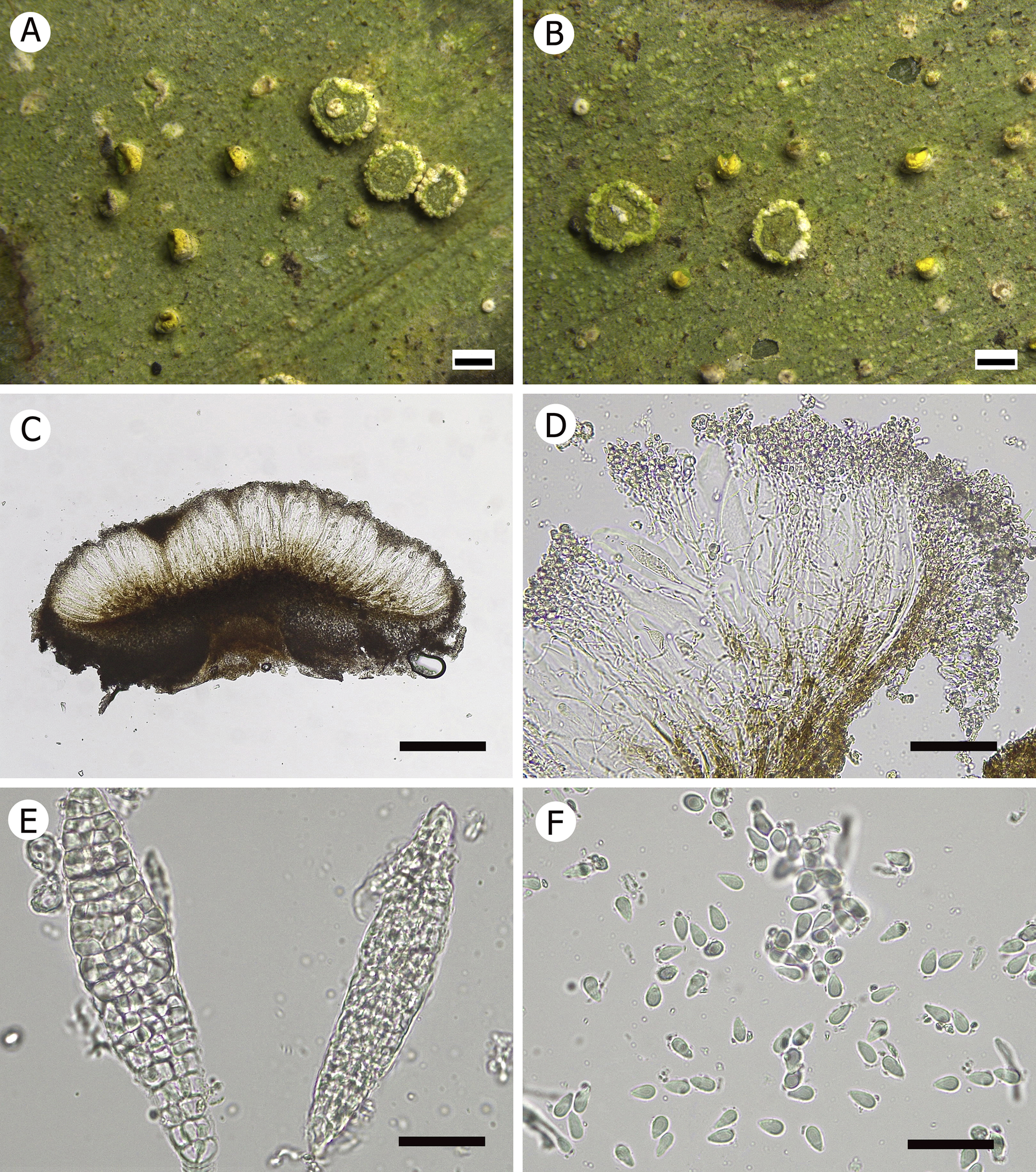
Figure 6. Long description
A multi-panel figure containing six micrographs labeled A through F.
* Panel A and B: Macroscopic photos of the lichen surface. The green thallus is dotted with circular, disc-like apothecia and smaller, bright yellow campylidia. Scale bars represent 500 micrometers.
* Panel C: A cross-section of an apothecium. It shows a dark, pigmented base and a lighter, fan-shaped upper layer with vertical structures. Scale bar represents 200 micrometers.
* Panel D: A high-magnification view of the internal structure showing epithecial algal cells and a network of branched, anastomosing paraphyses. Scale bar represents 50 micrometers.
* Panel E: Micrograph of two elongated, muriform ascospores with complex internal septation resembling a grid or mesh. Scale bar represents 20 micrometers.
* Panel F: Micrograph showing numerous small, translucent, drop-shaped conidia scattered across the field of view. Scale bar represents 20 micrometers.
Thallus epiphyllous, crustose, continuous, irregular in outline, 1.5–2.5 cm across, green, surface matt, finely verrucose; verrucae dense, tiny, 75–100 μm diam., of the same colour as the thallus. Hypothallus not observed. Photobiont a species of Chlorococcaceae; algal cells green, globose, 8–10 μm diam.
Apothecia numerous, sessile, rounded, scattered, distinctly constricted at the base, 0.7–1 mm diam. and 390–415 μm high; disc flat, covered by green epithecial algal cells, without pruina; margin distinct and persistent, dull, white, crenulate and somewhat swollen, 125–220 μm thick. Excipulum well developed, 55–85 μm thick, paraplectenchymatous, colourless but appears brown-black due to strong incrustation with numerous opaque granules which do not dissolve in K; hypothecium up to 110 μm high, brown, not significantly changing in K; epithecium indistinct because of numerous epithecial algal cells; epithecial alga abundant, algal cells smaller than those of the thallus, rounded, 5–7.5 μm diam.; hymenium 110–140 μm high, colourless; paraphyses colourless, thin, richly branched and anastomosing. Asci clavate, of the Sporopodium-type, 100–110 × 20–25 μm, with a well-developed, I+ dark blue tholus. Ascospores 1 per ascus, narrowly ellipsoid, richly muriform, colourless, 90–100 × 17–22 μm, 4–6 times as long as wide (n = 90).
Campylidia numerous, sessile; lobe well developed, 0.4–0.5 mm wide, mussel-shaped, upper part pale brown in young individuals, and becoming bright yellow when mature; socle present but not inflated, white. Conidia drop-shaped, non-septate, 5–6 × 2.5–3.5 μm, colourless, abundantly produced.
Chemistry
Didymic acid (major) and a didymic acid derivative (major) determined by TLC (Fig. 9-8) and HPLC.
Etymology
The epithet ‘chebalingensis’ refers to the place where it was initially collected, Cheba Ridge National Natural Reserve, China.
Habitat and distribution
Currently known only from Guangdong Province, China, found on the leaves of low shrubs along a river in subtropical rainforest.
Notes
Morphologically, Sporopodium chebalingensis is similar to S. aeruginascens Lücking & Lumbsch, S. citrinum, S. flavescens and S. subflavescens Lücking & Lumbsch, which all also have bright yellow campylidia and a grey to white apothecial margin (Elix et al. Reference Elix, Lumbsch and Lücking1995; Lücking & Lumbsch Reference Lücking and Lumbsch2001; Lücking Reference Lücking2008; Sérusiaux et al. Reference Sérusiaux, Lücking and Lumbsch2008). However, there are a number of differences which set the new species apart. Sporopodium aeruginascens has wider campylidial lobes without a socle and produces chlorinated xanthones and usnic acid. Sporopodium citrinum and S. flavescens have apothecia with greyish pruina and a whitish margin, lacking a yellowish tinge, and differ in their chemistry, S. citrinum producing dichlorolichexanthones and zeorin and S. flavescens an array of chlorinated xanthones, pannarin and zeorin; in addition, the campylidia in S. flavescens have a broadly inflated base. Finally, in addition to the different chemistry, S. subflavescens has a smooth thallus.
Additional specimens examined
China: Guangdong Province: Shixing County, Cheba Ridge National Natural Reserve, roadside from west gate to triangle pond, 24.718°N, 114.262°E, 350 m alt., on leaves, 19 iii 2019, W. C. Wang 20190501 (HMAS-L 163323), 20190508 (HMAS-L 163324), 20190509 (HMAS-L 163325).
Sporopodium flavomarginale W. C. Wang & Ek Sangvichien sp. nov.
Fungal Names No.: FN 570959
Similar to S. aurantiacum (Müll. Arg.) Lücking but differs in having a blue-grey thallus with dense, tiny verrucae, a distinct prothallus composed of white effuse hyphae and hyaline crystals in outer part of the excipulum.
Type: Thailand, Nakhon Ratchasima Province, Khao Yai National Park, Pha Kluay Mai Waterfall trail to Haew Suwat Waterfall, 14°23′N, 101°22′E, 800 m alt., on leaves, 5 May 2018, W. C. Wang KYW0589 (RAMK 31612—holotype!).
(Fig. 7)
Sporopodium flavomarginale (holotype, W. C. Wang KYW0589 RAMK). A, thallus with apothecia. B, thallus with a campylidium and an apothecium. C & D, section of apothecium showing (C) pigmentation in normal light and (D) crystals in exciple and apothecial base in cross-polarized light. E, muriform ascospore. F, drop-shaped conidia. (D, E & F observed using differential interference contrast microscopy) Scales: A = 0.5 mm; B = 200 μm; C & D = 50 μm; E & F = 10 μm.
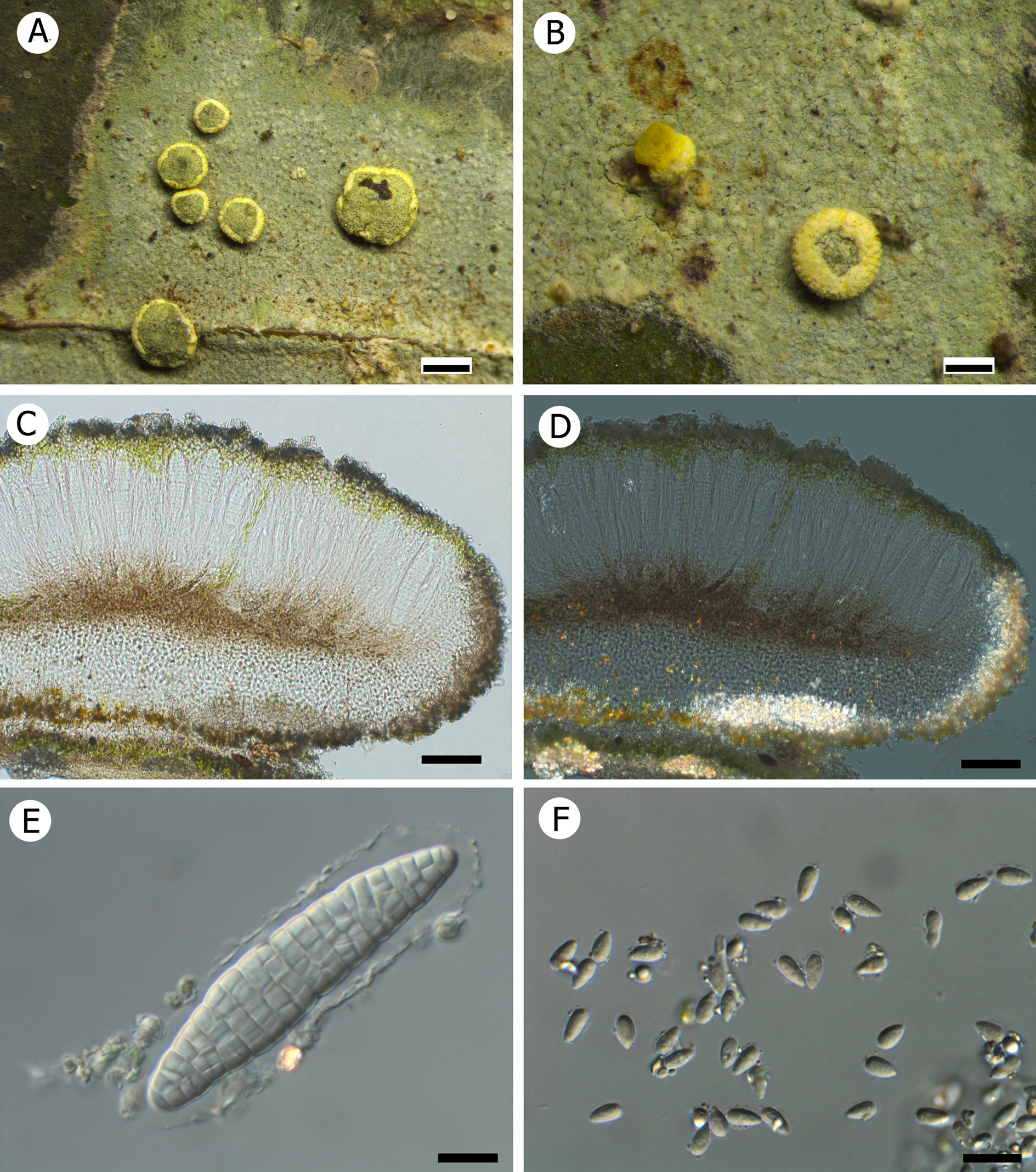
Figure 7. Long description
A six-panel figure labeled A through F.
* Panel A: A macroscopic photo of a pale green, crustose thallus featuring several circular, yellow-rimmed apothecia. A white scale bar at the bottom right represents 0.5 mm.
* Panel B: A closer view of the thallus showing a yellow campylidium and a single apothecium with a distinct yellow margin. A white scale bar at the bottom right represents 200 mu m.
* Panel C: A cross-section micrograph of an apothecium under normal light. It shows a layered structure with a dark upper hymenium, a brownish subhymenium, and a pale exciple. A black scale bar at the bottom right represents 50 mu m.
* Panel D: The same cross-section as panel C but viewed under polarized light using D I C microscopy. This view highlights bright, crystalline structures concentrated in the exciple and at the base of the apothecium. A black scale bar at the bottom right represents 50 mu m.
* Panel E: A high-magnification D I C micrograph of a single, elongated, muriform ascospore. The spore is divided into many small, rectangular internal cells. A black scale bar at the bottom right represents 10 mu m.
* Panel F: A D I C micrograph showing a cluster of numerous small, translucent, drop-shaped conidia scattered across the field of view. A black scale bar at the bottom right represents 10 mu m.
Thallus epiphyllous, crustose, continuous, slightly irregular in outline, 0.8–1.2 cm across, bluish grey, marginally yellowish green, surface matt, finely verrucose; verrucae dense, 0.06–0.08 mm diam., of same colour as the thallus. Hypothallus usually present, whitish, formed by free-living radiate hyphae. Photobiont a species of Chlorococcaceae; algal cells green, globose, irregularly arranged, 8–10 μm diam.
Apothecia usually numerous on thalli lacking campylidia, sessile, rounded to slightly irregular in outline, scattered, distinctly constricted at the base, 0.5–0.8 mm diam. and 240–270 μm high; disc flat when young, becoming distinctly convex at maturity, greyish green due to epithecial algal cells but completely covered by yellow pruina; margin distinct and persistent, dull, bright yellow, 60–80 μm thick. Excipulum well developed, 70–75 μm thick, typically paraplectenchymatous in its inner parts, colourless, 73–80 μm thick under the hypothecium; outermost parts grey-black with indistinct structure due to strong incrustation with colourless crystals; hypothecium up to 60 μm high, brown, not significantly changing in K; epithecium well developed, 20–25 μm high, granulose; epithecial alga abundant, algal cells small, rounded, 4–5.5 μm diam.; hymenium 80–90 μm high, colourless, I+ blue; paraphyses colourless, numerous, sparsely branched and anastomosing. Asci clavate, of the Sporopodium-type, 60–75 × 14–18 μm, with a well-developed, I+ dark blue tholus. Ascospores 1 per ascus, narrowly ellipsoid, richly muriform, colourless, with a distinct halo (easily seen in water), 55–65 × 11–15 μm, 4–6 times as long as wide (n = 30).
Campylidia sessile; lobe well developed, small, c. 200 μm wide, mussel-shaped, bright yellow; socle present but not inflated, pale yellow. Conidia drop-shaped, non-septate, 5–6 × 2.5–3 μm, colourless, abundantly produced.
Chemistry
Zeorin detected by TLC (Fig. 9-5). Due to the limited material of this species, we are unable to conduct HPLC experiments on it and can only conduct TLC. The TLC is based on a small amount of thallus and one apothecium. In the result, zeorin can be clearly seen, while other substances are relatively vague. Further experiments cannot be conducted at present due to material limitations.
Etymology
The epithet ‘flavomarginale’ refers to the yellow colour of the apothecial margin.
Habitat and distribution
Currently known only from Thailand and China, growing on leaf surfaces in the damp understorey of lowland rainforest, typically near rivers.
Notes
Sporopodium flavomarginale is a conspicuous species due to the bright yellow colour of the apothecial margins. Morphologically it is most similar to S. aurantiacum, but the latter species differs by the more orange tinge of the thallus and apothecial margins and by the excipulum being externally covered with yellowish brown crystals; it also produces chlorinated xanthones in addition to zeorin. Sporopodium flavomarginale is also close to S. lucidum Aptroot & Sipman (Aptroot & Sipman Reference Aptroot and Sipman1993), which has yellow apothecial margins and campylidia, but the colour of the latter is more bright yellow. Moreover, the apothecia of S. lucidum have a scabrose to crenulate margin and a pale brown, thinly white-pruinose disc, with scarce epithecial algal cells, and the excipulum is externally covered with yellow crystals. Besides containing chlorinated xanthones and pannarin, the yellow colour in S. lucidum is caused by vulpinic acid, a substance not found in the new species.
Sporopodium aurantiacum is known from the Neotropics and the eastern Paleotropics (Lücking Reference Lücking2008), whereas S. lucidum has been reported from Papua New Guinea (Aptroot & Sipman Reference Aptroot and Sipman1993). Sporopodium flavomarginale is so far known from Thailand.
Sporopodium flavomarginale is closely related to S. chebalingensis, which also has yellow campylidia and abundant epithecial algal cells. However, in S. chebalingensis, the thallus is green, the apothecia are non-pruinose and have a white margin, and it produces didymic acid.
Additional specimens examined
Thailand: Nakhon Ratchasima Province: Khao Yai National Park, Pha Kluay Mai Waterfall trail to Haew Suwat Waterfall, 14°23′N, 101°22′E, 800 m alt., on leaves, 5 v 2018, W. C. Wang KYW0646-1 (RAMK 31682), KYW0774 (RAMK 31704), KYW0471 (RAMK 31748).—China: Fujian Province: Yongtai County, Tianmen Mountain scenic spot, 25°49′N, 119°01′E, 500 m alt., on leaves, 9 xii 2021, W. C. Wang & X. L. Wang WWC682 (HMAS-L 163326), WWC687 (HMAS-L 163327); Dehua County, Stone Cow Mountain scenic spot, 25°38′N, 118°26′E, 600 m alt., on leaves, 12 xii 2021, W. C. Wang & X. L. Wang WWC1090 (HMAS-L 163328), WWC1095 (HMAS-L 163329).
Sporopodium pruinosum W. C. Wang & J. C. Wei sp. nov.
Fungal Names No.: FN 570960
Similar to S. lucidum but differs in having brown campylidia, and apothecia with a grey-green disc and a brown outer margin.
Type: China, Hainan Province, Wuzhishan City, Wuzhi Mountain National Forest Park, 18°54′N, 109°41′E, 800 m alt., on leaves, 12 December 2019, W. C. Wang 20192900 (HMAS-L 163330—holotype!).
(Fig. 8)
Sporopodium pruinosum (holotype, W. C. Wang 20192900 HMAS-L). A & B, thalli with apothecia and campylidia. C, sections of apothecium and campylidium under stereomicroscope. D, section of apothecium showing pigmentation. E, section of campylidium showing pigmentation. F & G, muriform ascospores. H, drop-shaped conidia. Scales: A = 1 mm; B & C = 500 μm; D & E = 200 μm; F–H = 20 μm.
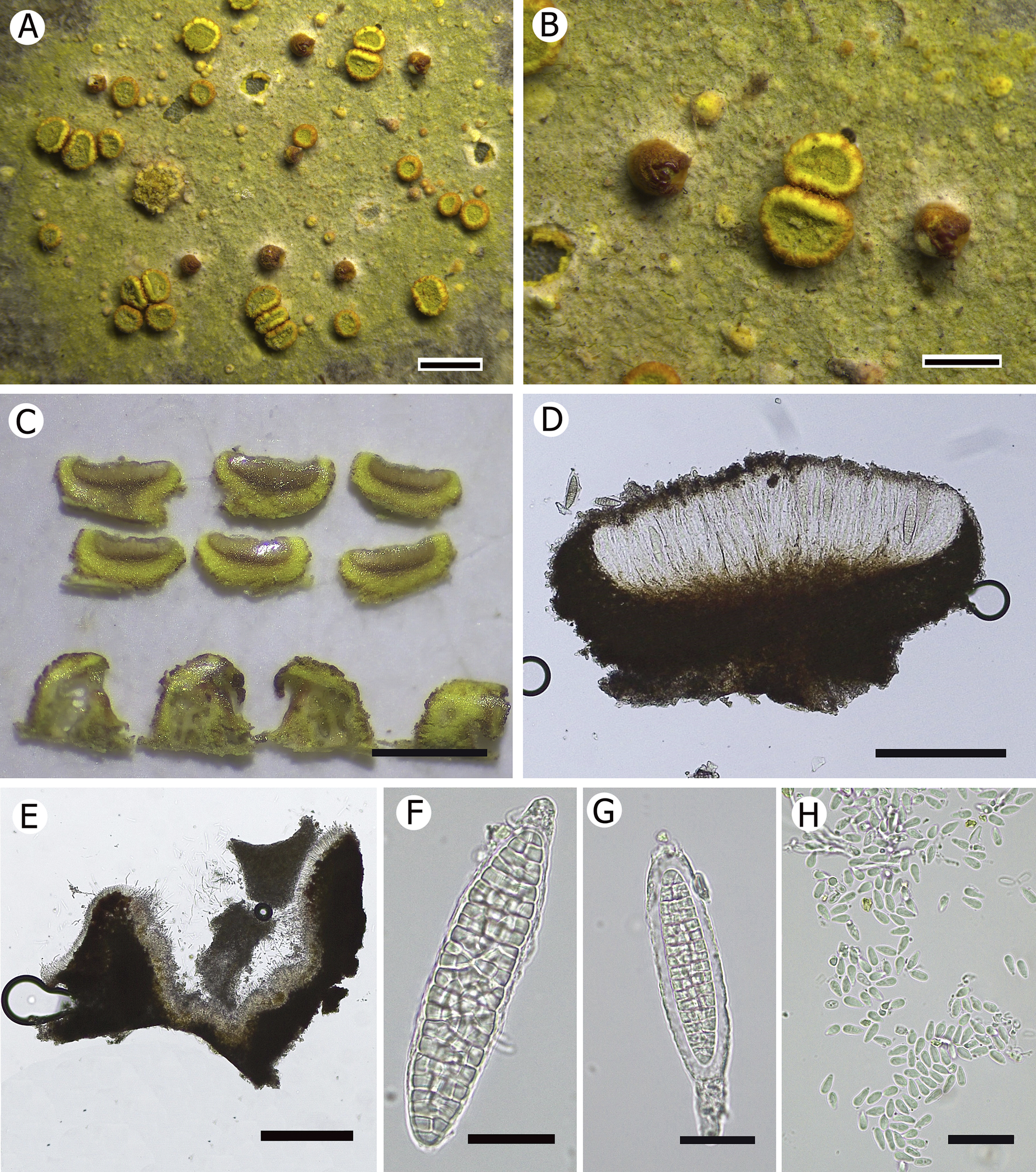
Figure 8. Long description
A multi-panel scientific photo of Sporopodium pruinosum.
* Panel A: Macroscopic view of the greenish-yellow granular thallus with scattered circular apothecia and campylidia. Scale bar is 1 mm.
* Panel B: Close-up of the thallus showing the raised, disc-like apothecia with yellow margins. Scale bar is 500 micrometers.
* Panel C: Stereomicroscope view of six cross-sections of apothecia and four sections of campylidia, showing internal layering. Scale bar is 500 micrometers.
* Panel D: Microscopic cross-section of an apothecium showing a dark brown pigmented base and a lighter, vertically oriented hymenium layer. Scale bar is 200 micrometers.
* Panel E: Microscopic cross-section of a campylidium showing dense dark pigmentation. Scale bar is 200 micrometers.
* Panels F and G: High-magnification views of individual muriform ascospores, which are elongated with a complex internal grid-like cellular structure. Scale bars are 20 micrometers.
* Panel H: Microscopic view of numerous small, translucent, drop-shaped conidia scattered across the field. Scale bar is 20 micrometers.
Thallus epiphyllous, crustose, continuous, nearly round, 0.8–1.2 cm across, yellowish green, surface matt, finely verrucose in the early stages of the thallus; verrucae dense, tiny, c. 75 μm diam., but not obvious in the later stage. White hyphae irregularly extend to the margin. Photobiont a species of Chlorococcaceae; algal cells green, globose, 8–10 μm diam.
Apothecia sessile, rounded, distinctly constricted at the base, 0.5–0.75 mm diam. and 280–320 μm high; disc flat, pruinose, greyish green, without epithecial algal cells; margin distinct and persistent, dull, outer surface brown when young, bright yellow when mature due to the brown outer layer partly peeling off, somewhat crenulate, 100–120 μm thick. Excipulum well developed, 75–90 μm thick, paraplectenchymatous, bright yellow under the dissecting microscope, but dark when examined with the compound microscope, incrusted with numerous yellow granules which do not dissolve in K; hypothecium up to 70 μm high, brown, K+ reddish brown; epithecium colourless, indistinct because of numerous colourless granules, K−; hymenium 120–140 μm high, colourless, K−; paraphyses colourless, thin, richly branched and anastomosing. Asci clavate, of the Sporopodium-type, 80–110 × 15–25 μm, with a well-developed, I+ dark blue tholus. Ascospores 1 per ascus, narrowly ellipsoid, strongly muriform, colourless, 75–100 × 15–18 μm, 4–5 times as long as wide (n = 60).
Campylidia sessile, surrounded by distinct white hyphae at the base; lobe well developed, 0.35–0.4 mm wide, mussel-shaped, outer surface brown, inner part bright yellow; socle present but not inflated. Conidia drop-shaped, non-septate, 5–6.5 × 2.5–3 μm, colourless, abundantly produced.
Chemistry
Zeorin (major) and calycin (major) determined by TLC (Fig. 9-9–12) and HPLC.
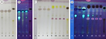
TLC (solvent C) results of the five new species; spot colour after acid and heating. A & C, natural light; B & D, UV light (365 nm). 1–3 didymic acid, Sporopodium asianum (HMAS-L 112799, RAMK 31679, HMAS-L 163279). 4 & 13 atranorin and norstictic acid (lower spots). 5 zeorin, Sporopodium flavomarginale (RAMK 31612). 6 & 7 didymic acid, Sporopodium album (HMAS-L 118415, 133043). 8 didymic acid, Sporopodium chebalingensis (HMAS-L 163323). 9–12 zeorin (purple) and calycin (yellow), Sporopodium pruinosum (HMAS-L 138627, 112825, 138635, 138629). In colour online.
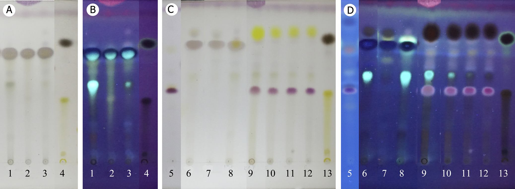
Figure 9. Long description
The T L C results are organized into four panels labeled A through D.
Panels A and B show lanes 1 through 4. Panel A is in natural light, showing light brown spots for didymic acid in lanes 1, 2, and 3, and a dark spot for atranorin with a lower yellow spot for norstictic acid in lane 4. Panel B shows the same lanes under 365 nm U V light, where lanes 1 to 3 exhibit bright blue fluorescent spots and lane 4 shows a dark quenching spot at the top.
Panels C and D show lanes 5 through 13. Panel C is in natural light. Lane 5 shows a single purple spot for zeorin. Lanes 6, 7, and 8 show light brown didymic acid spots. Lanes 9, 10, 11, and 12 show a distinct vertical pairing of a yellow calycin spot above a purple zeorin spot. Lane 13 repeats the atranorin and norstictic acid pattern.
Panel D shows lanes 5 through 13 under 365 nm U V light. Lane 5 appears as a bright blue-white spot. Lanes 6, 7, and 8 show varying intensities of blue fluorescence. Lanes 9 through 12 show dark quenching spots for calycin at the top and pinkish-purple fluorescent spots for zeorin below. Lane 13 shows a dark quenching spot at the top with a faint yellow-brown trail below.
Etymology
The epithet ‘pruinosum’ refers to its greyish green pruinose apothecial disc.
Habitat and distribution
Currently known only from tropical China (Hainan and Yunnan Provinces), growing on leaf surfaces in damp but somewhat illuminated understorey of rainforests.
Notes
Sporopodium pruinosum is similar to S. flavomarginale, which can be distinguished by its paler yellow apothecial margin and campylidia (without brown tinge), bluish grey thallus, and the presence of epithecial algal cells; chemically, it lacks calycin. The otherwise similar S. lucidum (Aptroot & Sipman Reference Aptroot and Sipman1993) has a more complex chemistry including chlorinated xanthones, and the yellow pigment is vulpinic acid instead of calycin.
Additional specimens examined
China: Hainan Province: Ledong County, Jianfeng Ridge, 18°44′N, 109°10′E, 900 m alt., on leaves, 3 iv 1993, Y. M. Jiang & S. Y. Guo H0682 (HMAS-L 138627), H0687 (HMAS-L 138629), H0655 (HMAS-L 138635); Ledong County, Jianfeng Ridge, hydrological station, 4 vi 2006, J. C. Wei s. n. (HMAS-L 112825); Wuzhishan City, Wuzhi Mountain National Forest Park, 18°54′N, 109°41′E, 800 m alt., on leaves, 2019, W. C. Wang 20192888-1 (HMAS-L 163331), 20192890-1 (HMAS-L 163332), 20192895 (HMAS-L 163333), 20192896 (HMAS-L 163334), 20193082 (HMAS-L 163335). Yunnan Province: Xishuangbanna City, Mengla County, ‘Wangtian’ Tree scenic spot, 21°37′N, 101°35′E, 689 m alt., on leaves, 29 i 2018, W. C. Wang 20180211 (HMAS-L 140678), 20180217 (HMAS-L 140684).
Acknowledgements
We are grateful to Mr Xue-Li Wang for his assistance in collecting specimens in the field in Fujian Province, China. This research was funded by the National Natural Science Foundation of China (32400009), and the Natural Science Foundation of Fujian Province, China (2023J05184).
Author ORCIDs
Wei-Cheng Wang, 0000-0002-8391-5120; Robert Lücking, 0000-0002-3431-4636.
Competing Interests
The authors declare none.