Introduction
Toxoplasma gondii is a widespread opportunistic protozoan capable of infecting a broad range of warm-blooded animals including humans, causing life-threatening toxoplasmosis in immunocompromised individuals and in congenitally infected foetuses (Saadatnia and Golkar, Reference Saadatnia and Golkar2012). Toxoplasma gondii is a member of the Apicomplexa, a phylum of parasites that is named for the apical complex containing several unique secretory organelles, such as rhoptries, micronemes and dense granules, which secrete effector molecules into the host cytosol and parasitophorous vacuole (PV) during parasite invasion and replication (Boothroyd and Dubremetz, Reference Boothroyd and Dubremetz2008; Morrison, Reference Morrison2009). Three predominant clonal lineages of T. gondii (types I, II, and III) have been identified in the majority of the isolates collected from North America and Europe, which differ genetically by 1% or even less (Sibley and Ajioka, Reference Sibley and Ajioka2008), but differ dramatically in a number of phenotypes, including growth, migration and differentiation (Radke et al., Reference Radke, Striepen, Guerini, Jerome, Roos and White2001; Barragan and Sibley, Reference Barragan and Sibley2002). The best-characterized phenotype is their virulence in laboratory mice (Mordue et al., Reference Mordue, Monroy, La Regina, Dinarello and Sibley2001), where the lethal dose (LD100) of type I strains is just a single parasite, whereas the median lethal dose (LD50) of types II and III strains range from 100 to more than 105 (Sibley and Boothroyd, Reference Sibley and Boothroyd1992; Saeij et al., Reference Saeij, Boyle, Coller, Taylor, Sibley, Brooke-Powell, Ajioka and Boothroyd2006; Sibley and Ajioka, Reference Sibley and Ajioka2008). Previous forward genetic mapping studies, in which genetic crosses between types I/II and III were used to determine the gene(s) responsible for virulence, identifying the highly polymorphic rhoptry protein ROP18 as a virulence determinant in both the I × III and II × III crosses (Saeij et al., Reference Saeij, Boyle, Coller, Taylor, Sibley, Brooke-Powell, Ajioka and Boothroyd2006; Taylor et al., Reference Taylor, Barragan, Su, Fux, Fentress, Tang, Beatty, Hajj, Jerome, Behnke, White, Wootton and Sibley2006). In murine cells, type I ROP18 (TgROP18I) has been shown to phosphorylate and inactivate immunity-related GTPases (IRGs) which are induced by interferon-γ (IFN-γ) and recruited to the parasitophorous vacuole membrane (PVM) to rupture the vacuole and kill the parasite (Fentress et al., Reference Fentress, Behnke, Dunay, Mashayekhi, Rommereim, Fox, Bzik, Taylor, Turk, Lichti, Townsend, Qiu, Hui, Beatty and Sibley2010; Steinfeldt et al., Reference Steinfeldt, Konen-Waisman, Tong, Pawlowski, Lamkemeyer, Sibley, Hunn and Howard2010). In contrast, in mouse cells, type II strains are less able to disrupt IRG recruitment to the PVM and avoid IFN-γ mediated clearance of parasites. However, this does not appear to be due to differences in the type I and type II ROP18 alleles but rather to differences in the ROP5 locus (where the Type II ROP5 locus is associated with reduced virulence (Reese et al., Reference Reese, Zeiner, Saeij, Boothroyd and Boyle2011)). Type III strains (such as CEP/CTG and VEG) are less virulent in mice compared to types I and II parasites since they lack expression of ROP18 because of a 2.1 kb insertion in ROP18 promoter (Saeij et al., Reference Saeij, Boyle, Coller, Taylor, Sibley, Brooke-Powell, Ajioka and Boothroyd2006; Taylor et al., Reference Taylor, Barragan, Su, Fux, Fentress, Tang, Beatty, Hajj, Jerome, Behnke, White, Wootton and Sibley2006; Boyle et al., Reference Boyle, Saeij and Boothroyd2007). Consequently, they fail to block IRGs and are readily cleared in IFN-γ activated cells (Hunter and Sibley, Reference Hunter and Sibley2012). While the IRGs have been extensively duplicated and diversified in rodents where there are ~20 IRG-encoding genes (Murillo-Leon et al., Reference Murillo-Leon, Muller, Zimmermann, Singh, Widdershooven, Campos, Alvarez, Konen-Waisman, Lukes, Ruzsics, Howard, Schwemmle and Steinfeldt2019), humans and most other non-rodent vertebrates have only a single full-length IRG-encoding gene, suggesting that TgROP18-mediated disruption of IRG activity only impacts mouse infection (Bekpen et al., Reference Bekpen, Hunn, Rohde, Parvanova, Guethlein, Dunn, Glowalla, Leptin and Howard2005). It is important to note that the type II ROP18 (TgROP18II) allele is functional and is able to enhance virulence in mice when heterologously expressed in a type III strain (Saeij et al., Reference Saeij, Boyle, Coller, Taylor, Sibley, Brooke-Powell, Ajioka and Boothroyd2006; Zhao et al., Reference Zhao, Ferguson, Wilson, Howard, Sibley and Yap2009; Walzer et al., Reference Walzer, Adomako-Ankomah, Dam, Herrmann, Schares, Dubey and Boyle2013). Complementation of a type III strain with either TgROP18I or TgROP18II increases Type III strain virulence by multiple orders of magnitude (Saeij et al., Reference Saeij, Boyle, Coller, Taylor, Sibley, Brooke-Powell, Ajioka and Boothroyd2006; Taylor et al., Reference Taylor, Barragan, Su, Fux, Fentress, Tang, Beatty, Hajj, Jerome, Behnke, White, Wootton and Sibley2006) and when compared head to head in mouse infections lead to similar in vivo proliferation profiles (Walzer et al., Reference Walzer, Adomako-Ankomah, Dam, Herrmann, Schares, Dubey and Boyle2013). Taken together these data suggest that the impact of Type I and Type II ROP18 during infection in the mouse model are similar. This is despite extensive polymorphism between these alleles as well as strong evidence for positive selection acting on this locus in Type I and II lineages (Saeij et al., Reference Saeij, Boyle, Coller, Taylor, Sibley, Brooke-Powell, Ajioka and Boothroyd2006). The impact of these polymorphisms is unknown.
Interestingly, in our previous high-throughput bimolecular fluorescence complementation (BiFC) based screening of TgROP18 (TgROP18I and TgROP18II) host targets in human cells, N-myc and STAT interactor (NMI) was found as a dominant target of TgROP18I, but not TgROP18II (Xia et al., Reference Xia, Kong, Zhou, Wu, Yao, He, He and Peng2018). Moreover, the interaction of TgROP18I with NMI was further confirmed by sensitized emission-fluorescence resonance energy transfer (s.e.-FRET) and co-immunoprecipitation (Co-IP) assays (Xia et al., Reference Xia, Kong, Zhou, Wu, Yao, He, He and Peng2018). NMI is an interferon-inducible protein and orthologues and paralogs for human NMI are found in all mammals and many non-mammalian vertebrates (e.g. in fish; Perera et al., Reference Perera, Godahewa, Nam and Lee2016; Liu et al., Reference Liu, Huang, Huang, Li, Ni, Yu and Qin2019). NMI plays an important role in interferon signalling and has been associated with immune responses against viral infections, including prototype foamy virus, foot-and-mouth disease virus, H3N2 swine influenza virus and Sendai virus infection (Wang et al., Reference Wang, Wang, Liu, Ding, Zhang, Li, Cao, Tang and Zheng2012, Reference Wang, Yang, Hu, Zheng, Zhou, Wang, Ma, Mao, Yang, Lin, Ji, Wu and Sun2013; Wu et al., Reference Wu, Wang, Yu, Zhang, Sun, Yan and Zhou2013; Hu et al., Reference Hu, Yang, Liu, Geng, Qiao and Tan2014). In response to IFN-γ stimulation, NMI potentiates transcription can modulate the activity of signal transducer and activator of transcription 1 (STAT1) (Zhu et al., Reference Zhu, John, Berg and Leonard1999). STAT1 is the central transcription factor for mediating the actions of IFN-γ in antimicrobial responses, cell growth and apoptosis (Rauch et al., Reference Rauch, Muller and Decker2013). When IFN-γ binds to its receptor, it induces receptor oligomerization and subsequent activation of receptor-associated Janus kinase 1 (JAK1) and JAK2, which recruits and phosphorylates STAT1 at tyrosine 701 (Platanias, Reference Platanias2005). The phosphorylated STAT1 forms homodimers and translocates to the nucleus, where it binds to gamma-activated sequence (GAS) elements, initiating transcription of IFN-γ inducible genes (Aaronson and Horvath, Reference Aaronson and Horvath2002; Stark, Reference Stark2007). One of the key early response genes is interferon regulatory factor 1 (IRF1), which is a transcription factor regulating multiple IFN-γ inducible genes (Taniguchi et al., Reference Taniguchi, Ogasawara, Takaoka and Tanaka2001). The NMI gene itself is also regulated by IRF1 (Xu et al., Reference Xu, Chai, Chen, Lin, Zhang, Li, Qiao and Tan2018) and STAT1 (Casper et al., Reference Casper, Zweig, Villarreal, Tyner, Speir, Rosenbloom, Raney, Lee, Lee, Karolchik, Hinrichs, Haeussler, Guruvadoo, Navarro Gonzalez, Gibson, Fiddes, Eisenhart, Diekhans, Clawson, Barber, Armstrong, Haussler, Kuhn and Kent2018). The NMI protein has been shown to directly interact with STAT1 in the host cell cytoplasm, augmenting IFN-γ-dependent STAT1-mediated transcription by enhancing recruitment of coactivator CREB-binding protein (CBP)/p300 to STAT1 (Zhu et al., Reference Zhu, John, Berg and Leonard1999).
Given the important role of NMI in IFN-γ/STAT1 pathway, it may not be surprising that Toxoplasma possesses effector(s) to interfere with NMI function as a strategy for evasion of IFN-γ-induced immunity. In this study, we report for the first time that TgROP18I, but not TgROP18II, disrupts NMI function and localization in human cells. This has the direct impact of reducing its binding to GAS in the IRF1 gene promoter. We have also identified the TgROP18I C-terminal domain as being required for TgROP18I effects on NMI localization in the host cell, providing new insight into the role of polymorphisms between ROP18 alleles.
Materials and methods
Parasite and cell culture
All T. gondii strains used in this study were maintained by serial passage of tachyzoites in human foreskin fibroblast (HFF) monolayers cultured at 37°C in a humidified 5% CO2 atmosphere. Strains used in this study are listed in Table S1. HFFs and human embryonic kidney 293T (HEK293T) cells were grown in Dulbecco's modified Eagle's medium (DMEM) supplemented with 10% fetal bovine serum (FBS), 2 mm glutamine and 50 mg mL−1 each of penicillin and streptomycin. Human monocytic leukaemia cell line THP-1s were grown in Roswell Park Memorial Institute (RPMI) 1640 medium supplemented with 10% FBS and 50 mg mL−1 each of penicillin and streptomycin.
Plasmid construction
For expression in mammalian cells, TgROP18I and TgROP18II genes were polymerase chain reaction (PCR)-amplified from genomic DNA of RH and ME49 strain, respectively, and FLAG-tagged with primers HindIII-TgROP18I-BamHI (Forward: 5′-CCCAAGCTTATGTTTTCGGTACAGCGGCC-3′; Reverse: 5′-CGGGATCCTTACTTGTCATCGTCATCCTTGTAATCGATGTCATGATCTTTATAATCACCGTCATGGTCTTTGTAGTCCATTTCTGTGTGGAGATG-3′) or HindIII-TgROP18II-NotI (Forward: 5′-ATTAAGCTTATGTTTTCGGTACAGCGGCCACCTC-3′; Reverse: 5′-AGCGGCCGCTTACTTGTCATCGTCATCCTTGTAATCGATGTCATGATCTTTATAATCACCGTCATGGTCTTTGTAGTCCATTTCTGTGTGGAGATG-3′). TgROP18I or TgROP18II gene was subcloned into pcDNA3.1 or pECFP-N1, and NMI gene was amplified and subcloned into pEYFP-C1 as described previously (Xia et al., Reference Xia, Kong, Zhou, Wu, Yao, He, He and Peng2018). For all of these constructs the full-length form of ROP18 (including the signal peptide and putative pro-domain) (Fentress et al., Reference Fentress, Steinfeldt, Howard and Sibley2012), with an expected size of ~60 KDa.
CEP strain parasites expressing either TgROP18I or TgROP18II were the same as those described previously (Walzer et al., Reference Walzer, Adomako-Ankomah, Dam, Herrmann, Schares, Dubey and Boyle2013) which had been transfected with either pGRA-HA-HPT-TgROP18I or pGRA-HA-HPT-TgROP18II. Each of these constructs was cloned using their endogenous promoter (Saeij et al., Reference Saeij, Boyle, Coller, Taylor, Sibley, Brooke-Powell, Ajioka and Boothroyd2006; Walzer et al., Reference Walzer, Adomako-Ankomah, Dam, Herrmann, Schares, Dubey and Boyle2013). As shown previously (Walzer et al., Reference Walzer, Adomako-Ankomah, Dam, Herrmann, Schares, Dubey and Boyle2013), the expected size of the fully processed forms of ROP18 is ~55 KDa, and the processed Type II form of ROP18 runs slightly faster on an SDS-PAGE gel than the processed Type I form of ROP18 (Walzer et al., Reference Walzer, Adomako-Ankomah, Dam, Herrmann, Schares, Dubey and Boyle2013). Point mutations in pGRA-HA-HPT-TgROP18I were introduced via site-directed mutagenesis using Q5 Site-Directed Mutagenesis Kit (NEB). To make pGRA-HA-ROP18-domain swap 1, TgROP18II N-terminus sequence, TgROP18I kinase domain sequence and pGRA-HA-HPT vector were assembled using Gibson Assembly Master Mix (NEB). pGRA-HA-HPT-ROP18-domain swap 2, which contained TgROP18I N-terminus and TgROP18II kinase domain in pGRA-HA-HPT vector, was constructed as described above. All constructs were verified by Sanger sequencing (Genewiz).
Immunoprecipitation
For the immunoprecipitation in HEK293T cells, cells were either left untransfected or transfected with pcDNA-TgROP18I-FLAG for 24 h using Lipofectamine 3000 reagent (ThermoFisher Scientific), followed by stimulation with 500 U mL−1 recombinant human IFN-γ (ThermoFisher Scientific) for 24 h. Cell lysates were prepared in Pierce IP lysis buffer (ThermoFisher Scientific) supplemented with protease inhibitor cocktail (Roche) and incubated with anti-NMI rabbit monoclonal antibody at 4°C for 1 h. Protein A agarose beads (Santa Cruz) were then added and incubated on a rotator at 4°C overnight. Beads were washed four times with phosphate-buffered saline (PBS) and eluted by boiling in LDS sample buffer (ThermoFisher Scientific). The eluates were subjected to western blotting with the indicated antibodies.
For the immunoprecipitation in HFFs, cells were left uninfected or infected with the indicated T. gondii strains at an MOI of 3 for 18 h and subsequently stimulated with 500 U mL−1 IFN-γ for 24 h. Cells were lysed as described above and cell lysates were incubated with Pierce anti-HA magnetic beads (ThermoFisher Scientific) at room temperature for 2 h. Beads were washed three times with PBS and boiled in SDS sample buffer for 10 min. The eluates were analysed by western blotting.
Western blotting
Whole-cell extracts were prepared as described above in Immunoprecipitation. Nuclear and cytoplasmic extracts were prepared using NE-PER Nuclear and Cytoplasmic Extraction Reagents (ThermoFisher Scientific) supplemented with protease inhibitor cocktail. Membranous extracts were prepared using the Subcellular Protein Fractionation Kit for Cultured Cells (ThermoFisher Scientific). Protein samples were separated by sodium dodecyl sulfate-polyacrylamide gel electrophoresis (SDS-PAGE) and subsequently electrotransferred to polyvinylidene difluoride (PVDF) membranes. The membranes were blocked in 5% bovine serum albumin (BSA) in PBS-0.05% Tween 20 (PBS-T) at room temperature for 2 h prior to incubation with primary antibodies at 4°C overnight. Membranes were then washed four times with PBS-T, followed by incubation with the corresponding secondary antibodies at room temperature for 2 h. After four PBS-T washes, blots were developed using SuperSignal™ West Pico PLUS Chemiluminescent Substrate (ThermoFisher Scientific). Antibodies used in these experiments: anti-NMI rabbit monoclonal antibody (abcam), normal rabbit control IgG (R&D Systems), anti-FLAG mouse monoclonal antibody (Abclonal), anti-β-actin rabbit monoclonal antibody (Cell Signaling Technology), anti-HA high-affinity rat monoclonal antibody (clone 3F10, Roche), anti-α-tubulin rabbit monoclonal antibody (Cell Signaling Technology), anti-histone H3 mouse monoclonal antibody (Cell Signaling Technology), anti-Na+/K+ ATPase mouse monoclonal antibody (abcam), goat anti-rabbit IgG-HRP (Southern Biotech), goat anti-mouse IgG-HRP (Southern Biotech; Schneider et al., Reference Schneider, Rasband and Eliceiri2012). Experiments were repeated at least three times.
Immunofluorescence assay (IFA) and microscopy
For IFAs in HFFs, cells were seeded on 8-well glass chamber slides (ThermoFisher Scientific) and infected with the indicated strains of T. gondii at an MOI of 0.5, or left uninfected, for 18 h. Subsequently, cells were stimulated with 500 U mL−1 IFN-γ, or not, for 24 h. Cells were then fixed with ice-cold methanol for 15 min and blocked/permeabilized with blocking buffer (5%BSA and 0.1% Triton X-100 in PBS). Cells were immunostained with the indicated primary antibodies at room temperature for 2 h, followed by incubation with the corresponding secondary antibodies at room temperature for 1 h. Cells were mounted in Vectashield mounting medium with 4′,6-diamidino-2-phenylindole (DAPI) (Vector Laboratories) and sealed with a cover glass. Cells were visualized and images were captured using an IX83 inverted fluorescence microscope (Olympus). Images were processed with ImageJ.
For the IFA in HEK293T cells, cells were seeded on coverslips in 24-well plates and cultured overnight before transfection. At 24 h after transfection, cells were stimulated with 500 U mL−1 IFN-γ for another 24 h. Cells were then fixed with 4% paraformaldehyde in PBS for 15 min and blocked/permeabilized with the blocking buffer described above. Cells were immunostained and visualized as described above.
Generation of transgenic parasites
A measure of 50 μg of HindIII-linearized plasmid was transfected into parental strain CEPΔhxgprt::Luciferase::GFP tachyzoites by electroporation for the generation of transgenic CEP parasites. Electroporation was performed in a 0.2 cm cuvette (Bio-Rad) with 2 × 10⁷ tachyzoites in Cytomix (120 mm KCl, 0.15 mm CaCl2, 10 mm KPO4, 25 mm Hepes, 2 mm EDTA, 5 mm MgCl2, pH = 7.6), 250 mm GSH, and 100 mm ATP. Tachyzoites were electroporated with 1.6 kV and 25 μFD capacitance and subjected to mycophenolic acid (MPA)/xanthine selection after overnight growth. Stable lines were cloned by limiting dilution in 96-well plates and individual clones were confirmed for transgene expression by IFA for HA staining. A control transgenic strain was generated by transfecting HindIII-linearized pGRA-HA-HPT empty vector into CEPΔhxgprt::Luciferase::GFP tachyzoites. Electroporation, drug selection and stable clones isolation were performed as described above.
RNA isolation and quantitative reverse transcription PCR (qRT-PCR)
HEK293T cells were left untransfected or transfected with the indicated plasmid for 24 h, and subsequently stimulated with 500 U mL−1 IFN-γ, or not, for 24 h. Total RNA was isolated using RNeasy Mini Kit (QIAGEN) with DNase treatment, and the first-strand cDNA was synthesized using SuperScript III Reverse Transcriptase (ThermoFisher Scientific). Real-time PCR was carried out on QuantStudio 3 Real-Time PCR System (ThermoFisher Scientific) using iQ SYBR Green Supermix (Bio-Rad). Each sample was run in triplicate and relative quantitation was determined using comparative Ct method with data normalized to the housekeeping gene, β-actin. Primers used and product sizes were as follows: NMI (Forward: 5′- CAGGAGTCAGATTCCAGGTTTAT-3′; Reverse: 5′- GCTCAGCTCTAGTTTGTCTCTC-3′; 110 bp), IRF1(Forward: 5′- GATGAGGATGAGGAAGGGAAAT-3′; Reverse: 5′- GGACTCCAGGTTCATTGAGTAG-3′; 115 bp) and β-actin (Forward: 5′-AGCACTGTGTTGGCGTACAG-3′; Reverse: 5′-TCCCTGGAGAAGAGCTACGA-3′; 194 bp). Real-time PCR results were expressed as fold change compared to the untransfected and unstimulated sample, and were derived from three independent experiments.
Chromatin immunoprecipitation (ChIP)
HFFs were infected with the indicated strains of T. gondii at an MOI of 6, or left uninfected, for 18 h. Subsequently, cells were stimulated with 500 U mL−1 IFN-γ, or not, for 24 h. Cells were cross-linked with 1% formaldehyde in culture medium for 10 min, followed by quenching with 1.25 m glycine for 5 min. Cells were then lysed in SDS lysis buffer (Millipore Sigma) and sonicated four times for 10 s with 40 s intervals using a Fisher Scientific sonicator at 40 amplitude. The ChIP assay was performed using a One-Step ChIP Kit (abcam), anti-NMI rabbit monoclonal antibody (abcam) was used for immunoprecipitation, and normal rabbit IgG (R&D Systems) was used as a negative control. Cross-links were reversed by heating the samples at 65°C overnight and DNA was purified using a GeneJET PCR Purification Kit (ThermoFisher Scientific). Input samples were subjected to electrophoresis on 1.5% agarose gels to check the sheared DNA fragment size (Fig. S1). ChIPed and input samples were analysed by real-time PCR conducted as described above using the published primers (Olias et al., Reference Olias, Etheridge, Zhang, Holtzman and Sibley2016) to amplify the GAS in the IRF1 gene promoter. Each sample was run in triplicate and relative quantitation was determined using comparative Ct method with data normalized to the input samples. Real-time PCR results were expressed as fold change compared to the negative control and were derived from three independent experiments.
Parasite replication assay
For the parasite replication assay in HFFs, cells were grown on 8-well glass chamber slides and infected with equal numbers (8 × 10⁴ tachyzoites) of the indicated strains of T. gondii for 18 h, followed by stimulation with 500 U mL−1 IFN-γ for 24 h. Cells were observed for any adverse effects due to IFN-γ treatment and none were observed. Cells were then fixed with 4% paraformaldehyde in PBS for 15 min and mounted in Vectashield mounting medium with DAPI (Vector Laboratories). The number of vacuoles containing parasites at different replication stages (i.e. 1, 2, 4, 8, 16, 32, or more than 32 tachyzoites) was counted by epifluorescence microscopy from 20 separate field of view. Experiments were repeated three times.
For the parasite replication assay in monocytes, THP-1s were grown on 8-well glass chamber slides and pretreated with 100 ng mL−1 phorbol 12-myristate 13-acetate (PMA) for 24 h. 2 × 10⁵ tachyzoites of the indicated strains were allowed to infect the PMA-pretreated cells for 18 h prior to stimulation with 500 U mL−1 IFN-γ for 24 h. Cells were observed for any adverse effects of IFN-γ treatment and none were observed. Cells were fixed and mounted, and parasite replication was examined as described above. Experiments were repeated three times.
Sequence alignment
For selection analysis, ROP18 coding sequences for 11 T. gondii strains were downloaded from ToxoDB.org and aligned using Muscle as implemented in MEGA6 (Tamura et al., Reference Tamura, Stecher, Peterson, Filipski and Kumar2013). Phylogenetic trees of amino acid alignments were made using maximum likelihood and bootstrapped using 1000 permutations. Pairwise selection analyses were made using the Codon-Based Fisher's Exact Test for selection. In both cases, the default settings as implemented in MEGA6 were used.
Results
TgROP18I, but not TgROP18II, interacts with NMI in human cells
To identify the potential host proteins targeted by TgROP18 (TgROP18I and TgROP18II), we previously applied a genome-wide BiFC-based protein–protein interaction (PPI) screening in which a human ORFeome library (Yang et al., Reference Yang, Boehm, Yang, Salehi-Ashtiani, Hao, Shen, Lubonja, Thomas, Alkan, Bhimdi, Green, Johannessen, Silver, Nguyen, Murray, Hieronymus, Balcha, Fan, Lin, Ghamsari, Vidal, Hahn, Hill and Root2011) containing more than 18 000 human cDNA clones was used as prey (Xia et al., Reference Xia, Kong, Zhou, Wu, Yao, He, He and Peng2018). In this dual-expression system, NMI was shown to interact with TgROP18I, but not TgROP18II and NMI had the highest total reads among TgROP18I-interacting proteins (Xia et al., Reference Xia, Kong, Zhou, Wu, Yao, He, He and Peng2018). The impact of this interaction on endogenous NMI protein levels, subcellular localization and NMI downstream function was unknown. Moreover, it was not known if the ROP18I-NMI interaction was relevant during parasite infection (rather than only during ectopic expression in host cells).
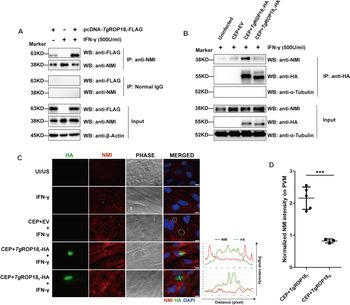
To determine whether TgROP18I interacted with endogenous NMI in human cells (rather than only during dual-overexpression of TgROP18I and NMI; Xia et al., Reference Xia, Kong, Zhou, Wu, Yao, He, He and Peng2018), we transfected an expression construct encoding TgROP18I-FLAG into HEK293T cells and treated the cells with IFN-γ to induce expression of NMI. After immunoprecipitation using anti-NMI rabbit monoclonal antibody, TgROP18I-FLAG was readily detected in the immunoprecipitates, but not after using rabbit normal IgG (Fig. 1A). To determine whether the interaction occurred during infection with parasites expressing TgROP18I, HFFs were infected for 18 h with T. gondii CEP (a type III strain), or the CEP strain complemented with HA-tagged TgROP18I (CEP + TgROP18I) or TgROP18II (CEP + TgROP18II) and were then stimulated with IFN-γ for 24 h. After anti-HA immunoprecipitation, endogenous NMI was found to significantly co-immunoprecipitate with TgROP18I, but not TgROP18II (which run at slightly different apparent molecular weights as shown previously; Walzer et al., Reference Walzer, Adomako-Ankomah, Dam, Herrmann, Schares, Dubey and Boyle2013; Fig. 1B). When NMI was localized in infected cells, IFN-γ induced NMI was found to localize to the parasite-containing vacuole membrane in CEP + TgROP18I parasites (Fig. 1C), whereas this was not found to be present in HFFs infected with CEP or CEP + TgROP18II parasites (Fig. 1C). We quantified this difference across multiple coverslips and parasite-containing vacuoles (Fig. 1C and D) and found that cells infected with CEP + TgROP18I had significantly higher NMI staining on the PVM compared to those infected with CEP + TgROP18II (Fig. 1D). Collectively, these data demonstrate a TgROP18I-specific interaction (whether direct or indirect) with NMI during ectopic expression in mammalian cells and during Toxoplasma infection in IFN-γ-stimulated cells, and that one potential impact of this interaction is relocalization of IFN-γ-stimulated NMI to the PVM.
TgROP18I, but not TgROP18II, co-precipitates with NMI in human cells. (A) HEK293T cells were transfected with pcDNA-TgROP18I-FLAG, or left untransfected, for 24 h and subsequently treated with IFN-γ for 24 h. (B) HFFs were infected with the indicated strains at an MOI of 3, or left uninfected, for 18 h and subsequently treated with IFN-γ for 24 h. Note the efficacy of the HA-pulldown of TgROP18I or TgROP18II and the fact that these proteins run at distinct apparent molecular weights as described previously (Walzer et al., Reference Walzer, Adomako-Ankomah, Dam, Herrmann, Schares, Dubey and Boyle2013). (A and B) Cells were then lysed for immunoprecipitation and western blotting. (C) HFFs were infected with the indicated strains at an MOI of 0.5 or left uninfected, for 18 h and subsequently stimulated or not, with IFN-γ for 24 h. Immunofluorescence was performed with rat monoclonal anti-HA (green) and rabbit monoclonal anti-NMI (red). UI/US, uninfected and unstimulated HFFs (scale bar = 10 μm). Dotted circles on the image indicate the location of the parasite-containing vacuole for strain CTG transfected only with empty vector (EV). White lines on the merged images represent the regions used for the fluorescence intensity analysis shown in the panel to the right of the image. (D) Mean fluorescence intensity of NMI on the PVM, determined by immunofluorescence assay and quantification using ImageJ. Immunofluorescence assay was performed 5 times for CEP + TgROP18I and 3 times for CEP + TgROP18II infection. Each data point indicates the mean fluorescence intensity of NMI on the PVM normalized to the mean fluorescence intensity of NMI in cytosol in the same cell, combined from a minimum of 10 vacuoles and a maximum of 34 vacuoles. ***, student's t-test, P < 0.001.

The C-terminal kinase domain of TgROP18I is required for allele-specific immunoprecipitation of NMI
As for all ROP2 family members, the ROP18 protein contains an N-terminal signal peptide (residues 1–27, numbering based on the first methionine codon predicted to be a part of the CDS), which is subjected to proteolytic cleavage during intracellular trafficking (Sadak et al., Reference Sadak, Taghy, Fortier and Dubremetz1988; Qiu et al., Reference Qiu, Wernimont, Tang, Taylor, Lunin, Schapira, Fentress, Hui and Sibley2009). The signal peptide is followed by an arginine-rich helical region that has been identified as a vacuole-targeting domain required for PVM association (Reese and Boothroyd, Reference Reese and Boothroyd2009). The C-terminus (residues 187–554) of ROP18 encodes a conserved serine/threonine kinase domain, which is functional and its catalytic activity is required for conferring virulence in mice when expressed in a TgROP18 null background (Taylor et al., Reference Taylor, Barragan, Su, Fux, Fentress, Tang, Beatty, Hajj, Jerome, Behnke, White, Wootton and Sibley2006). Given that TgROP18I and NMI were co-localized on PVM, we sought to identify which part of the TgROP18I protein was required for the observed allele-specific NMI localization. To do this we made chimeric sequences consisting of the N-terminus of TgROP18II and kinase domain of TgROP18I (TgROP18-domain swap 1, or TgROP18-DS1), and the reverse (TgROP18-domain swap 2, or TgROP18-DS2) using Gibson Assembly (Gibson et al., Reference Gibson, Young, Chuang, Venter, Hutchison and Smith2009) (Fig. 2A). We found that cells infected with parasites expressing TgROP18-DS1 had similar levels of vacuole-associated NMI compared to WT TgROP18I, while the TgROP18-DS2 chimera resulted in vacuoles with significantly reduced vacuolar NMI localization compared to WT TgROP18I, implicating the C-terminal region (including the protein kinase domain) of TgROP18I as being important for the NMI-associating phenotype of TgROP18I (Fig. 2A and B).
The type I kinase domain and catalytic residue of TgROP18I is required for allele-specific NMI association. (A) HFFs were infected with the indicated strains at an MOI of 0.5 or left uninfected, for 18 h and subsequently stimulated or not, with IFN-γ for 24 h. Double immunofluorescence was performed with rat monoclonal anti-HA (green) and rabbit monoclonal anti-NMI (red) (scale bar = 10μm). (B) Normalized fluorescence intensity of NMI on PVM, determined by immunofluorescence assay using ImageJ software. Mean ± s.d. indicates the fluorescence intensity of NMI on PVM normalized to the fluorescence intensity of NMI in cytosol in the same cell, combined from a minimum of 10 vacuoles and a maximum of 18 vacuoles. ***, one-way ANOVA, P < 0.001.

Kinase-dead TgROP18I mutant fails to drive vacuolar NMI association
The catalytic activity of TgROP18I is essential for the impact of ROP18I on virulence when expressed in less virulent and ROP18null genetic background (Saeij et al., Reference Saeij, Boyle, Coller, Taylor, Sibley, Brooke-Powell, Ajioka and Boothroyd2006; Taylor et al., Reference Taylor, Barragan, Su, Fux, Fentress, Tang, Beatty, Hajj, Jerome, Behnke, White, Wootton and Sibley2006; Yamamoto et al., Reference Yamamoto, Ma, Mueller, Kamiyama, Saiga, Kubo, Kimura, Okamoto, Okuyama, Kayama, Nagamune, Takashima, Matsuura, Soldati-Favre and Takeda2011). Given that the kinase domain of TgROP18I is required for localization of NMI at the PVM, we asked whether the kinase activity of TgROP18I was required for this association. To directly test this, we generated a kinase-dead TgROP18I mutant (TgROP18I-KD) by mutating Asp-427, which is required for TgROP18I kinase activity (El Hajj et al., Reference El Hajj, Lebrun, Arold, Vial, Labesse and and Dubremetz2007), to alanine. When expressed in a type III genetic background, wild-type TgROP18I mediated NMI-vacuolar association but both the TgROP18I-KD and wild-type TgROP18II did not (Fig. 2A and B). These data indicate that either (a) TgROP18I kinase activity is required to mediate relocalization of host NMI on the PVM or (b) that D427 is involved in interactions with NMI or other proteins required for NMI vacuolar localization.
TgROP18I, but not TgROP18II, reduces endogenous NMI protein levels after induction by IFN-γ
In the input samples prepared from the HEK293T cells transfected with TgROP18I expression construct (Fig. 1A, Lanes 1 and 3) and the HFFs infected with CEP + TgROP18I parasites (Fig. 1B, Lane 3), we noticed a decrease in NMI protein level compared to control experiments and those with TgROP18II. To determine whether the decreased level of NMI protein is caused by a suppressive effect of TgROP18I on NMI gene transcription, we quantified NMI transcript abundance in HEK293T cells ectopically expressing TgROP18I or TgROP18II (Fig. 3A). No significant change in NMI mRNA level by TgROP18I or TgROP18II was observed, suggesting that reduced NMI levels associated with TgROP18I (and not TgROP18II) were not due to changes in NMI transcription (Fig. 3A). To verify the impact of TgROP18I on IFN-induced NMI levels, we measured NMI protein abundance in HEK293T cells transfected with a constant amount (2μg) of pEYFP-NMI and an increasing amount (0, 0.5, 1, or 2 μg) of pECFP-TgROP18I (Fig. 3B). As with the experiments in Fig. 3B, NMI protein abundance decreased with increasing amounts of pECFP-TgROP18I plasmid (Fig. 3B). This suggests that the impact of TgROP18I on NMI protein level is associated with its ability to drive localization of NMI at the PVM.
TgROP18I, but not TgROP18II, reduces NMI protein induction by IFN-γ. (A and B) HEK293T cells were transfected with the indicated plasmid or not, for 24 h and subsequently stimulated with IFN-γ for 24 h. (A) NMI mRNA were assessed by qRT-PCR, normalized to β-actin. UT, untransfected HEK293 T cells. US, unstimulated HEK293T cells. (B) Lysates of cells were analyzed through western blotting. Densitometric quantification (fold change) of NMI/β-Actin in the blot using ImageJ software. One-Way ANOVA followed by Dunnet's multiple comparison post-test using Group 8 as the control; **, P < 0.01; ***, P < 0.001.

TgROP18I disrupts IFN-γ induced NMI IRF1-GAS binding
In the presence of IFN-γ, NMI binds to STAT1 and translocates to the nucleus, where it binds to GASs with STAT1, modulating transcription of various IFN-γ inducible genes by facilitating recruitment of CBP/p300 (Zhu et al., Reference Zhu, John, Berg and Leonard1999). Recently, it has been reported that a human cytomegalovirus protein, UL23 interacts with NMI and disrupts its nuclear translocation and modulation of IFN-γ inducible gene transcription (Feng et al., Reference Feng, Sheng, Vu, Liu, Foo, Wu, Trang, Paliza-Carre, Ran, Yang, Sun, Deng, Zhou, Lu, Li and Liu2018). As our data above shows, TgROP18I co-immunoprecipitates with NMI (Fig. 1), and therefore we hypothesized that reduced overall NMI protein levels in cells expressing TgROP18I or infected with TgROP18I-expressing parasites (as Fig. 3 shows) could also lead to decreased nuclear-associated NMI. To assess whether the distinct subcellular localization of NMI mediated by TgROP18I results in attenuated DNA association of NMI, we examined the binding of NMI to GAS in the IRF1 gene promoter using ChIP assay. IFN-γ treatment increased the recruitment of NMI to IRF1-GAS, which was diminished globally by pre-infection with Toxoplasma (Fig. 4A). NMI association with IRF1-GAS was significantly reduced in the presence of CEP + TgROP18I parasites, compared to CEP or CEP + TgROP18II parasites, indicating that the reduced nuclear-associated NMI by TgROP18I leads to impaired NMI association with GAS at the IRF1 locus (Fig. 4A). Since NMI was shown to be recruited to the PVM surrounding CEP + TgROP18I parasites (Fig. 1C and D), we, therefore, wondered whether the decreased NMI protein levels in nucleus and cytoplasm was due to TgROP18I sequestration of NMI on the PVM. To test this hypothesis, HEK293T cells were preinfected with wild-type CTG strain, a type III strain that lacks expression of TgROP18, at an MOI of 1 for 18 h, followed by transfection of pcDNA-TgROP18I, pcDNA-TgROP18II, or pcDNA3.1 empty vector for 24 h. Preinfection of CTG parasites enabled the formation of PVM in HEK293T cells, and the distribution of NMI protein was then examined by analysing NMI protein levels in cytoplasmic, membranous and nuclear fractions isolated from these cells. The purity of the fractions was confirmed by western blotting for α-tubulin (cytoplasmic marker), Na+/K+ ATPase (membranous marker) and histone H3 (nuclear marker). As shown in Fig. 4B, cytoplasmic, membranous and nuclear levels of NMI were diminished in the presence of TgROP18I (Fig. 4B) but not TgROP18II. The fact that the membrane fraction also contained reduced NMI suggests that visible accumulation of NMI on the PV during infection with TgROP18I-expressing parasites occurs despite the fact that overall levels of this host factor are lower. Collectively, these data indicate that TgROP18I affects overall NMI protein levels in infected cells, and this includes nuclear NMI which is the most likely cause for the observed overall reduction in NMI binding to GAS in the IRF1 gene promoter (Fig. 4A).
TgROP18I disrupts IFN-γ induced NMI IRF1-GAS binding by decreasing nuclear-associated NMI. (A) Chromatin Immunoprecipitation (ChIP) followed by qPCR for of anti-NMI from HFFs infected with the indicated strains ± IFN-γ (500 U mL−1). Normalized qPCR analysis for the GAS site in promoter elements of IRF1 is shown. UI/US, uninfected and unstimulated HFFs. **, one-way ANOVA, followed by Dunnet's post-hoc test; P < 0.01; ***, P < 0.001. (B) HEK293T cells were infected with CTG parasites at an MOI of 1 for 18 h, followed by transfection of pcDNA-TgROP18I, pcDNA-TgROP18II, or pcDNA3.1 empty vector for 24 h. Cells were subsequently treated with 500 U mL−1 IFN-γ for 8 h, and cytoplasmic, membranous and nuclear extracts were prepared and analysed by western blotting.

TgROP18I is not sufficient to confer parasite resistance to IFN-γ
The ability of TgROP18I to inhibit NMI IRF1-GAS binding prompted us to investigate whether TgROP18I played a role in inhibiting IFN-γ induced IRF1 transcript expression since NMI is a known potentiator of STAT1 activity. To test this possibility, we transfected pcDNA-TgROP18I, pcDNA-TgROP18II, or the empty plasmid pcDNA3.1 into HEK293T cells. At 24 h post-transfection, cells were treated with IFN-γ, or not, for another 24 h. IRF1 transcription was remarkably augmented by IFN-γ stimulation as expected, regardless of whether cells were transfected or not (Fig. S2). Similar to NMI transcript abundance seen in Fig. 3A, neither TgROP18I nor TgROP18II significantly impacted IRF1 transcript abundance (Fig. S2A) or nuclear IRF1 in infected (Fig. S2B and C) or transfected (Fig. S2D and E) cells. Interestingly, both TgROP18I and TgROP18II showed a significant inhibitory effect on the transcription of another IFN-γ inducible gene, IFP35 (Fig. S3). However, this effect is unlikely to be due to defective NMI signalling since TgROP18I and TgROP18II equally inhibit IFP35 transcription, and impairment of NMI DNA association is specific to TgROP18I (Fig. 4A).
To determine if TgROP18I could alter IRF1 transcription during parasite infection, we infected HFFs with CEP, CEP + TgROP18I or CEP + TgROP18II parasites for 18 h, or left cells uninfected and subsequently stimulated the cells with IFN-γ or normal medium for 24 h and then visualized IRF1 nuclear accumulation by IFA (Fig. S2B). As expected, IRF1 expression was induced by IFN-γ treatment and strong nuclear staining of IRF1 was observed in IFN-γ treated cells. This induction was significantly inhibited by prior infection with either CEP, CEP + TgROP18I, or CEP + TgROP18II parasites, while the neighbouring uninfected cells showed normal induction of IRF1 under IFN-γ stimulation (Fig. S4B and C). Given that TgIST has been shown to block STAT1-mediated gene expression in all three clonal types of T. gondii infected cells (Gay et al., Reference Gay, Braun, Brenier-Pinchart, Vollaire, Josserand, Bertini, Varesano, Touquet, De Bock, Coute, Tardieux, Bougdour and Hakimi2016; Olias et al., Reference Olias, Etheridge, Zhang, Holtzman and Sibley2016), we postulated the involvement of TgIST in inhibiting IRF1 nuclear accumulation in the CEP strains infected HFFs. To determine whether TgROP18I alone played a role in IRF1 expression, we, therefore, expressed FLAG-tagged TgROP18I or TgROP18II in HEK293T cells and subsequently treated the cells with IFN-γ, and then visualized IRF1 expression in nucleus by IFA (Fig. S2D). Cells expressing TgROP18I or TgROP18II showed an equally high level of IRF1 expression in the nucleus to those seen in untransfected cells and similar results were found in control cells expressing GFP (Fig. S4D and E). Thus, TgROP18I is not required for inhibition of IFN-γ induced IRF1 expression, and the TgROP18I-NMI interaction is not sufficient for IRF1 repression, even though the binding of NMI to GAS in IRF1 gene promoter is remarkably disrupted by TgROP18I (Fig. 4A).
IFN-γ is the major mediator of host immune defence against Toxoplasma infection (Suzuki et al., Reference Suzuki, Orellana, Schreiber and Remington1988; Yap and Sher, Reference Yap and Sher1999) and it is thought that blocking IFN-γ signalling is required for parasite immune evasion and intracellular survival. To test whether TgROP18I facilitates parasite resistance to IFN-γ treatment via interference with NMI, we compared parasite replication by counting a number of parasites per vacuole in HFFs that were pre-infected with either CEP, CEP + TgROP18I or CEP + TgROP18II parasites for 18 h and then stimulated, or not, with IFN-γ for 24 h (Fig. 5A). In the absence of IFN-γ, a statistically significant increase in parasite replication was shown in CEP + TgROP18II parasites, with most vacuoles containing 8 tachyzoites, while most vacuoles contained 4 tachyzoites in CEP + TgROP18I infected cells (Fig. 5A). IFN-γ treatment showed a slightly inhibitory effect on parasite replication of all of the three strains, where more vacuoles containing a single tachyzoite and less vacuoles containing 16 tachyzoites were observed (Fig. 5A). However, no significant difference was identified in the distribution of vacuoles with different numbers of tachyzoites among the three strains in the presence of IFN-γ (Fig. 5A). To see whether TgROP18I plays a role in parasite resistance to IFN-γ in the context of monocytes, we examined parasite replication of CEP, CEP + TgROP18I and CEP + TgROP18II strains in PMA-transformed monocytes (Fig. 5B). For all of the three strains infected monocytes, more vacuoles (52–55%) with a single tachyzoite observed in IFN-γ treated cells than those observed in untreated cells indicated a lag of parasite replication state upon IFN-γ treatment, however, there was no significant difference in parasite replication among the three strains in the presence of IFN-γ (Fig. 5B). Collectively, these data show that TgROP18I does not greatly affect parasite replication or parasite resistance to IFN-γ treatment in HFFs or monocytes.
TgROP18I is not sufficient to confer parasite resistance to IFN-γ during growth in vitro (A) HFFs were infected with the indicated strains (8 × 10⁴ parasites) for 18 h and subsequently stimulated with IFN-γ for 24 h, then the number of vacuoles containing 1, 2, 4, 8, 16, 32, or more than 32 parasites was determined by epifluorescence microscopy. (B) THP-1 monocytes were infected with the indicated strains (2 × 10⁵ parasites) for 18 h and subsequently stimulated with IFN-γ for 24 h, then the number of vacuoles containing 1, 2, 4, 8, 16, 32, or more than 32 parasites was determined by epifluorescence microscopy. Mean ± s.d. was combined from 20 separate fields of view, counting a minimum of 125 vacuoles and a maximum of 298 vacuoles. Experiments were repeated three times. ***, χ 2 test, P < 0.001.

Discussion
Toxoplasma gondii ROP18 is a potent virulence factor and is highly divergent between strains likely due to selection-driven diversification (Saeij et al., Reference Saeij, Boyle, Coller, Taylor, Sibley, Brooke-Powell, Ajioka and Boothroyd2006; Boyle et al., Reference Boyle, Saeij and Boothroyd2007). The type I and type II TgROP18 alleles have 30 nucleotide polymorphisms between them, resulting in 28 amino acid polymorphisms (dN/dS ratio = 4.6). These observations are consistent with the alignments of TgROP18 alleles from 11 different strains found on ToxoDB, which also show strong patterns of polymorphism that are indicative of selection-driven diversification (Fig. S4A). Importantly, while there is no statistical evidence for selection in determining differences in the type III clade of TgROP18 alleles (most typified by TgVEG; Fig. S4B), the type I and type II clades show strong evidence for selection-driven diversification between them. The lack of selection in the type III background is consistent with a neutral phenotype for this particular allele since the gene in this genetic background is predicted to encode a full-length protein but an indel in the promoter has effectively inactivated TgROP18III transcription (Boyle et al., Reference Boyle, Saeij and Boothroyd2007). This provides further evidence for the impact of selection-driven diversification at the TgROP18 loci belonging to the type I and type II clades (Fig. S4B). The present study represents the first that has tied any functional significance to these sequence differences. Given the critical role played by ROP18 during infection in murine hosts based on its interaction with members of the IRG protein family, it is possible that some of these differences evolved in response to selective pressures in rodent hosts. However, here we show evidence for allele-specific targeting of NMI, a single copy gene that is conserved across all vertebrate phyla.
Mechanistic studies in human and murine cells clearly demonstrate a strong affinity of ROP18 for the host cell cytoplasm side of the PVM (Fentress et al., Reference Fentress, Behnke, Dunay, Mashayekhi, Rommereim, Fox, Bzik, Taylor, Turk, Lichti, Townsend, Qiu, Hui, Beatty and Sibley2010; Steinfeldt et al., Reference Steinfeldt, Konen-Waisman, Tong, Pawlowski, Lamkemeyer, Sibley, Hunn and Howard2010; Du et al., Reference Du, An, Chen, Shen, Chen, Cheng, Jiang, Zhang, Yu, Chu, Shen, Luo, Chen, Wan, Li, Xu and Shen2014). This interaction (which is conserved across all T. gondii strain types which express TgROP18 (Saeij et al., Reference Saeij, Boyle, Coller, Taylor, Sibley, Brooke-Powell, Ajioka and Boothroyd2006; Taylor et al., Reference Taylor, Barragan, Su, Fux, Fentress, Tang, Beatty, Hajj, Jerome, Behnke, White, Wootton and Sibley2006) and also in H. hammondi (Walzer et al., Reference Walzer, Adomako-Ankomah, Dam, Herrmann, Schares, Dubey and Boyle2013)) is mediated by amphipathic helices found within the ROP18 N-terminus (Reese and Boothroyd, Reference Reese and Boothroyd2009). This localization is less obvious during infection (although can be quantified using image analysis as shown in Fig. 1C; see also Behnke et al., Reference Behnke, Fentress, Mashayekhi, Li, Taylor and Sibley2012; Fentress et al., Reference Fentress, Steinfeldt, Howard and Sibley2012), but is quite obviously visible during ectopic expression in host cells followed by infection (Reese and Boothroyd, Reference Reese and Boothroyd2011). Consistent with this localization for ROP18 and its interactions with NMI, TgROP18I-expressing parasites infected cells treated with IFN-γ show relocalization of NMI around the PVM (Fig. 1C). The strain-specificity of this relocalization is driven by the kinase domain of TgROP18I (Fig. 2). However, we also found that the catalytic residue of TgROP18I is also required to localize NMI at the PV (Fig. 2). The D427A mutation in TgROP18I-KD leads to the loss of catalytic activity of TgROP18I, and also its capacity of NMI association (Fig. 2). The mechanism for how mutating the catalytic TgROP18I residue abrogates interactions with NMI is unclear, although it suggests that specific polymorphisms unique to the Type I allele of ROP18 mediate interactions with NMI (either directly or indirectly) and to do this requires active kinase.
Overall, these studies reveal a potential new host pathway targeted by ROP18 (and specifically TgROP18I) in non-rodent cells. While data are lacking in T. gondii, it is clear from studies in other host-pathogen systems that the existence of multiple divergent targets for host-interacting effectors is the rule rather than the exception. This is most remarkably illustrated by the pleiotropic SV-40 effector large T-antigen which has at least ten distinct targets in the host cell (Ahuja et al., Reference Ahuja, Saenz-Robles and Pipas2005). While the mechanisms by which TgROP18I suppresses the induction of NMI protein by IFN-γ remain unknown, our data indicate that this inhibitory impact relies on protein binding. Pathways that mediate protein degradation, such as proteasomal or lysosomal proteolysis (Huber and Teis, Reference Huber and Teis2016; Budenholzer et al., Reference Budenholzer, Cheng, Li and Hochstrasser2017), might be possibly involved in TgROP18I/NMI interaction and would be worth testing. While we did not detect any impact of TgROP18I inhibition of NMI on the magnitude of IFN-γ-induced IRF1 expression (Fig. S2) or parasite growth in the presence of IFN-γ (Fig. 5), which might be due to the important but dispensable role of NMI in IFN-γ/STAT1 pathway, our data show that by interacting with NMI, TgROP18I has the capacity to directly modulate the cellular response to IFN-γ in a cell-autonomous fashion. It is difficult to imagine how this would not have an impact during infection, given the known role for this factor in potentiating IFN-γ responses, although we have not investigated this directly. This might be particularly important in hosts (including humans) that lack a well-developed repertoire of immune-related GTPases which are a major target in TgROP18I and TgROP18II (among other effectors) in mice (Feng et al., Reference Feng, Sheng, Vu, Liu, Foo, Wu, Trang, Paliza-Carre, Ran, Yang, Sun, Deng, Zhou, Lu, Li and Liu2018). Investigating the impact of suppression of NMI by TgROP18I in different cell contexts will be worthwhile. It will also be interesting to see if key residues in TgROP18I that are required for NMI suppression are also critical for interactions with other known ROP18 targets such as ATF6β (Yamamoto and Takeda, Reference Yamamoto and Takeda2012).
Supplementary material
The supplementary material for this article can be found at https://doi.org/10.1017/S0031182020001249.
Acknowledgements
The authors would like to thank members of the Boyle lab for critical reading of the manuscript.
Financial support
This research was supported by National Key Research and Development Program of China (2017YFD0500400) to HJP; National Institutes of Health (NIH) grant NIH-R01AI114655 and NIH-R01AI116855 to JPB; National Natural Science Foundation of China (No. 81572012, 81772217, 20180907), Guangdong Provincial Natural Science Foundation Project (2016A030311025, 2017A030313694) to HJP; and State Scholarship Fund from China Scholar Council (201708440340) to JX.
Conflicts of interest
None to declare.
Ethical standards
No animal studies.