Introduction
Lower lip squamous cell carcinoma (SCC) is a common tumour, found mostly in middle-aged elderly males.Reference Czerninski, Zini and Sgan-Cohen1 It has a variable incidence around the world, with the highest rates being reported in southern Australia, and in some regions of Canada and Spain.Reference Moore, Johnson, Pierce and Wilson2, Reference Perea-Milla Lopez, Miñarro-Del Moral, Martínez-García, Zanetti, Rosso and Serrano3 Primary malignant lip tumours account for 2.06 per cent of all malignancies and for approximately 30 per cent of malignant neoplasias located in the oral cavity. The lower lip is affected at a much higher rate (80 per cent), while the upper lip and commissures are involved in 5–8 per cent and in 7–15 per cent of cases, respectively.Reference Czerninski, Zini and Sgan-Cohen1–Reference Perea-Milla Lopez, Miñarro-Del Moral, Martínez-García, Zanetti, Rosso and Serrano3 The most common pathology is SCC, while basal cell carcinomas and adenocarcinomas occur more rarely.Reference Moore, Johnson, Pierce and Wilson2, Reference Perea-Milla Lopez, Miñarro-Del Moral, Martínez-García, Zanetti, Rosso and Serrano3
The aetiology of lower lip SCC is highly related to sun exposure. The worldwide available data reveal that this type of cancer is much more frequent in White males and in those persons working under conditions of prolonged sun exposure.Reference Zini, Czerninski and Sgan-Cohen4 However, the definitive pathogenic pathway remains unclear. Carcinogenesis does not seem to be limited to a single agent, but rather to a complex multistep process of interactions between putative risk factors.Reference Perea-Milla Lopez, Miñarro-Del Moral, Martínez-García, Zanetti, Rosso and Serrano3, Reference Zini, Czerninski and Sgan-Cohen4 The prognosis depends mainly on the clinical staging of the tumour, especially with regard to its size and to lymph node status.Reference Rodolico, Daniele, Leonardi, Marra, Luciani and Settineri5
This study aimed to provide information about the clinicopathological features of 46 lower lip SCC patients admitted to our tertiary hospital during the past 6 years.
Materials and methods
Study design
This study was approved by the local Institutional Review Board. A total of 46 consecutive patients with lower lip SCC, admitted to the otorhinolaryngology department at our tertiary centre, between June 2009 and June 2014, constituted the study group. Demographic, clinical and pathological data were collected retrospectively.
Tumours were divided by size into two categories, T1 and T2, according to the tumour–node–metastasis system. Lymph node involvement was clinically and histologically assessed without consideration of their number and size. None of the patients examined presented with distant metastases.
All patients were treated with primary surgical resection. Ipsilateral or bilateral elective neck dissection was performed on all patients except those who had T1 disease. Patients with T1 disease underwent supraomohyoid neck dissection when lymph node metastases were suspected based on palpation, ultrasound or computed tomography findings.
The patients were followed up for at least five years after surgery; they had routine follow-up sessions at four-month intervals for the first year, at six-month intervals for the second and third year, and annually thereafter.
For the measurement of microscopic thickness, an optical microscope was used. The measurements were recorded using a 2.5 objective lens. The resulting data were expressed in millimetres. Maximal thickness of each tumour was determined by measuring vertically from the surface to the bottom of the tumour; surface keratin and exudates were excluded from the measurement.
Statistical analysis
Data were analysed using the Statistical Package for Social Sciences for Windows software, version 20.0 (SPSS, Chicago, Illinois, USA). Parametric tests were applied to data of normal distribution and non-parametric tests were applied to data of questionably normal distribution. To calculate correlation coefficients, a partial correlation analysis was used. The distribution of categorical variables in both groups was compared using a Pearson chi-square test. Continuous data were presented as means ± standard deviations, or medians and ranges, as appropriate. All differences associated with a chance probability of 0.05 or less were considered statistically significant.
Results
Of the 46 patients, 36 (78.3 per cent) were male and 10 (21.7 per cent) were female. The mean age was 63.6 ± 11.8 years (range, 39–83 years). The follow-up period for all patients ranged from 12 to 70 months, with a mean of 32.0 ± 18.9 months.
With regard to tumour size, 95.7 per cent (44 out of 46) of the tumours were staged as T1 and 4.3 per cent (2 out of 46) were T2. Of the 46 patients, 1 patient (2.2 per cent) had histologically confirmed lymph node metastases (N1) at the time of primary surgery. A total of 10 patients with T1 disease underwent supraomohyoid neck dissection because of suspected lymph node metastases based on palpation, ultrasound or computed tomography findings. Two patients with T2 disease also underwent neck dissection.
Histological grading of the surgical specimens revealed that 73.9 per cent (34 out of 46) of the tumours were well differentiated (low grade), classified as G1, and 26.1 per cent (12 out of 46) were moderately differentiated (intermediate grade), classified as G2. The mean depth of neoplastic invasion in the study group was 3.6 ± 2.6 mm (range, 1–15 mm); the depth was less than 3 mm in 47.8 per cent of these tumours (22 out of 46), between 3 and 6 mm in 43.5 per cent (20 out of 46), and more than 6 mm in 8.7 per cent (4 out of 46). There was one case each of perineural invasion and lymphovascular invasion. Post-operative radiotherapy was performed on 4.3 per cent (2 out of 46) of the study group.
In the group of N0 patients, two (4.3 per cent) presented with neck metastasis (N+) in the follow-up period. The metastatic spread in these two cases was classified as level I and level II. These cases were managed with neck dissection and radiotherapy. Four patients (8.7 per cent) had local recurrence, which was treated with revision surgery and neck dissection. Two patients died in the follow-up period for reasons other than lip cancer.
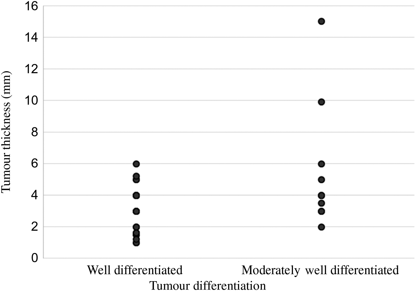
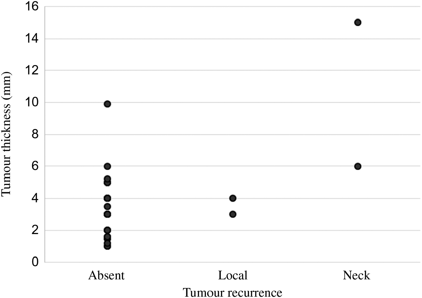
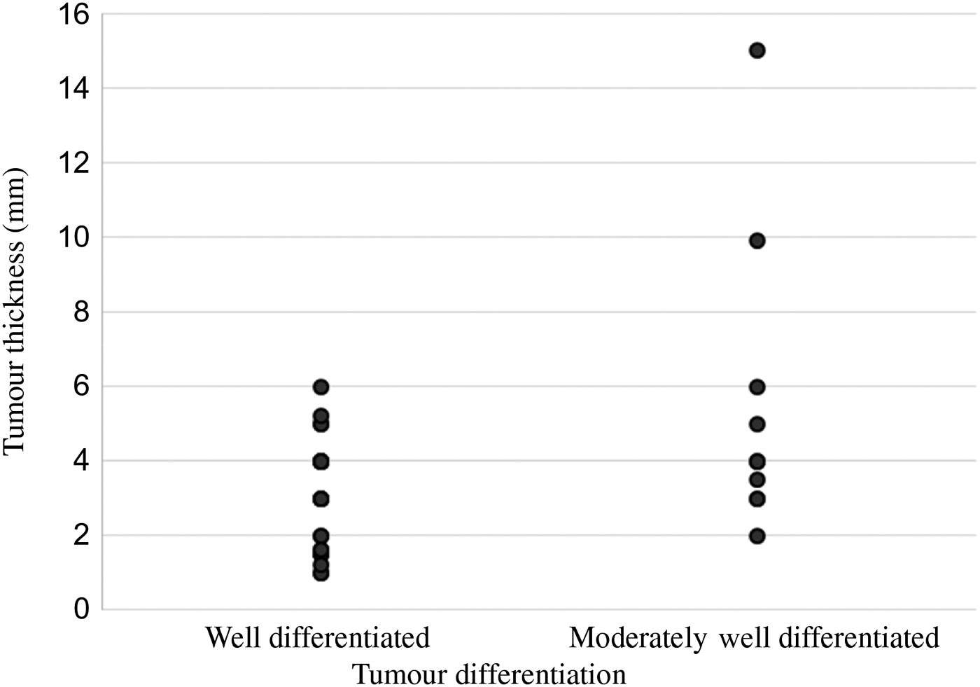
Correlation analysis of the data revealed that microscopic thickness was significantly associated with tumour differentiation (r = 0.394, p = 0.014) and tumour recurrence (r = 0.328, p = 0.045) (Figures 1 and 2). Tumour thickness was positively correlated with tumour recurrence.
Correlation analysis of tumour thickness and tumour differentiation.
Correlation analysis of tumour thickness and tumour recurrence.

Discussion
In this study, we evaluated the clinicopathological features of 46 lower lip SCC patients retrospectively, and found that tumour thickness was correlated with tumour recurrence and tumour differentiation.
This disease almost exclusively affects males (80–90 per cent of cases), with a frequency that increases progressively with age and reaches its peak during the seventh and eighth decades of life. In the present report, we found that the proportion of lip cancer cases recorded in women (21.7 per cent) is higher than that reported in the world literature, although some studies recognise that the incidence has increased slightly in women during recent years.Reference Moore, Johnson, Pierce and Wilson2 Our results may be explained, at least partially, by the fact that a high percentage of women perform tasks under prolonged solar exposure, such as agricultural activities, and therefore have the same risk as men regarding the effects of solar radiation.
Lip cancer is a slow-developing tumour, with lymph node metastases reported at rates between 3 and 29 per cent. The metastases are initially located at level I nodes (submental and submandibular sites) and only later extend to lower levels.Reference Santos, Silva, Cavalcante, Alves, Godoy and Nonaka6 The diagnosis of lymphatic cervical metastases in this study was initially made in 4.3 per cent of those patients without previous treatment, which is similar to reports from other series (2–12 per cent).Reference Zitsch, Lee and Smith7, Reference McGregor, Davis and Hay8 Lymphatic cervical metastases that developed after treatment were found in 4.3 per cent of the patients, which is also similar to reports from other series (3–13 per cent).Reference Zitsch, Lee and Smith7
Surgery is the treatment of choice, despite the fact that, for less extensive lesions, the results obtained with radiotherapy may compare favourably. The surgical techniques to approach the primary tumour are relatively standardised.Reference Vukadinovic, Jezdic, Petrovic, Medenica and Lens9 In lip cancer patients, some authors advocate neck dissection only in cases of palpable nodes, while others recommend that patients with a high risk for neck metastasis should undergo elective neck dissection.Reference Kowalski, Magrin, Waksman, Santo, Lopes and de Paula10 In such cases, supraomohyoid neck dissection is the preferred option.Reference Byers, Clayman, McGill, Andrews, Kare and Roberts11 Several reports in the literature have described factors associated with the risk of neck metastasis from lip cancer. These include T stage, histological degree of differentiation and commissure involvement; however, the results are not uniform in all series.Reference Carvalho, Kowalski, Borges, Aguiar and Magrin12 Considering the distribution of lymph node metastasis, recent reports have suggested the use of selective neck dissection as an option in selected patients with limited neck disease.Reference Kowalski and Carvalho13 In the present report, surgery was the treatment of choice in all patients, and only 4.3 per cent of the study group underwent neck dissection.
Most of the tumours in our series of SCC cases were well differentiated (73.9 per cent), and only a minority of tumours were moderately well differentiated (26.1 per cent). This is in stark contrast to the 47.6 per cent frequency of poorly differentiated SCCs of the lip in a recent series from Nigeria.Reference Effiom, Adeyemo, Omitola, Ajayi, Emmanuel and Gbotolorun14 This may be attributed to an earlier detection of tumours in primary care services in Western countries compared with developing nations, which in turn allows earlier treatment of lip cancer.
In our series, perineural invasion was found post-operatively in only 2.2 per cent of cases. This value is much lower than the 5 per cent described by Dinehart and Pollack.Reference Dinehart and Pollack15 However, perineural invasion may not be apparent until the tumour has spread extensively.16 Further, it has been demonstrated that if special staining for perineural invasion is not routinely performed on histological specimens, the true incidence may be underestimated.Reference Geist, Garcia-Moliner, Fitzek, Cho and Rogers17 Therefore, it is hardly surprising that studies specifically investigating the incidence of perineural invasion in patients with lip cancer have produced inconsistent results, varying from 2.1 to 5 per cent.Reference Leibovitch, Huilgol, Selva, Paver and Richards18 Consequently, we believe that the differences in the rate of perineural invasion in the different series, and between our series and others quoted above, may be due mainly to patient heterogeneity and methodological differences in the search for this parameter.
In a recent series of 899 lip cancer patients, Papadopoulos et al. found that surgical excision followed by direct repair was the type of surgery associated with the highest 5-year recurrence rate.Reference Papadopoulos, Konofaos, Tsantoulas, Chrisostomidis, Frangoulis and Karakitsos19 Our data suggest that microscopic tumour thickness is directly related to the possibility of tumour recurrence.
The prognosis of patients with lower lip SCC is good when the disease is diagnosed at an early stage, with five-year survival rates ranging from 85 to 99 per cent; this drops to 25–50 per cent in cases with cervical node involvement.Reference Sargeran, Murtomaa, Safavi, Vehkalahti and Teronen20, Reference Gutierrez-Pascual, Vicente-Martin, Fernandez-Alvarez, Martin-Lopez, Pinedo-Moraleda and Lopez-Estebaranz21 In our group of patients, who were followed up for a mean of 32.0 ± 18.9 months, 2 patients died in the follow-up period for reasons other than lip cancer.
The limitations of our study include the retrospective design and the relatively small number of patients in our series. In addition, some details of patient history and factors that may influence the outcome may not have been fully documented. Given these restrictions, the reported associations should be interpreted with caution.
• Clinicopathological features of 46 lower lip squamous cell carcinoma patients were evaluated retrospectively
• Tumour thickness was correlated with tumour recurrence and tumour differentiation
In summary, our results indicate that tumour thickness is directly related to recurrence of lower lip SCC. In addition, neck metastasis is rare in early stage lower lip SCC. A ‘wait and see’ policy might be preferred in early stage T1 lower lip SCC cases.