I. INTRODUCTION
The dye-sensitized solar cell (DSSC) is a promising candidate for low-cost photovoltaic devices. Porous TiO2 film and its huge internal surface area play an important role in both charge separation and transport, and the efficiency of DSSCs is strongly dependent on the existence of surface defects on the porous TiO2 network.Reference Barbé, Arendse, Comte, Jirousek, Lenzmann, Shklover and Grätzel1 Long relaxation times and charging effects provide evidence for the existence of a large number of surface defects in the TiO2/electrolyte system.Reference Schwarzburg and Willig2, Reference Jongh and Vanmaekelbergh3 Because of the large surface-to-volume ratio, these surface defects are expected to affect the cell performance significantly. Although TiO2 has high lattice energy, a photoelectron-conducting TiO2 film usually suffers from small oxygen loss,4 which results in the formation of dominant surface defect states. These surface defect states (also known as oxygen vacancies) are located in the interband region and cause slow charge transport in a TiO2 film owing to the trapping and detrapping of electrons.Reference Nelson5 Oxygen vacancies, which act as electron scavengers, facilitate the recombination of injected conduction-band electrons with triiodide ions (I3−) in the electrolyte. Interband surface defects can also increase the amount of light absorption in the visible region. The oxygen vacancy states exist at approximately 0.75–1.18 eV below the conduction band edge of TiO2Reference Cronemeyer6 and can be excited from the valence band by visible wavelengths of ∼506 and 614 nm. However, the optical absorption by these surface states does not contribute to the photocurrent in a DSSC. Therefore, to reduce the recombination of injected electrons and the light absorption by the surface states, it is essential to reduce the number of oxygen vacancies. It was known that the oxygen vacancies in TiO2 can be reduced by exposing to an oxygen atmosphere.Reference Weidmann, Dittrich, Konstantinova, Lauermann, Uhlendorf and Koch7
Ion beam treatments are quite effective in modifying the surface properties of materials and have been applied to the surface modification of thin films because the ion fluence and ion energy can be varied independently and used to tailor the chemical and physical properties of the film.Reference Takeuchi, Onozaki, Matsumura, Uchida and Kuji8, Reference Narita, Iida, Ogawa, Mizuno, So, Kondo, Yoshida, Itoh, Nonomura and Tanaka9 In this study, we report the effect of ion beam irradiation on TiO2 and on the photoconversion efficiencies of DSSCs. To decrease the oxygen vacancies and to reduce the electron recombination reactions between the injected conduction band electrons of TiO2 and I3− ions in the electrolyte, TiO2 films were irradiated with an oxygen ion beam of 500 and 1000 eV beam energy. Also, the reduction mechanism of oxygen vacancies by ion beam irradiation was explained.
II. EXPERIMENTAL
A. Preparation of TiO2 film and its oxygen ion-beam irradiation
Eight-μm-thick TiO2 paste (Ti-Nanoxide D; Solaronix SA, Aubonne, Switzerland) film with an active area of 0.25 cm2 was prepared on cleaned fluorine-doped tin oxide (FTO) (TCO30-8, ~8 Ω/sq.; Solaronix SA, Aubonne, Switzerland) glass substrates using the doctor blade method followed by sintering at 450 °C for 30 min in air. The resulting TiO2 film was irradiated with an oxygen ion beam at 500 and 1000 eV under a pressure of ~2.5 × 10⁻⁴ Torr. The acceleration voltage of the ion gun was 100 V, and the oxygen flow rate to the ion beam gun was 10 sccm. The details of the ion gun optics and operation principles can be found elsewhere.Reference Koh, Park, Kim, Choi, Jung and Pae10 Each TiO2 film was treated with different ion fluences of the ion beam by varying the irradiation time, that is, 5, 10, and 15 min.
B. Dye sensitization of TiO2 film and DSSC fabrication
The resulting oxygen ion beam–treated film was sensitized with Ru dye (0.5 mM, Ru 535-bis TBA, N719; Solaronix SA) for ∼24 h. Platinum-coated FTO glass was used as the counter electrode. The electrolyte was composed of 0.5 M lithium iodide, 0.05 M I2, 0.5 M 4-tert-butylpyridine, and 0.5 M 1,2-dimethyl-3-propylimidazolium iodide in a mixed solvent of acetonitrile and propylene carbonate (4:1). DSSCs without an ion beam–treated TiO2 film were also prepared for comparison.
C. Characterization of TiO2 film and DSSCs
The TiO2 film was irradiated with an oxygen ion beam using an ion beam system (KVIBAD; Korea Vacuum Tech., Ltd., Gimpo, Korea). The ion fluence was measured using a custom-built Faraday Cup. The surface morphology of the TiO2 films was observed by field emission scanning electron microscopy (FE-SEM; JSM-6700, JEOL Ltd., Tokyo, Japan). The Brunauer-Emmett-Teller (BET)-specific surface area was determined by the level of nitrogen (N2) adsorption (BELSOPR II, BEL Japan Inc., Osaka, Japan) at 77 K, and the pore size distribution was calculated using the Barrett, Joyner, and Halenda method. The x-ray photoelectron spectroscopy (XPS) spectra of the TiO2 films were obtained using ESCALAB 210 (VG Scientific Ltd., West Sussex, England). The contact angle on the TiO2 surface was measured using a Phoenix 300 apparatus, and the UV–visible spectra of the adsorbed dye on the TiO2 surface were obtained using a UV–visible Spectrophotometer (Hewlett Packard 8453). The DSSCs were illuminated with 100 mW/cm2, air mass (AM) 1.5 using a 450 W Xe high power arc lamp housing (LH 151, Spectral Energy Co., Chester, NY). The light intensity was calibrated using a Radiometer Photometer (ILT 1400-A). The impedance spectra were measured using an impedance analyzer (CH Instruments, Inc., Austin, TX) and scanned over the frequency range 10-1–106 Hz.
III. RESULTS
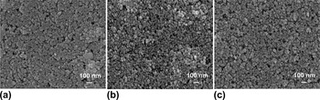
Figure 1 shows FE-SEM images of the TiO2 film surfaces before and after oxygen ion beam irradiation. Nanoparticles in a TiO2 paste tend to agglomerate to reduce the surface energy.Reference Kim, Parvez and Chhowalla11 The oxygen ion beam irradiation at 500 and 1000 eV resulted in a significant decrease in the sizes of the TiO2 particle agglomeration. A TiO2 film surface with smaller agglomeration would have a large surface area, which may allow more dye adsorption.
Field emission scanning electron microscopy images of (a) untreated, (b) 500 eV (2.79 × 10¹⁷ ions/cm2), and (c) 1000 eV (3.19 × 10¹⁷ ions/cm2) oxygen ion beam–treated TiO2 film surface.

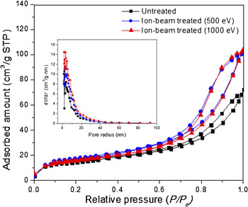
To further investigate the effect of ion beam on TiO2 film surface area, BET measurements were performed. Figure 2 shows the N2 adsorption–desorption isotherm and pore size distribution of the oxygen ion beam–treated TiO2 film. The surface area of the untreated TiO2 film was 49.3 m2/g with a pore volume and average pore diameter of 0.10 cm3/g and 8.9 nm, respectively. After an oxygen ion beam treatment at an ion fluence of 2.79 × 10¹⁷ ions/cm2 at 500 eV, the surface area of the TiO2 increased to 58.5 m2/g with pore volume of 0.15 cm3/g and pore diameter of 10.5 nm. Also, after ion beam treatment at 3.19 × 10¹⁷ ions/cm2 at 1000 eV, the surface area of the TiO2 obtained was 57.4 m2/g, and the pore volume and pore diameter were 0.15 cm3/g and 10.7 nm, respectively. These values were about the same at 500 eV. These increased surface area of the TiO2 film after oxygen ion beam treatment was expected to adsorb more dye than untreated TiO2 film.
Nitrogen gas adsorption and desorption isotherms and pore size distribution (inset) of untreated and oxygen ion beam–treated TiO2 films.

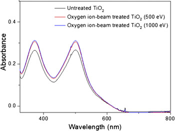
The water contact angle of the untreated TiO2 film was 21.6°, which was decreased to 7.5° and 8.8° with the oxygen ion beam treatment at 500 and 1000 eV, respectively. Moreover, the low contact angles were stable even after 72 h compared to the untreated TiO2. This suggests that the surface characteristics of oxygen ion beam–treated TiO2 particles were retained for a considerable amount of time. The increased hydrophilic nature of the TiO2 film is expected to interact more strongly with the carboxylic functional groups of the dye, resulting in more dye adsorption.Reference Kim, Yoon, Kim and Lee12Figure 3 presents the absorption spectra of the N719 dye, obtained by desorbing it from the oxygen ion beam–treated and –untreated TiO2 films (dimensions of 0.5 cm × 0.5 cm). Figure 2(b) clearly shows that there was an increase in N719 dye adsorption on the TiO2 film after the oxygen ion beam treatment at 500 and 1000 eV.
Ultraviolet–visible absorption spectra of N719 dye desorbed from the untreated and oxygen ion beam–treated TiO2 films.

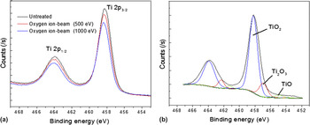
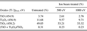
XPS analysis was carried out to characterize changes in surface charge states of the TiO2 film. Figure 4(a) shows the overlay XPS spectra of untreated, 500 and 1000 eV oxygen ion beam–treated TiO2 films. The binding energies of Ti 2p3/2 and Ti 2p1/2 peaks of TiO2 were observed at 458.2 and 463.9 eV, respectively. Both oxygen ion beam irradiation of TiO2 film at 500 and 1000 eV showed a little difference in XPS results. Here, we present the XPS at 1000 eV spectrum obtained from oxygen ion beam–treated TiO2 as shown in Fig. 4(b). The subregions of Ti 2p1/2 and Ti 2p3/2 were deconvoluted to three peaks. The nonstochiometric nature of the TiO2 (Ti4+) surface was indicated by the presence of TiO (Ti2+) and Ti2O3 (Ti3+) peaks. The surface stochiometry was determined by calculating the relative peak areas of TiO, Ti2O3, and TiO2 as at.% with respect to the total area of the Ti 2p peak. By deconvoluting the Ti 2p3/2 peak region of TiO2 film and calculating the ratios of the TiO2 to TiO + Ti2O3 relative peak areas, it was found that the relative peak area ratio of the TiO and Ti2O3 decreased and the ratio of TiO2 increased after 500 and 1000 eV oxygen ion beam treatment as shown in Table I. The ratio of TiO2 with respect to the sum of TiO and Ti2O3 increased after ion beam treatment. It implies that the lower oxidation states of titanium (i.e., Ti2+ and Ti3+) in the TiO2 film were oxidized to Ti4+, and the oxygen vacancies were reduced.
(a) Overlayed x-ray photoelectron spectroscopy (XPS) spectra of three conditions; (b) deconvoluted XPS spectra of 1000 eV (15 min) oxygen ion beam–treated TiO2 film.

X-ray photoelectron spectroscopy relative peak areas in percentage with respect to the total area of Ti 2p region for the untreated and oxygen ion beam treated at 500 eV and 1000 eV.

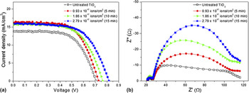
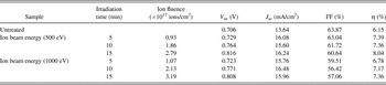
Figure 5(a) shows the photocurrent–voltage characteristics of the DSSCs based on the untreated and oxygen ion beam–treated TiO2 at 500 eV, and Table II summarizes the photovoltaic parameters of DSSC using untreated and oxygen ion beam–treated TiO2 electrodes. DSSCs with ion beam–treated TiO2 showed higher open-circuit voltages (V oc’s) and short-circuit currents (J sc’s) with increasing ion fluence. As a result, the photoconversion efficiencies (η%) were increased significantly. Oxygen ion beam irradiation with an ion dose of 2.79 × 10¹⁷ ions/cm2 (15 min) showed a maximum 110 mV higher V oc than the untreated TiO2. The oxygen vacancies are due to the presence of suboxides in TiO2, such as TiO (Ti2+) and Ti2O3 (Ti3+). Because the oxygen ion beam contains mostly O+ and O2+ ions,Reference Geer, Van, Fraser, Coburn and Graves13 these ions can effectively oxidize the Ti2+ and Ti3+ oxidation states to Ti4+ in TiO2, decrease the number of oxygen vacancies, and increase the V oc. The Nyquist plots [Fig. 5(b)] measured in the dark under a forward bias of −0.7 V showed an increase in the radius of the middle semicircle with the ion beam irradiation time, indicating that the electron recombination resistance is augmented with the oxygen ion beam treatment.Reference Kuang, Uchida, Baker, Zakeeruddin and Grätzel14, Reference Wu, Han, Tai, Zhang, Xu, Zhou, Yang, Hu, Chen and Zhao15 DSSCs with 1000 eV ion beam–treated TiO2 films showed a similar pattern in Nyquist plots (not shown here).
(a) J–V characteristic curves and (b) impedance spectra (Nyquist plots) of the dye-sensitized solar cells incorporated with untreated and 500 eV oxygen ion beam–treated TiO2 films with different irradiation time.

Characteristics of the DSSCs of the untreated and oxygen ion beam–treated TiO2 film with different irradiation time.

DSSC, dye-sensitized solar cell; V oc, open-circuit voltage; J sc, short-circuit current; FF, fill factor.
The increase in J sc with the oxygen ion beam treatment was attributed to the higher surface energy and increased dye adsorption of the TiO2 film.Reference Park and Dhayal16 The electrostatic interaction between the Ti4+ and –COO− groups of the dye is expected to be stronger than that for Ti2+ and Ti3+.Reference Kim, Yoo, Vittal, Lee, Park and Kim17 The oxygen ion beam–treated TiO2 films showed a small decrease in fill factors (FFs) compared to the untreated TiO2. The conductivity of the TiO2 decreases and the internal resistance of the cell increases with decreasing concentration of oxygen vacancies.Reference Weidmann, Dittrich, Konstantinova, Lauermann, Uhlendorf and Koch7 This might result in low FFs after an ion beam treatment.
IV. CONCLUSIONS
TiO2 film electrodes were irradiated with oxygen ion beam energy of 500 and 1000 eV to reduce the number of oxygen vacancies. Different ion fluences (0.93 × 10¹⁷ to 3.19 × 10¹⁷ ions/cm2) were obtained by varying the irradiation time. With the oxygen ion beam treatment, the TiO2 film showed a significant decrease in the agglomeration of the TiO2 particles and an increase in the surface area and pore volume. The XPS spectra revealed a significant decrease in oxygen vacancies after ion beam treatment, and the ratio of TiO2 to TiO + Ti2O3 increased from 3.18 to 4.37%. In addition, the increased hydrophilicity of the TiO2 surface led to more dye adsorption than that of the untreated TiO2 film. The increase in V oc and J sc with the oxygen ion beam treatment resulted in an improved TiO2 film electrode with reduced oxygen vacancies and fewer recombination reactions. The oxygen ion beam–treated TiO2 film at 500 eV and 2.79 × 10¹⁷ ions/cm2 showed a maximum 110 mV increase in the V oc and a 30% increase in efficiency (η%) compared to the untreated TiO2 film. Therefore, it can be concluded that oxygen ion beam treatment was effective to improve the performance of DSSCs.
ACKNOWLEDGMENTS
This research was financially supported by the Ministry of Education, Science Technology of Korea through the Human Resource Training Project for Regional Innovation. This work was also supported by National Research Foundation of Korea Grant (2010-0006808) funded by the Korean Government. The authors thank J. Kim and G. Yoo for ion beam experiments.