INTRODUCTION
Infectious disease is listed among the top five causes of global species extinctions (Young, Reference Young1994; Smith et al. Reference Smith, Sax and Lafferty2006). Historically, diseases of wildlife have only attracted public attention when domestic animals or human health were involved. However, there is a growing awareness of the role of pathogens in the population dynamics of wildlife and, in particular, endangered species (Daszak et al. Reference Daszak, Cunningham and Hyatt2000; Köndgen et al. Reference Köndgen, Kühl, N'Goran, Walsh, Schenk, Ernst, Biek, Formenty, Mätz-Rensing, Schweiger, Junglen, Ellerbrok, Nitsche, Briese, Lipkin, Pauli, Boesch and Leendertz2008). Indeed, disease has become a major concern in wildlife conservation, both inside and outside of park and reserve boundaries (Daszak et al. Reference Daszak, Cunningham and Hyatt2000; Lafferty, Reference Lafferty2003; Smith et al. Reference Smith, Sax and Lafferty2006). As a result of the human population explosion, combined with illegal hunting, and the fragmentation and degradation of formerly natural habitats, wild animals are increasingly subjected to contact with humans. This situation may facilitate disease transmission among humans, livestock and wildlife (Bengis et al. Reference Bengis, Kock and Fischer2002; Woodford et al. Reference Woodford, Butynski and Karesh2002).
Although humans have always shared habitats with non-human primates the dynamics of human-primate interactions are changing radically (Chapman and Peres, Reference Chapman and Peres2001; Wolfe et al. Reference Wolfe, Prosser, Carr, Tamoufe, Mpoudi-Ngole, Torimiro, LeBreton, McCutchan, Birx and Burke2004). Non-human primates are now commonly forced to live in an anthropogenically disturbed landscape comprising farmland, human settlements, forest fragments and isolated protected areas (Chapman and Peres, Reference Chapman and Peres2001). Gillespie and Chapman (Reference Gillespie and Chapman2006) found that the index of forest patch degradation and the presence of humans strongly influenced the prevalence of parasitic gastrointestinal nematodes in red colobus monkeys. This suggests that, as forest fragments become smaller and frequency of contact between humans and non-human primates becomes higher, transmission of diseases and pathogens is increasingly likely.
Due to the genetic similarity between humans and non-human primates, the transmission of pathogens between or among primates is of particular significance (Wolfe et al. Reference Wolfe, Escalante, Karesh, Kilbourn, Spielman and Lal1998; Chapman et al. Reference Chapman, Gillespie and Goldberg2005). Indeed, recent work has shown that non-human primate populations harbour a variety of pathogens similar to those causing significant disease in humans (Leendertz et al. Reference Leendertz, Yumlu, Pauli, Boesch, Couacy-Hymann, Vigilant, Junglen, Schenk and Ellerbrok2006; Chi et al. Reference Chi, Leider, Leendertz, Bergmann, Boesch, Schenk, Pauli, Ellerbrok and Hakenbeck2007; Perpens et al. Reference Perpens, Kreuzer, Leendertz, Nitsche and Ehlers2007) that may be zoonotic. Such pathogens have already led to major epidemic problems for human health, such as the transfer of HIV viruses to the human population (Daszak et al. Reference Daszak, Tabor, Kilpatrick, Epstein and Plowright2004). Cross-transmission of parasites to non-human primates has been inferred in a number of African primates (Mudakikwa et al. Reference Mudakikwa, Sleeman, Foster, Meader, Patton and Baer1998; Graczyk et al. Reference Graczyk, DaSilva, Cranfield, Nizeyi, Kalema and Pieniazek2001; Lilly et al. Reference Lilly, Mehlman and Doran2002).
However, finding morphologically identical nematodes in human and wild primates should be treated with caution. For example, Oesophagostomum bifurcum from humans were genetically distinct from those harboured by some non-human primates, and these genetic differences were predicted to be associated with distinct transmission patterns (de Gruijter et al. Reference de Gruijter, Gasser, Polderman, Asigri and Dijkshoorn2005; Gasser, Reference Gasser, de Gruijter, Polderman, Huffman and Chapman2009). Still, very little information is available on the extent of transfer of pathogens between humans and non-human primates. In particular, limited information is available on the prevalence of intestinal parasitism in feral neotropical primates (Michaud et al. Reference Michaud, Tantaleán, Ique, Montoya and Gozalo2003; Phillips et al. Reference Phillips, Haas, Grafton and Yrivarren2004). Almost nothing is known about the influence of human settlements bordering the natural habitats of non-human primates on the gastrointestinal parasites of primates. The primary objective of this study was to determine the influence of human contact and habitat modification on the presence of gastrointestinal parasites in two tamarin species from a tropical rainforest area in Peru, the saddleback tamarin (Saguinus fuscicollis) and the moustached tamarin (Saguinus mystax).
MATERIALS AND METHODS
Study site
The study was conducted in the Amazonian lowlands of northeastern Peru around the Estación Biológica Quebrada Blanco (EBQB, 4°21′S and 73°09′W), a field site located on the right bank of the Quebrada Blanco, a tributary of the Rio Tahuayo. The study area comprised ~100 hectares of undisturbed primary forest and contained a 100 m×100 m trail system (for further details see Heymann, Reference Heymann1995). Additionally, samples were collected in and around the village Diamante on the opposite bank of the Quebrada Blanco. Diamante is the home of ~40–50 people. The village centre of Diamante includes 7 houses and a school. This population has no adequate medical treatment for intestinal parasites. The only other humans occurring in the area are members of the scientific establishment with access to anthelmintics.
Study animals
The subjects of the study were 8 wild groups from 2 species of tamarins: 4 groups of moustached tamarins (Saguinus mystax mystax) and 4 groups of saddleback tamarins (Saguinus fuscicollis nigrifrons). Both species lived in mixed-species troops. Some of the groups have been under observation for up to 8 years (Müller, Reference Müller2007). All group members are known individually by natural markings (Löttker et al. Reference Löttker, Huck and Heymann2004). The groups investigated differed in their proximity to humans and their facilities. One of the mixed-species troops lived around the village Diamante. The home range of this group reached to the houses of the villagers and included the village toilet, the rubbish dump and fields planted with yucca, plantains and other fruit. In addition, parts of the tamarin homerange consist of secondary forest. Villagers cross the homerange every day to get to their crops and to collect wood. This tamarin group is designated group D. The second human contact troop had its homerange around the EBQB, which is situated in a clearing, with a minimum distance of 5 metres to the field station. There are no crops in this area, but the tamarin's range includes the station rubbish dump and the toilet. The number of occupants of the EBQB varies between 2 and up to 20 people, and some of the villagers from Diamante work at the station. The sampled tamarin group is designated group 1. The other groups lived deeper in the forest with no contact to humans or their facilities except for the observers watching them for a variable number of days each month (groups 2 and 3). For size and species composition of the groups see Table 2.
Collection and examination of faecal samples
Fresh faecal droppings from all group members were collected during the dry season between July and September 2007 by following the groups during their whole activity period (06·00–16·30). Faecal samples were gathered directly after defecation. Locality, date, group, species, sex, individual and time of defecation were recorded. After collection, the faecal samples were immediately preserved in separate 5·0 ml vials containing 10% buffered formalin (solution of 10% formaldehyde and sodium phosphate buffer, pH 7·0). Samples were stored at ambient temperature. From each of the 43 individuals at least 3 samples were collected on non-consecutive days. In total, we have 356 faecal samples from the 8 study groups. Additionally, faecal samples from 27 people between the ages of 1 year and 66 years in Diamante were taken, at least 3 samples per individual from different days. In total, 85 human stool samples were collected. The finding of a parasite in any one of multiple samples for a given individual resulted in a positive report of that parasite.
To process the preserved faecal samples in the laboratory, a modified formalin-ethyl acetate sedimentation technique was used (Ash et al. Reference Ash, Orihel and Savioli1994). Samples were microscopically examined for the presence of eggs and larvae of different intestinal helminths. Eggs and larvae were identified by shape, size and other visible structures as far as possible. However, identification to species level was not always possible based on microscopical examination of eggs and larvae without the adult worms or coprological cultures (Ash and Orihel, Reference Ash and Orihel1987; Gillespie et al. Reference Gillespie, Greiner and Chapman2005). Measurements were made to the nearest 0·1 μm±s.d. using an ocular micrometer fitted to a compound microscope. Photographs of representative specimens were taken and the egg (larvae) output per 100 μl of concentrated sediment was counted. These data come with a caveat: many external and internal factors may account for variability in parasite infection patterns (Stuart and Strier, Reference Stuart and Strier1995). Host immunity, density-dependent factors and environmental cues can depress worm ovulation (Christensen et al. Reference Christensen, Barnes, Nansen, Roepstorff and Slotved1995; Stear et al. Reference Stear, Bishop, Doligalska, Duncan, Holmes, Irvine, McCririe, McKellar, Sinski and Murray1995; Roepstorff et al. Reference Roepstorff, Bjørn, Nansen, Barnes and Christensen1996). In addition, some parasite species release eggs or larvae intermittently (Anderson and Schad, Reference Anderson and Schad1985; Warnick, Reference Warnick1992; Cabaret et al. Reference Cabaret, Gasnier and Jacquiet1998).
Eggs and larvae of each parasite excreted per 100 μl of faecal sediment (EPS) were measured both for infected individuals only (intensity) as well as for all sampled individuals (abundance). The egg output of 3 different samples per individual was counted and for each sample the count of 3 slides was averaged.
Statistical analysis
All statistical tests were carried out using SPSS 11.5 for Windows. In all tests, significance levels were set to α=0·05. To test statistical differences in prevalence of parasites between species, sexes and groups Fisher's exact test was used. To test differences in intensity and abundance of each parasite species, the parasite count from each of the 3 samples per individual was summed and averaged. Mann-Whitney U-test was used to look for variation in parasite egg output of all individuals between troops and between sexes. No statistical test on the egg output of infected individuals was carried out because the sample sizes of positive samples were too small. Egg output in infected animals was determined by the mean output per species and group.
RESULTS
Human faecal samples
The human faeces contained 4 different helminth species: Ascaris lumbricoides, Trichuris trichiura, Strongyloides stercoralis and hookworm eggs (Necator americanus/Ancylostoma duodenale). The prevalence of Ascaris was extremely high (Table 1), whereas Trichuris and hookworms were moderately represented. Strongyloides infections were rare (Table 1).
Total prevalences in percentage and mean intensity of infection for male and female humans
(N indicates number of samples; PSR indicates parasite species richness. Intensity represents mean egg output per 100 μl of faecal sediment of the infected individuals with standard deviation (SD).)

In humans, the mean intensity was moderate except for Ascaris (Table 1). In 1 sample we counted more than 6000 Ascaris eggs in 100 μl of faecal sediment. The maximum for Trichuris was 33 eggs, for hookworms 49 and for Strongyloides larvae it was 4.
Tamarin faecal samples
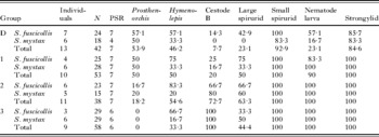
None of the parasites from the human samples were found in tamarins. We recovered 7 parasite taxa – 1 acanthocephalan species, 2 cestodes and 4 different nematode species (Table 2). The helminth taxa were identified as: the acanthocephalan species Prosthenorchis elegans, one of the cestodes belonged to the Hymenolepididae, probably being Hymenolepis cebidarum, the other cestode species could not be further identified and will be referred to as ‘cestode B’. We could also identify the eggs of 2 nematode taxa from the order Spirurida: one will be referred to as ‘large spirurid’, probably Gongylonema sp. or Trichuspirura leptostoma and the other as ‘small spirurid’. In addition to these spirurids we found the eggs of ‘strongylids’ from the orders Rhabditida and Tylenchida, probably Molineus sp. or Strongyloides cebus. Furthermore, we found nematode larvae belonging either to the order Rhabditida (Strongyloides sp.) or to the superfamily Metastrongiloidea (i.e. Filaroides sp. and Angiostrongylus sp.).
Total prevalences in percentage parasite infestation for the different tamarin groups and species
(N indicates number of samples; PSR indicates parasite species richness.)

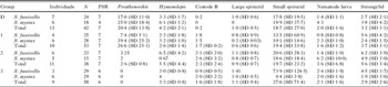
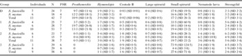
All of these taxa were prevalent in all of the mixed species troops, except Prosthenorchis elegans, which was absent from group 3, a non-contact group. Cestode B and the large spirurid were missing in Saguinus mystax from group D around the village. An overview of the different parasites in groups and species can be found in Table 2. There were no significant differences in the prevalence of parasites between the two tamarin species within the groups and we could not find significant differences between the sexes (Fisher's exact test, P>0·05). Thus we pooled species and sexes for further analyses. We also measured the eggs and larvae excreted per 100 μl of faecal sediment in tamarin samples. Here we found the highest EPS for Prosthenorchis and small spirurids (Tables 3 and 4). The maximum count of Prosthenorchis eggs in 100 μl of faecal sediment was 158, and for small spirurids 1506.
Mean intensities of parasite infestation for the different tamarin groups and species
(N indicates number of samples; PSR indicates parasite species richness. Intensity represents mean egg output per 100 μl of faecal sediment of the infected individuals with standard deviation (SD).)

Mean abundances of parasite infestation for the different tamarin groups and species
(N indicates number of samples; PSR indicates parasite species richness. Abundance represents mean egg output per 100 μl of faecal sediment of all sampled individuals with standard deviation (SD).)

Differences in prevalence among tamarin groups
There were significant differences in the prevalence of parasites between the tamarin groups. Prosthenorchis was absent in the sylvatic group 3 leading to a significant difference compared to the human contact groups D (Fisher's exact test, P=0·017) and 1 (Fisher's exact test, P=0·03). There was also a trend to lower prevalences in the forest group 2 than in the human contact groups D and 1, but this was not significant. Nematode larvae had a significantly lower prevalence in the village group D than in all other groups (Fisher's exact test D+1, P=0·029; D+2, P=0·002; D+3, P=0·006). The sylvatic groups 2 and 3 had a significantly higher prevalence of cestode B eggs than the human contact groups D and 1 (Fisher's exact test, D+2, P=0·002; D+3, P=0·001; 1+2, P=0·009; 1+3, P=0·001). None of the other parasite taxa showed significant differences in prevalence between the tamarin groups.
There were also significant differences in the abundance of Prosthenorchis, nematode larvae and cestode B. For Prosthenorchis, the mean abundance in the human contact groups D and 1 was significantly higher than for the sylvatic group 3 (MWU D+3, P=0·011; 1+3, P=0·018). Additionally, the mean abundance in group D was significantly higher than in group 2 (MWU D+2, P=0·03). The differences between group 1 and 2 were nearly significant (MWU P=0·085).
For nematode larvae the mean abundance was significantly lower in the village group D than in the forest groups 2 and 3 (MWU D+2, P=0·013; D+3, P=0·009). For cestode B eggs the mean abundance was significantly lower in group D and group 1 than in groups 2 and 3 (MWU D+2, P<0·001; D+3, P<0·001; 1+2, P=0·014; 1+2, P=0·007).
Differences in mean intensity were also recorded for Prosthenorchis. The mean outputs of the human contact groups D (20·2 eggs /100 μl) and 1 (22·0 eggs/100 μl) were considerably higher than in the sylvatic groups 2 (2·6 eggs /100 μl) and 3 (0 eggs). All other parasite species had similar mean egg outputs in the positive samples (see Table 3). Abundance and intensity values showed the high variability common in parasite species (Engels et al. Reference Engels, Sinzinkayo and Gryseels1996, Reference Engels, Sinzinkayo, DeVlas and Gryseels1997b; Ross et al. Reference Ross, Yuesheng, Sleigh, Williams and McManus1998).
DISCUSSION
It is widely accepted that there is a relationship between the emergence of zoonotic diseases and human influenced landscapes (Bradley and Altizer, Reference Bradley and Altizer2007; Jones et al. Reference Jones, Patel, Levy, Storeygard, Balk, Gittleman and Daszak2008), but a potential relationship between human influenced landscapes and wildlife diseases is less well understood (St-Amour et al. Reference St-Amour, Wong, Garner and Lesbarrères2008). From the present results, we conclude that the presence of humans and an environment modified by humans can lead to substantial changes in the community structure of intestinal helminths in wild non-human primates. Patterns of parasitism in wildlife populations are thought to be influenced by host-ranging patterns, density, intra- and interspecific contact rates and diet, as well as increases in stress levels (Hudson et al. Reference Hudson, Rizzoli, Grenfell, Heesterbeek and Dobson2002; Nunn et al. Reference Nunn, Altizer, Jones and Sechrest2003; Nunn and Altizer, Reference Nunn and Altizer2006). Studies of a variety of species have demonstrated that these characteristics can be affected by changes in forest structure (Olupot et al. Reference Olupot, Chapman, Brown and Waser1994; Heydon and Bulloh, Reference Heydon and Bulloh1997; Patriquin and Barclay, Reference Patriquin and Barclay2003). Areas of land unaffected by human encroachment or habitat degradation are increasingly rare (Lilly et al. Reference Lilly, Mehlman and Doran2002). The planting of crops is one important way in which humans modify the habitat of wildlife species and affect the behaviour and diet of primates (Weyher et al. Reference Weyher, Ross and Semple2006). Comparisons of wild foraging and crop-raiding baboons have shown significant differences in the parasite burdens between the troops. Wild foraging troops showed higher prevalences in Trichuris sp. and Physaloptera sp. infections (Hahn et al. Reference Hahn, Proulx, Muruthi, Alberts and Altmann2003; Weyher et al. Reference Weyher, Ross and Semple2006). Weyher et al. (Reference Weyher, Ross and Semple2006) suggested that crop-raiding baboons are in better physical condition due to their diet and therefore able to ‘fight off’ the helminth infections more readily. Another result of the studies was that crop-raiding baboons had higher prevalences of Streptopharagus sp. As both Streptopharagus and Physaloptera spp. are spirurid nematodes with indirect life cycles, having arthropods as intermediate hosts, the authors suggested that, in the distinct troop environments, the arthropod community is a different one (Weyher et al. Reference Weyher, Ross and Semple2006). Vitazkova and Wade (Reference Vitazkova and Wade2007) also assumed a connection with environmental factors when they found the strongest association of Controrchis biliophilus infection with the troop membership in Alouatta pigra, because C. biliophilus has an indirect life cycle which excludes direct infection by group members. Unfortunately, none of these studies examined the role of arthropod ingestion in parasite transmission.
In the present study, the tamarins which foraged in or near the human settlement also differed markedly from the groups which had less overlap with humans and human-modified habitat, showing significantly lower prevalences and abundances of a cestode species, but significantly higher levels of the pathological acanthocephalan P. elegans (see Taraschewski, Reference Taraschewski2000).
In our study, the prevalences of P. elegans in the groups around the village and the station showed a similar high level of parasite infestation (50–58%), whereas the forest groups were much less infected (0–18%). In addition, the number of Prosthenorchis eggs in the tamarin faeces was considerably higher in the groups with contact to human facilities (groups D and 1) than in the forest groups (groups 2 and 3). The number of parasite eggs in host faecal material is affected by many factors. Numbers of eggs excreted on a daily basis differ due to variations in egg production, differences in stool consistency and clumping of eggs (heterogenous mixing of eggs in stools) as shown by studies in Schistosoma mansoni and S. japonicum (see Engels et al. Reference Engels, Sinzinkayo and Gryseels1996, Reference Engels, Nahimana, DeVlas and Gryseels1997a, Reference Engels, Sinzinkayo, DeVlas and Gryseelsb; Ross et al. Reference Ross, Yuesheng, Sleigh, Williams and McManus1998). Host immunity, density-dependent factors and environmental cues can depress worm ovulation (Christensen et al. Reference Christensen, Barnes, Nansen, Roepstorff and Slotved1995; Stear et al. Reference Stear, Bishop, Doligalska, Duncan, Holmes, Irvine, McCririe, McKellar, Sinski and Murray1995; Roepstorff et al. Reference Roepstorff, Bjørn, Nansen, Barnes and Christensen1996). The propagule shedding depends on the density of adult female worms at the same location within the host individual; some parasite species release their eggs or larvae intermittently, and pre-patent adults, larvae and adult males do not excrete propagules at all (Anderson and Schad, Reference Anderson and Schad1985; Warnick, Reference Warnick1992; Cabaret et al. Reference Cabaret, Gasnier and Jacquiet1998). However, a roughly linear positive relationship between numbers of eggs/gramme of faeces and parasite burden has been observed for a number of parasite species (e.g., Keymer and Hiorns, Reference Keymer and Hiorns1986; Pritchard et al. Reference Pritchard, Quinnell, Slater, McKean, Dale, Raiko and Keymer1990; Sithithaworn et al. Reference Sithithaworn, Tesana, Pipitgool, Kaewkes, Pairojkul, Sripa, Paupairoj and Thaiklar1991), suggesting that the intensities of infestation by adult P. elegans were higher in the groups with human contact compared with the sylvatic ones.
P. elegans is an acanthocephalan parasite of carnivores and primates occurring naturally in South and Central America, including tamarins in Peru (Tantaleán et al. Reference Tantaleán, Sánchez, Gómez and Huiza2005). Numerous studies on captive Old and New World primates have demonstrated that these thorny-headed worms are highly pathogenic leading to morbidity and mortality (Taraschewski, Reference Taraschewski2000) and this species has been associated with sudden die-offs of entire colonies of monkeys, lemurs and chimpanzees in captivity (Moore, Reference Moore1970; Schmidt, Reference Schmidt and Fiennes1972). Attachment of the hooked proboscis may cause pain, inflammation, haemorrhaging and secondary infections. Often a complete penetration of the intestinal wall occurs (Schmidt and Roberts, Reference Schmidt and Roberts1981), with clinical signs in primates including anorexia, depression and emaciation (Cubas, Reference Cubas1996). However, most authors concluded that the parasite does not directly cause the animal's death, but rather causes lesions which enable secondary pathogens to become established, resulting in debilitation and death of the primate (Cubas, Reference Cubas1996; Taraschewski, Reference Taraschewski2000; Tantaleán et al. Reference Tantaleán, Sánchez, Gómez and Huiza2005).
Known intermediate hosts of P. elegans are cockroaches (Blatella germanica, Blabera fusca, Rhyparobia madera) and certain coprophagous beetles (Lasioderma serricorne, Stegobium paniceum) (Schmidt, Reference Schmidt and Fiennes1972). Captive primates in zoological gardens acquired their infections with P. elegans by ingesting cockroaches (Moore, Reference Moore1970). Cockroaches are very common in the canopy of tropical rainforests (Basset, Reference Basset2001; Gurgel-Gonçalves et al. Reference Gurgel-Gonçalves, Palma, Motta, Bar and Cuba2006). Their presence is related to environmental parameters among which human activity is a decisive factor (Boyer and Rivault, Reference Boyer and Rivault2006). Furthermore, cockroaches frequently feed on human wastes as well as on human faeces (Burgess et al. Reference Burgess, McDermott and Whiting1973; Graczyk et al. Reference Graczyk, Knight and Tamang2005). As mentioned above, the home range of the tamarins around Diamante included the village toilet, the rubbish dump and fields planted with yucca, plantains and other fruit. The tamarins were not observed to feed on this produce, but they did cross the fields and ingested insects on the ground. In addition, part of the tamarin's homerange consists of secondary forest. The second human contact troop had its homerange around the EBQB where the influence of human is less marked. There are no crops, but the tamarin's range also included the dump and the toilet.
Currently, it is uncertain how the tamarins contact these parasites and thus far we do not have information on which intermediate hosts occur in the area of study. Our tamarin groups are not known to feed on beetles and only ingest cockroaches very rarely (Heymann, personal observation). If cockroaches serve as a source of infection, as they do in captive tamarins, the prevalence of P. elegans infection of cockroaches must be extremely high. In this case, even the very rare event of cockroach ingestion by tamarins would be sufficient for parasite transmission. Another option would be the presence of unknown intermediate hosts in this area. Crompton (Reference Crompton1970) noted, that acanthocephalans use crickets and other adult orthopterans as intermediate hosts. Tettigoniid orthopterans are the most common prey items of tamarins (Nickle and Heymann, Reference Nickle and Heymann1996; Smith, Reference Smith2000). The prevalence of infection found in our human contact tamarins reaches similar values as in captive monkeys (Middleton, Reference Middleton1966; Vickers, Reference Vickers1969). Therefore, it is important to determine the complete life cycle of P. elegans in our study area.
Beside the higher prevalence of P. elegans we found a significantly lower prevalence of a cestode species in both groups living in proximity to human facilities. These could not be identified to species, highlighting the lack of taxonomic work on the parasites of tropical primates. Nevertheless, our findings support the hypothesis of a change in the composition of possible intermediate hosts in human-modified habitats and therefore in the diet of the different tamarin groups. To date, however, there is no information available of the potential pathogenic effect of these parasites on their host species.
The divergences observed in parasite community structure in the tamarins are not related to the transfer of parasites between humans and primates. In the human samples the major intestinal geohelminths of temperate and tropical latitudes (A. lumbricoides, T. trichiura, N. americanus/A. duodenale and S. stercoralis) were present. This can be traced back to the practice of humans defecating in the forest around the village rather than in toilet facilities (Wenz, personal observation). The very high prevalences and abundances of these parasites in the human population indicate that they could potentially be transferred to the tamarins. Philipps and colleagues (Reference Phillips, Haas, Grafton and Yrivarren2004) detected T. trichiura in 2 wild S. fuscicollis in southeastern Peru, demonstrating that tamarins can act as suitable hosts for this whipworm. However, studies demonstrating identical nematodes in humans and wild animals based on morphological examination alone have to be treated with caution. Oesophagostomum bifurcum from humans were found to be genetically distinct from those harboured by some non-human primates, and these genetic differences were predicted to be associated with distinct transmission patterns (de Gruijter et al. Reference de Gruijter, Gasser, Polderman, Asigri and Dijkshoorn2005; Gasser, Reference Gasser, de Gruijter, Polderman, Huffman and Chapman2009). In our study, none of the helminths found in the villagers, even those which were highly prevalent, could be found in the faeces of the tamarins. Thus, no transmission of human parasites to tamarins takes place in Diamante or, if transmission does occur, the low suitability of tamarins as hosts for human intestinal nematodes means that these worms cannot establish (short longevity, no excretion of eggs). A previous study in the area showed that primates were infected with a similar spectrum of parasites during dry and wet seasons (Müller, Reference Müller2007), nevertheless, possible variation in parasite infection between wet and dry seasons should be investigated further.
Two hypotheses may explain the lack of successful transfer of human parasites to tamarins. First, humans and tamarins may vary in their life styles and have different habitat preferences. Tamarins are tree-dwelling primates which only spend 0·5% to 1·6% of their time on the ground (Müller, Reference Müller2007). The possibility that they contact contaminated soil or human faeces is therefore very low. All of the human parasites recovered by us were geohelminths, i.e. they have a direct life cycle and infective stages of the parasites are excreted via the faeces or other body fluids and accumulate on or in the ground (Holland and Kennedy, Reference Holland and Kennedy2002). Consequently, terrestrial species may be at greater risk of aquiring parasites with a direct life cycle through faecal contamination of soil (Nunn et al. Reference Nunn, Gittleman and Antonovics2000).
The second, non-mutually exclusive, hypothesis involves the host specificity of the parasites. Tamarins belong to the New World primate family Callitrichidae, which separated about 40 million years ago from the ancestors of humans (Kageyama, Reference Kageyama2000; Neusser et al. Reference Neusser, Stanyon, Bigoni, Wienberg and Müller2001), suggesting a long divergence time between the parasites communities infesting these respective hosts. Because of their phylogenetically closer relationship to humans, great apes should be more susceptible to human parasites. We know that captive orangutans can carry patent infections of Giardia lamblia and A. lumbricoides (Mul et al. Reference Mul, Paembonan, Singleton, Wich and van Bolhuis2007).
Although we could not find direct transmission of parasites from humans to primates, the tamarins foraging in an area of human altered habitat showed substantial changes in their parasite communities with potential pathogenic consequences. In terms of conservation, this indicates that areas designated for the protection of primate species should be placed far enough from human settlements to prevent foraging on disturbed land.
ACKNOWLEDGEMENTS
The presentation of these data would not have been possible without the invaluable assistance of Ney Shahuano Tello and Migdonio. We thank the Instituto Nacional de Recursos Naturales (INRENA) in Lima for permission to carry out fieldwork at the Estacion Biologica Quebrada Blanco (authorization no. 106-2007-INRENA-IFFS-DCB). This study is part of a Ph.D. project of the senior author. Field work was carried out under a memorandum of understanding between the Universidad Nacional de la Amazonía Peruana (UNAP) in Iquitos and the Deutsches Primatenzentrum and complied with Peruvian laws. Financial support was provided by the German Academic Exchange Service (DAAD) and the Landesgraduiertenförderung Baden–Württemberg.