Introduction
Breast cancer is the second most common cancer among both sexes worldwide. About 2 million new cases of breast cancer were diagnosed worldwide in 2018.Reference Bray, Ferlay, Soerjomataram, Siegel, Torre and Jemal1 Increasing breast cancer incidence rates, improved diagnosis and management modalities and growing life expectancy have resulted in increasing numbers of women at risk of developing contralateral primary breast cancer.Reference Kheirelseid, Jumustafa and Miller2 Breast cancer patients have had a two- to six-fold increased risk of developing a new primary cancer in the contralateral breast during their life,Reference Chaudary, Millis and Hoskins3, Reference Soerjomataram, Louwman, Lemmens, de Vries, Klokman and Coebergh4 corresponding to a risk of 0·3–1·0% per year.Reference Hill-Kayser, Harris, Hwang and Solin5, Reference Gao, Fisher and Emami6 Bilateral breast cancers are stratified according to the time of their presentation: synchronous (within 6 months of primary breast cancer) and metachronous (after 6 months of primary breast cancer).Reference Kuo, Yen and Lee7
This study analyses three cases of bilateral breast cancer treated at our oncology department, of which two were metachronous while one developed cancer in the second breast during the course of management.
Material and Methods
This study was conducted to identify the clinicopathological profile of the two breast cancers occurring in the same patient and analyse the various factors associated with the occurrence of bilateral breast cancer.
Data collection
The medical records of 58 patients of breast cancer who underwent radiation treatment during March 2018 to March 2019 in our department were studied. Three patients of bilateral breast cancer were identified and their case records were analysed. The data were collected about the patient’s age at both the first and the second diagnosis, weight, the tumour localisation (breast side and quadrant) and the specific finding at the first and the second diagnosis. Then, we retrieved information about the treatment received for the first and the second tumour and type of surgery performed. The presence of multiple tumours and the time of presentation of second cancer were noted. Tumour pathological characteristics including size, histological type, nuclear/histologic grading, multicentricity, eventual presence of peritumoural vascular invasion, extensive in situ component, axillary lymph node status, lymph nodes extracapsular invasion, oestrogen (ER) and progesterone receptor (PR) status, and HER-2 Neu expression of both breast cancers were noted. Data about familial history were also collected. The treatment details about the type of surgery, chemotherapy and radiation treatment were also noted.
Case one
A 44-year-old female presented with a fungating left breast lump cT4bN1M0 in 2007 in our oncology outdoor patient department (OPD). She underwent left modified radical mastectomy, and her histopathological report revealed it to be medullary carcinoma of breast. The tumour was negative for ER and PR. The patient received adjuvant chemotherapy with 5-fluorouracil-adriamycin-cyclophosphamide regimen (FAC) and paclitaxel, followed by adjuvant radiation to the chest wall and drainage area to a dose of 50 Gy in 25 fractions. The patient tolerated treatment well but was lost to follow-up after 1 year.
After 11 years, the patient again attended our OPD with the complaint of right breast lump which was proven as carcinoma by fine needle aspiration cytology. The disease was clinically staged as cT3N1M0. The patient underwent right modified radical mastectomy, and her histopathological report revealed the tumour as invasive ductal carcinoma. Hormonal status was positive for ER and PR. The patient received adjuvant chemotherapy with FAC and taxane followed by post-mastectomy radiation to chest wall and drainage area to a dose of 40 Gy in 15 fractions.
The patient was advised hormonal therapy with letrozole. The patient was again lost to follow-up after 6 months of treatment.
This metachronous presentation had a negative to positive change in hormonal status. Inspite of undergoing rigorous treatment for primary breast cancer and knowing about the prognosis, the patient presented late with her second breast cancer. This was due to lack of proper health awareness and socio-economic factors.
Case two
A 35-year-old female presented with a palpable lump in right breast in our oncology OPD. Clinical examination revealed a hard lump in the central quadrant of size 7 cm × 5 cm. The overlying skin of breast along with nipple and areolae were involved. Multiple lymph nodes were palpable in axilla. The patient was clinically staged as cT4N2M0. The patient underwent right modified radical mastectomy at surgery department in December 2017.
Histopathological examination revealed infiltrative ductal carcinoma of right breast. The patient was ER and PR positive.
Post-operatively, the patient was on adjuvant chemotherapy with FAC regimen in our department. After three cycles of chemotherapy, the patient complained of lump in left breast. On examination, the patient had a mobile lump of 7 cm × 4 cm in the upper outer quadrant of left breast with no palpable axillary nodes. The disease was clinically staged as cT3N0M0. The patient underwent left modified radical mastectomy with axillary dissection in our surgery department. Histopathological report revealed infiltrating lobular carcinoma of left breast with lymph node infiltration.
Post-operatively, the patient underwent adjuvant chemotherapy with one cycle of FAC and four cycles of paclitaxel.
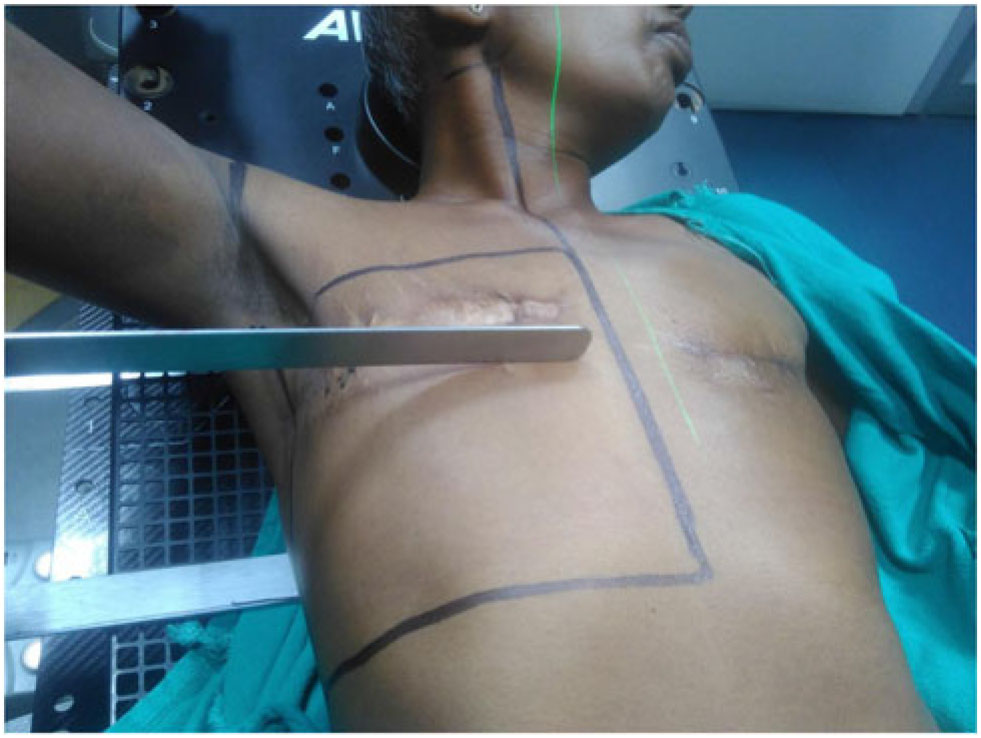
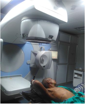
The patient was then treated with post-mastectomy radiation to chest wall and drainage area bilaterally with a dose of 40 Gy in 15 fractions. The chest wall was treated with tangential fields, and the drainage area was treated with antero-posterior field as shown in Figures 1 and 2. Humeral head was shielded bilaterally.
Field marking for post-mastectomy radiation.

Patient undergoing post mastectomy radiotherapy (PMRT) in ELEKTA linear accelerator.

The patient tolerated treatment well. The patient is on regular follow-up and is disease-free till date.
This synchronous presentation taught us the importance of examination and imaging of contralateral breast even during the treatment.
Case three
A 41-year-old female presented at our oncology OPD after undergoing right modified radical mastectomy for her right breast cancer. Histopathological report revealed invasive ductal carcinoma of right breast and was staged as pT2N1M0. Immunohistochemistry was negative for ER, PR and HER-2 Neu receptor.
The patient had a history of left breast cancer 3 years back for which she had undergone breast conserving surgery after receiving neoadjuvant chemotherapy. The primary left breast cancer was invasive ductal carcinoma with positive ER and PR. The primary breast cancer was staged as pT3N0M0, for which she underwent adjuvant chemotherapy and radiation in 2016. She received adjuvant radiation to the intact breast to a dose of 50 Gy in 25 fractions.
The patient had no positive familial history. The patient received hypofractionated radiotherapy to right chest wall and drainage area for her second breast cancer to a dose of 40 Gy in 15 fractions.
This case of metachronous bilateral breast cancer had advanced stage of presentation for both her tumours. Hormonal status was positive for primary breast cancer, while her second breast cancer had triple negative hormonal status. The patient was not on regular follow-up after the treatment of her primary breast cancer which resulted in delayed diagnosis of her second breast cancer.
Discussion
Bilateral breast cancer is a rare clinical presentation. Majority of bilateral breast cancers are metachronous. Synchronous breast cancers constitute about 0·2–3% incidence of all newly diagnosed breast cancer.Reference Chaudery, Mills and Hoskins8 As per our study, we identified two cases of metachronous and one case of synchronous bilateral breast cancer.
Primary synchronous bilateral breast cancers are thought to be independent tumour rather than secondary to metastatic spread from one of the primary lesions. The characteristics that imply independent tumours are the presence of an intraductal component, different histologies or different degrees of differentiation between the tumours.Reference De la Rochefordiere, Asselain and Scholl9 All three patients had different histologies for their bilateral breast cancers.
The risk factors associated with bilateral occurrence are familial or hereditary breast cancer, young age at primary breast cancer diagnosis, lobular invasive carcinoma, multicentricity and radiation exposure.Reference Dawson, Chow and Goss10, Reference Chuba, Hamre and Yap11 Although our patients had no familial history of breast cancer, but they had the risk factors of young age at primary breast cancer and lobular carcinoma histology.
Synchronous breast cancer has a poorer prognosis than metachronous or unilateral breast cancer.Reference Bailey, Royce, Sloane, Ford, Powles and Gazet12 For patients with metastatic breast cancer (MBC), tumour characteristics of previous cancer including stage, grade and hormone receptors are important to predict survival outcomes of the second breast cancer.Reference Baretta, Olopade and Huo13
The concordance in hormone receptor status of first and second breast cancers affecting the same patient could suggest that the two tumours may arise in a common milieu and that their subtypes are predetermined in the early stage of breast cancer development.Reference Whittemore, Gong and John14, Reference Kakarala and Wicha15 One of our MBC patients showed positive to negative change in hormonal status, while the other showed negative to positive change.
In our country, females are still very shy about talking about their health problems. Poor awareness on breast cancer care and the lack of national screening guidelines and programmes, and poor infrastructure, all contribute to late presentation and difficult breast cancer management. Lack of regular follow-up resulted in the advanced stage of presentation of second breast cancer in the metachronous cases reported in the study. Regular examination of opposite breast is very important and necessary in all cases.
Conclusion
Any female with unilateral breast cancer must be examined and fully evaluated for contralateral breast cancer and metastatic disease. Patients with a positive family history of breast cancer should have early screening for breast cancer. Proper clinical examination and imaging of opposite breast should be done to ensure adequate and timely management of bilateral breast cancer.
Acknowledgements
I would like to thank my patients and entire department of radiotherapy for extending their help to carry out this study.