Introduction
Dacryocystorhinostomy (DCR) surgery involves creating a ‘fistula’ in the bony nasal cavity to allow the tears to bypass a blocked or narrow nasolacrimal duct. Air ‘blow back’ is a known sequelae due to air reflux from the nose to eye, with the sensation of cold air or bubbles at the inner canthus whilst nose-blowing or performing a Valsalva manoeuvre. The passage of air beneath the subcutaneous tissue, termed surgical emphysema, is also possible.
Peri-orbital emphysema has been described in several procedures, including external DCR, functional endoscopic sinus surgery, dental surgery and even after an endoscopic bile duct papillotomy.Reference Ghosheh and Kathuria1–Reference Colemont, Pelckmans, Moorkens and Van Maercke5 Peri-orbital surgical emphysema is most commonly seen in the context of orbital trauma where air also enters the orbit, indicating a fracture of the orbital wall into a sinus.Reference Zimmer-Galler and Bartley6,Reference Abdesslem, Sondes, Wael, Hajer, Morched and Fathi7 In such cases, the protection of the optic nerve is paramount, and urgent treatment is required if clinical signs demonstrate the presence of optic neuropathy. If no signs are present then treatment can be expectant.
This study reports the occurrence of surgical emphysema after endoscopic endonasal DCR. To our knowledge, this has never been reported in association with endoscopic DCR.
Materials and methods
Institutional ethics review board approval was obtained to carry out a retrospective, non-comparative case series of patients who developed peri-orbital surgical emphysema following an endoscopic DCR procedure over a six-year period. The principles of the Helsinki declaration were followed. All procedures were performed in a single unit by two surgeons using identical techniques described elsewhere.Reference Malhotra, Norris, Sagili, Al-Abbadi and Avisar8
Briefly, a vertical tram-track, inferiorly based nasal mucosal flap is fashioned, with one line 3 mm posterior to the maxillary line and the second line 8 mm anterior to this. In revision DCR surgery, where a soft-tissue ostium or internal ostium is not visible (that is to say, the soft-tissue ostium has closed completely), a similar tram-track is made over the maxillary line; however, care is taken to dissect a plane between the location of the soft-tissue ostium and the lacrimal sac. Sharp submucosal dissection is carried out using a posteriorly directed number 15 blade, to create a plane between the lacrimal sac and the nasal mucosa. The nasal mucosal flap is then reflected backwards with relieving incisions above the axilla of the middle turbinate and below the level of the middle turbinate. Subsequently, the closed lacrimal sac is exposed.
In primary and revision surgery, bone is mechanically removed using a 40° Kerrison antrum bone punch (Storz, St Louis, Missouri, USA) and Malhotra endonasal DCR nibbler (Altomed, Boldon, UK) or with powered assistance. Thus a large osteotomy can be created that fully exposes the lacrimal sac, from its fundus down to the nasolacrimal duct junction and the agger nasi air cells posterior to the sac.Reference Patel, Ross and Malhotra9 This allows the lacrimal sac to be fully marsupialised and flaps opened flat, both anteriorly and posteriorly, using a standard crescent blade.
Results
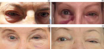
A total of 356 endoscopic DCR cases (primary surgery, n = 316; revision surgery, n = 40) were performed over a six-year period (author RM, n = 294; author ASL, n = 62). We identified seven cases of post-operative surgical emphysema. One case occurred after a primary endoscopic DCR with concomitant Lester Jones tube insertion (Table 1). Four surgical emphysema cases occurred following primary and two following revision endoscopic DCR. The surgical emphysema occurred within the first week post-operatively, was painless in nature and lacked any visual symptoms (Figure 1).
Details of patients experiencing surgical emphysema after endoscopic dacryocystorhinostomy

Pt. no. = patient number; DCR = dacryocystorhinostomy; post-op = post-operatively
Representative images of peri-orbital surgical emphysema in cases of endoscopic dacryocystorhinostomy (DCR). (a) Patient one with right-sided surgical emphysema at day 1 post-operatively after violent coughing. (b) Patient two with left-sided surgical emphysema at day 2 after uncontrolled sneezing. (c) Patient six with left-sided surgical emphysema (following bilateral endoscopic DCR) at day 1 after uncontrolled sneezing stifled with a closed mouth. (d) Patient seven with left-sided surgical emphysema (following bilateral endoscopic DCR) on the same day as the operation and without an obvious preceding event.

Examination of the globe and optic nerve revealed no compromise, with full range of movement, normal vision, normal intra-ocular pressure and normal optic nerve function. Resolution occurred within 48 hours of onset and no residual impairment of vision was detected.
Discussion
This is the first study to highlight the occurrence of peri-orbital surgical emphysema in patients undergoing endoscopic DCR. It occurred in primary and revision procedures, and with Lester Jones tube insertion. This complication had a comparable trigger in all but one of our cases; namely, sneezing, coughing or vigorous nose-blowing within one week of surgery, but none of the cases were sight-threatening and all patients had spontaneous resolution within 48 hours.
Although all of the surgical emphysema cases appeared predominantly limited to the eyelid, there is the potential for orbital compartment syndrome. Peri-orbital surgical emphysema related to trauma can create a ball–valve effect, whereby air can enter the orbit but not leave; this ophthalmic emergency must be excluded when faced with surgical emphysema. Clinical features of compartment syndrome include painful peri-orbital swelling, in contrast to our cases of painless surgical emphysema.Reference Lima, Burt, Leibovitch, Prabhakaran, Goldberg and Selva10 Another important differential diagnosis is gas-producing necrotising fasciitis; however, the history is not instantaneous like post-operative surgical emphysema. Instead, the patient tends to be unwell, with other signs of infection including a temperature.Reference Sultan, Malik, Li, Chevez-Barrios and Lee11 The relatively benign nature of eyelid surgical emphysema, coupled with the fact that patients are routinely seen at week three for tube removal, rather than week one when this tends to occur, may mean that the condition is underreported.
It is presumed that these cases have arisen because of new micro-passages created during endoscopic DCR that are vulnerable to the transmission of air to the skin during the early post-operative period. Advising patients not to carry out manoeuvres such as sneezing, coughing or vigorous nose-blowing within the first two weeks post-operatively may negate surgical emphysema whilst these connections close and heal. Warning patients of the symptoms and signs to look out for, and consenting them for the risk of its occurrence, may be warranted, along with advice on reducing triggering events.
• Surgical emphysema has been reported following external dacryocystorhinostomy (DCR)
• This is the first case series highlighting its occurrence in endonasal DCR patients
• Formation of a large and high osteotomy exposing the agger nasi may predispose to surgical emphysema following endoscopic DCR
• The triggers of uncontrolled sneezing, nose-blowing or coughing within the first week of surgery were common in all six cases identified
• Clinical examination revealed peri-orbital swelling and crepitus on the affected side, without optic neuropathy, in all cases
• Advising patients to sneeze with an open mouth within the first post-operative week may reduce the likelihood of surgical emphysema
Sneezing was the most common trigger in our case series, a reflex involving air speeds of 100 mph. Stifling of the sneeze reflex (which occurred in two of our patients) will force the air through the osteotomy and may be one mechanism of surgical emphysema. Opening of the mouth during the sneeze reflex may help to reduce the occurrence of surgical emphysema. In addition, sneezing can be mitigated by reducing allergic triggers or taking antihistamines, along with methods to reduce the risk of a repetitive sneezing attack by stimulating the trigeminal nerve. This includes pressure or pulling of the upper lip, an ear lobe pinch or pull, pulling or pinching the tip of the nose, or sticking the tongue out. Moreover, reducing sneezing, coughing or nose-blowing during the early post-operative period will help to reduce the risk of epistaxis; thus, two complications can be prevented with such advice.
In conclusion, surgical emphysema is another complication of endoscopic DCR. In this study, it occurred at a rate of 7 in 356 over six years, equating to a 2 per cent risk. The potential occurrence of surgical emphysema warrants discussion with the patient as a possible risk of surgery.
Competing interests
None declared