Ventricular septal defect is one of the most common congenital cardiac defects.Reference Hoffman and Kaplan1 Doubly committed subarterial ventricular septal defect, also referred to as supracristal, subpulmonary, or infundibular ventricular septal defect, is a unique type of ventricular septal defect with a high incidence in Asian populations, accounting for as many as one-quarter of defects requiring surgical closure.Reference Sim, Grignani and Wong2 Because of the lack of muscular support and the Venturi pulling effect on the leaflet, the aortic valve tends to prolapse into the defect, resulting in aortic insufficiency.Reference Chauvaud, Serraf and Mihaileanu3,Reference Bonhoeffer, Fabbrocini and Lecompte4 Whereas spontaneous closure of other types of ventricular septal defect is estimated to be between 25 and 50% in patients followed up from birth, spontaneous closure of doubly committed subarterial ventricular septal defect is extremely rare. Early closure is recommended for this type of ventricular septal defect.Reference Layangool, Kirawittaya and Sangtawesin5,Reference Komai, Naito and Fujiwara6 Therefore, most of the previous studies of surgical closure reported the outcomes of children. Little is known about the outcomes of surgical closure in adult patients with doubly committed subarterial ventricular septal defects.
Given that the development of aortic insufficiency in patients with doubly committed subarterial ventricular septal defects is significantly dependent on age,Reference Cho, Jang and Sun7 and the aorta and cardiac chambers grow only during childhood and stop expanding significantly after 15 years of age,Reference Nidorf, Picard and Triulzi8 the prevalence of the aortic insufficiency, the management, and hence the outcomes in adult patients with doubly committed subarterial ventricular septal defects may differ from those in children. Also, the outcome of adult patients with an unoperated ventricular septal defect is not uniformly favourable. Adult patients with unrepaired small ventricular septal defects carry a 11- to 15-fold increased risk of infective endocarditis compared to the general populationReference Lee, Uy, Foo and Tan9,Reference Berglund, Johansson and Dellborg10 and a non-negligible incidence of late complications such as double-chambered right ventricle and left ventricle dysfunction.Reference Karonis, Scognamiglio and Babu-Narayan11 Furthermore, most surgical series of doubly committed subarterial ventricular septal defects closure included a small number of adult patients, making contemporary outcome data on which to base recommendations very limited. We gathered the largest contemporary single centre experience with surgical doubly committed subarterial ventricular septal defects closure in adults.
Materials and methods
Study population
Approved by the Institutional Review Board at Union Hospital, Fujian Medical University, we reviewed the medical records of patients >18 years of age who underwent doubly committed subarterial ventricular septal defect closure between June 2010 and June 2017. Patients with the following conditions were excluded: complex cardiac lesions such as transposition of the great arteries or double-outlet right ventricle, incomplete medical records. On the basis of the surgical procedures performed, the patients were categorised into two groups for comparison. The valve intervention group consisted of 31 patients who underwent aortic valvuloplasty, aortic valve replacement, or repair of sinus Valsalva aneurysm in addition to closure of the defect. Non-valvular intervention group comprised 58 patients who underwent simple closure of the defect. Informed consent was obtained from each patient or the legal guardian before any of the procedures.
Telephone conversations were done by three physicians using a standard questionnaire containing questions on symptoms and signs, laboratory reports made elsewhere, and hospitalisations for such problems as endocarditis, cardiac arrhythmia, and reoperation. To collect the information regarding echocardiography performed elsewhere, we asked the patients or family to send the report to us by cell phone.
Echocardiographic assessment
Cardiac chamber dimensions, and left ventricular ejection fraction, were evaluated according to the recommendations of the American Society of Echocardiography. Valvular regurgitation, assessed by comprehensive echocardiographic evaluation, was graded as mild, moderate, and severe.Reference Lang, Badano and Mor-Avi12 In the absence of right ventricular outflow tract obstruction or pulmonic stenosis, the pre-operative pulmonary artery systolic pressure was calculated by subtracting the pressure gradient across ventricular septal defect from the systolic blood pressure. Post-operative pulmonary artery systolic pressure was derived from right ventricular pressure estimated from the tricuspid regurgitation velocity added to a qualitative assessment of right atrial pressure. Patients without tricuspid regurgitation and a normal right ventricular and interventricular septal configuration were assumed not to have pulmonary artery hypertension. Pulmonary artery hypertension was classified as mild (40–49 mmHg), moderate (50–mmHg), or severe (>60 mmHg). The largest diameter, measured on echocardiogram of the left ventricular long axis or the right ventricular outflow view, was taken as the ventricular septal defect diameter.Reference Tomita, Arakaki, Ono, Yamada, Yagihara and Echigo13
Operative technique
Surgical repair for ventricular septal defect was performed through a median sternotomy or right anterolateral minithoracotomy. Cardiopulmonary bypass with moderate hypothermia and aorta cross-clamping was used during the procedure. Cold blood cardioplegia was performed for myocardial preservation. The transpulmonary approach was the preferred approach for ventricular septal defect closure. The ventricular septal defect was closed with a pericardial patch.Reference Chen, Ma and Pan14,Reference Zhao Yang, Hua and Yuan Ji15 An aortotomy was made when aortic valve exploration was required. Aortic valvuloplasty or aortic valve replacement was performed when more than moderate aortic insufficiency was confirmed or at the discretion of operator. Resuspension of prolapsing leaflets and commissuroplasty, techniques of aortic valvuloplasty, were utilised. Excessive valve tissue was plicated and sutured to the commissures close to the aortic annulus. Commissuroplasty involved repair of commissures with mattress sutures. Aortic valve replacement was performed when poor valvular tissue quality was noted. After the disease valve was excised and the annulus carefully decalcified, the annulus was sized. A prosthesis with appropriate size was used. Simple interrupted suture or non-everting mattress suture was used to sew the prosthesis sewing cuff to the annulus. Twelve to 19 sutures were performed. Sinus Valsalva aneurysm was repaired by direct suture or patch closure. Follow-up assessments were carried out at the approximate time points of the 3rd and 12th months in the first year and once yearly thereafter.
Statistical analysis
Data were expressed as mean ± standard deviation for continuous variables and frequencies and percentages for categorical variables. Statistical analysis consisted of the unpaired Student's t-test for continuous variables and the chi-square test or Fisher's test for categorical variables. A p-value <0.05 was considered statistically significant.
Results
Baseline clinical characteristics
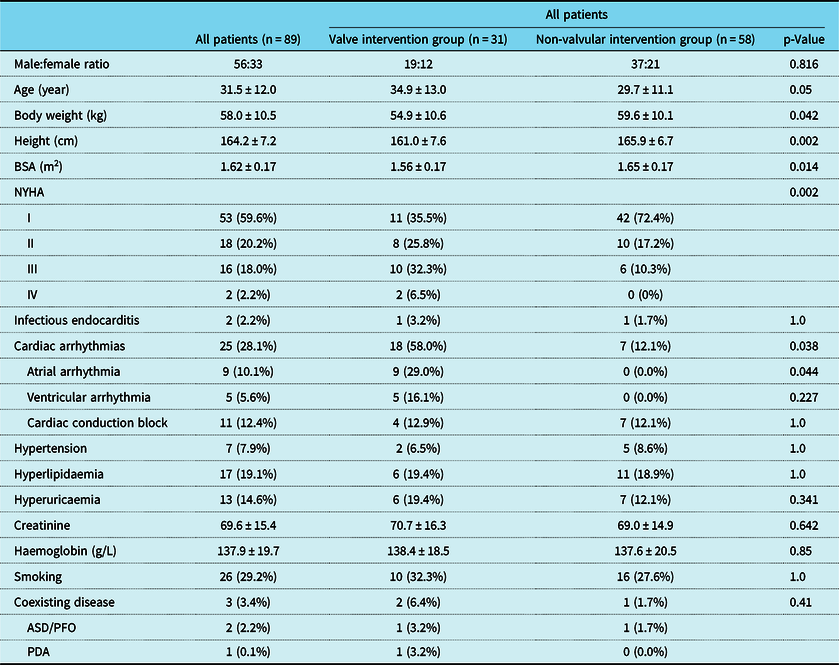
The median age at ventricular septal defect closure was 28 years (range 18–67 years). Among the 31 patients with valve intervention, 16 had aortic valve replacement, 2 had aortic valvuloplasty, and 16 had repair of sinus Valsalva aneurysm. The groups did not differ by sex and age. Patients with valve intervention were shorter and tended to be lighter, and had a lower body surface area than those without valve intervention. More patients in the valve intervention group were with NHYA class II/III/IV and suffered from cardiac arrhythmias. Baseline characteristics are shown in Table 1.
Baseline clinical characteristics

ASD = atrial septal defect; BSA = body surface area; PDA = patent ductus arteriosus; PFO = patent foramen ovale
Pre-operatively echocardiographic characteristics
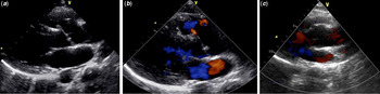
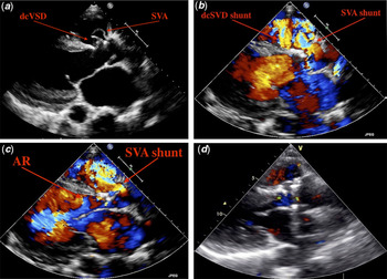
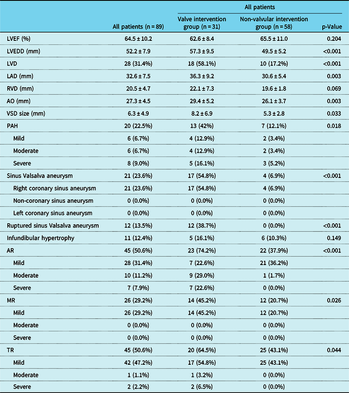
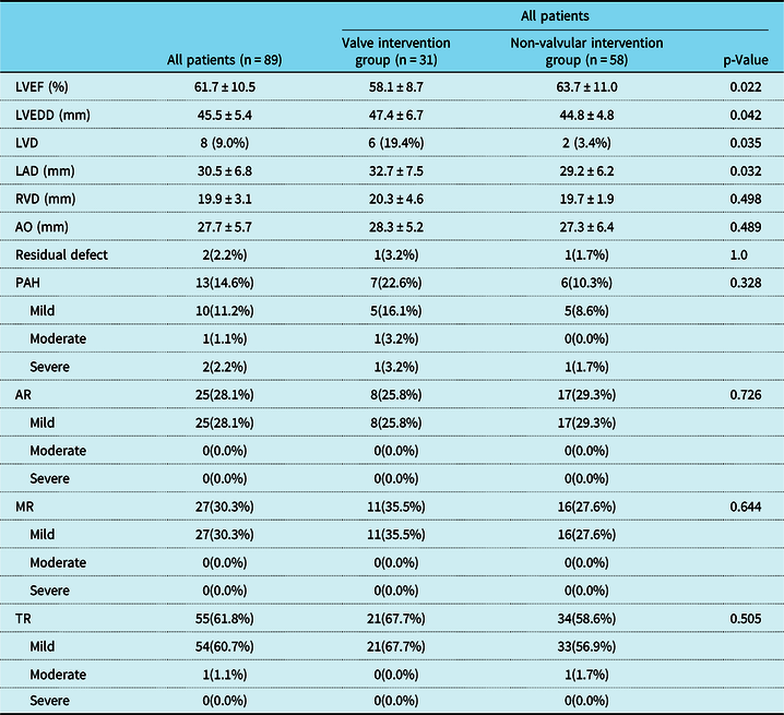
For the entire cohort, aortic regurgitation was noted in 45 (50.6%) patients, sinus Valsalva aneurysm in 21 (23.6%) patients, and ruptured sinus Valsalva aneurysm in 12 (13.5%) patients (Figs 1 and 2). Among 21 patients with sinus Valsalva aneurysm, 14 had aortic regurgitation (7 in mild, 6 in moderate, and 1 in severe). Patients with valve intervention had a larger ventricular septal defect size, and thus lager cardiac chamber sizes and aorta diameter. Pre-operatively, echocardiographic characteristics are shown in Table 2.
(a) preoperative echocardiography showing SVA. (b) color doppler showing no flow in SVA. (c) postoperative echocardiography after repair of unruptured SVA.

(a) preoperative echocardiography showing dcVSD and SVA. (b) color doppler showing shunt flow of dcVSD and ruptured SVA. (c) color doppler showing SVA shunt and AR. (d)postoperative echocardiography after repair of ruptured SVA and dcVSD, and aortic valve replacement.

Pre-operatively echocardiographic characteristics

AO = aorta; AR = aortic regurgitation; LAD = left atrial diameter; LVD = left ventricular dilation; LVEDD = left ventricular end-diastolic diameter; LVEF = left ventricular ejection fraction; MR = mitral regurgitation; PAH = pulmonary artery hypertension; RVD = right ventricular diameter; TR = tricuspid regurgitation; VSD = ventricular septal defect
Complications
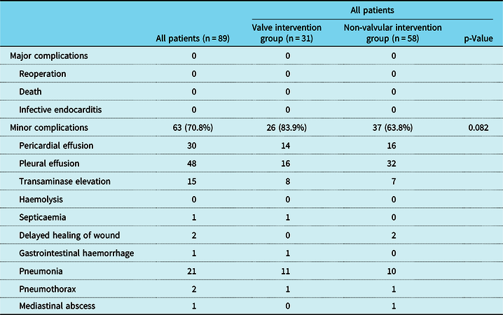
No major complications were noted in the two groups. The valve intervention group had more patients with pneumonia (p = 0.044). There was no significant difference in the rate of minor complications between the two groups (Table 3). In non-valvular intervention group, one patient had a mediastinal abscess requiring debridement.
Complications

Post-operative echocardiography
Post-operatively, patients with valve intervention had a significantly lower left ventricular ejection fraction. There was no significant difference in cardiac chamber sizes except left ventricular end-diastolic and left atrial diameter. The incidence of aortic regurgitation, mitral regurgitation, and tricuspid regurgitation were similar between the groups. One patient in valve intervention group and one patient in non-valvular intervention group had residual shunt, respectively. Post-operatively, echocardiographic characteristics are shown in Table 4.
Post-operatively echocardiographic characteristics

AO = aorta; AR = aortic regurgitation; LAD = left atrial diameter; LVD = left ventricular dilation; LVEDD = left ventricular end-diastolic diameter; LVEF = left ventricular ejection fraction; MR = mitral regurgitation; PAH = pulmonary artery hypertension; RVD = right ventricular diameter; TR = tricuspid regurgitation; VSD = ventricular septal defect
Follow-up
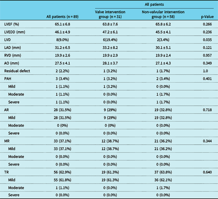
The follow-up echocardiographic data were listed in Table 5. The median follow-up period was 36 months, ranging from 9 to 82 months. No death and infective endocarditis were noted during the study period. No reoperation was performed during the follow-up period. Atrial fibrillation developed in two patients. In patients without pre-operative aortic regurgitation, 12 (27.3%) had new mild aortic regurgitation at last follow-up.
Echocardiographic characteristics at follow-up

AO = aorta; AR = aortic regurgitation; LAD = left atrial diameter; LVD = left ventricular dilation; LVEDD = left ventricular end-diastolic diameter; LVEF = left ventricular ejection fraction; MR = mitral regurgitation; PAH = pulmonary artery hypertension; RVD = right ventricular diameter; TR = tricuspid regurgitation; VSD = ventricular septal defect
Discussion
For the outcomes of doubly committed subarterial ventricular septal defect closure in adult patients, as indicated by our literature review, no special study has been conducted yet. We report the largest contemporary series of adults who underwent doubly committed subarterial ventricular septal defect closure at a single institution. The main findings of this study were these: first, 34.8% of adult patients with doubly committed subarterial ventricular septal defect required concurrent intervention on aortic valve or sinus Valsalva aneurysm; second, closure of doubly committed subarterial ventricular septal defect in adult patients was safe without serious complications; third, 12 (27.3%) patients developed new mild aortic regurgitation; fourth, no infective endocarditis, reoperation, and other late complication were recorded; and fifth, at last follow-up, 91 and 96.6% of the studied patients were free from left ventricle dilation and pulmonary hypertension.
The doubly committed subarterial ventricular septal defect is a unique type of ventricular septal defect because of the particular aspects of the clinical presentation. First, the aortic sinus may prolapse into the doubly committed subarterial ventricular septal defect resulting in aortic regurgitation. Second, sinus Valsalva aneurysm may develop in association with doubly committed subarterial ventricular septal defect. In a Japanese study including both children and adult patients with doubly committed subarterial ventricular septal defect, the incidence of aortic regurgitation, sinus Valsalva aneurysm, and ruptured sinus Valsalva aneurysm was 24, 9.1 and 8.1%, respectively.Reference Momma, Toyama and Takao16 In a Taiwanese study including 209 doubly committed subarterial ventricular septal defect patients with an operation age of 11.2 ± 10 years, the rate of aortic regurgitation, ruptured sinus Valsalva aneurysm, and aortic valvuloplasty or aortic valve replacement was 15.8, 8.1, and 14.8%, respectively.Reference Chiu, Wang and Lin17 Because the development of aortic insufficiency and sinus Valsalva aneurysm is significantly associated with patient age,Reference Cho, Jang and Sun7 the incidence of aortic insufficiency and sinus Valsalva aneurysm in adult patients will be much higher than that of younger patients. In present study, aortic regurgitation was noted in 45 (50.6%) patients, sinus Valsalva aneurysm in 21 (23.6%) patients, ruptured sinus Valsalva aneurysm in 12 (13.5%) patients, and aortic valvuloplasty or aortic valve replacement in 18 (20.2%) patients.
Heart failure, a large shunt, poor growth, or pulmonary artery hypertension would all be indications for ventricular septal defect closure. A lower threshold for closure of doubly committed subarterial ventricular septal defects can be justified because of the increased risk for development of aortic insufficiency, and the fact that spontaneous closure is unlikely. Therefore, surgical intervention for patients with doubly committed subarterial ventricular septal defect is recommended immediately after the detection of trivial aortic regurgitation or as soon after the diagnosis of doubly committed subarterial ventricular septal defect as possible, regardless of shunt volume.Reference Devlin, Russell and Mongé18,Reference Backer, Idriss and Zales19 Furthermore, the outcome of adult patients with an unoperated small ventricular septal defect is not uniformly favourable. Karonis et alReference Karonis, Scognamiglio and Babu-Narayan11 reported the long-term outcomes of 231 patients with unoperated small ventricular septal defect. They found that double-chambered right ventricle occurred in 29 (13%) patients, more than mild aortic regurgitation in 6 (3%) patients, infective endocarditis in 24 (10%) patients, reduced left ventricular ejection fraction in 10 (6%) patients, and left ventricle dilation in 34 (21%) patients.Reference Karonis, Scognamiglio and Babu-Narayan11 In the study by Cho et al, among the 60 patients with small doubly committed subarterial ventricular septal defect followed up conservatively, 5 had sinus Valsalva aneurysm rupture requiring urgent surgical treatment, 3 had infective endocarditis, and 2 developed heart failure symptoms.Reference Cho, Jang and Sun7 In our study, although a considerable number of patients had cardiac chamber dilation, pulmonary hypertension, and concurrent operation on aortic valve, no serious procedure-related complication was observed, and no reoperation and infective endocarditis during the follow-up.
Some studies reported that closure of the defect in itself has been shown to be sufficient to arrest progression of aortic insufficiency in patients with mild insufficiency.Reference Cheung, Chiu, Yung and Chau20,Reference Moreno-Cabrol, Mamiya, Nakamura, Brainard and McNamara21 However, other investigators have not found that surgery performed before the onset of aortic regurgitation is beneficial.Reference Karpawich, Duff, Mullins, Cooley and McNamara22 Recently, Amano et al reported that among patients with doubly committed subarterial ventricular septal defect, aortic regurgitation progression occurred in seven (7.7%) patients at a mean of 13.7 ± 7.2 years after ventricular septal defect closure, and re-do aortic valve surgery was performed in three (3.3%) patients. They found post-operative leakage may be a significant risk factor for late aortic regurgitation progression.Reference Amano, Izumi and Imamura23 In our study, among those patients with newly developed aortic regurgitation, only two had residual shunt which disappeared during the follow-up.
Our results are limited by the retrospective survey design of the study. A second limitation is that the study was limited to one institution, and data for other institutions may show different results. Third, although the midterm results of present study were satisfactory, we did find a number of new mild aortic regurgitation; so longer follow-up times are needed in future research.
In conclusion, closure of doubly committed subarterial ventricular septal defect in adult patients was safe, and the midterm results were favourable. However, the incidence of new mild aortic regurgitation after ventricular septal defect closure alone was high (27.3%). Long-term follow-up of aortic regurgitation progression is needed.
Acknowledgement
The authors thank all the staff of Department of Cardiology and Cardiovascular Surgery at Union Hospital for their kind assistance with this study.
Financial Support
This research received no specific grant from any funding agency, commercial, or not-for-profit sectors.
Conflicts of Interest
None.
Ethical Standards
This study was approved by the Ethics Committee of the Union Hospital Affiliated to Fujian Medical University and complied with the 1964 Helsinki Declaration and its later amendments.